Sultan Muhammad H, Moni Sivakumar S, Alqahtani Saad S, Ali Bakkari Mohammed, Alshammari Abdulrahman, Almoshari Yosif, Alshahrani Saeed, Madkhali Osama A, Mohan Syam
Department of Pharmaceutics, College of Pharmacy, Jazan University, Jazan, Saudi Arabia.
Department of Clinical Pharmacy, College of Pharmacy, Jazan University, Jazan, Saudi Arabia.
Saudi Pharm J. 2023 Jun;31(6):861-873. doi: 10.1016/j.jsps.2023.04.005. Epub 2023 Apr 13.
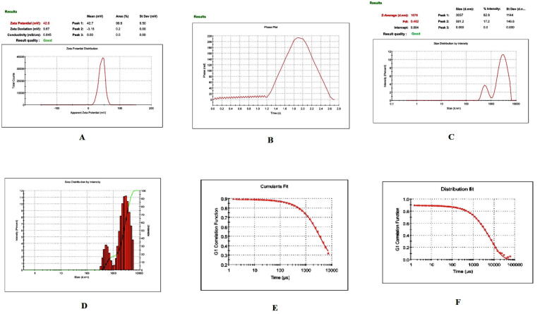
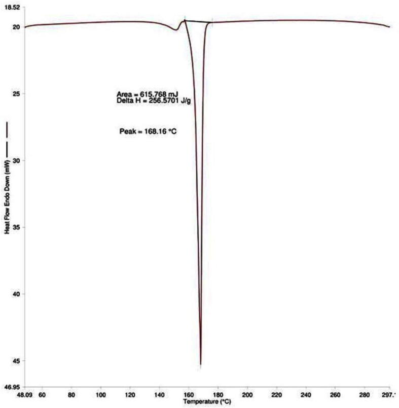
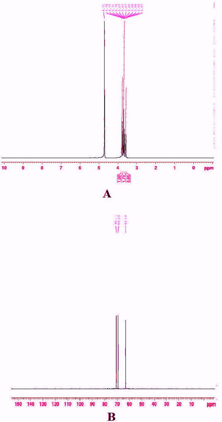
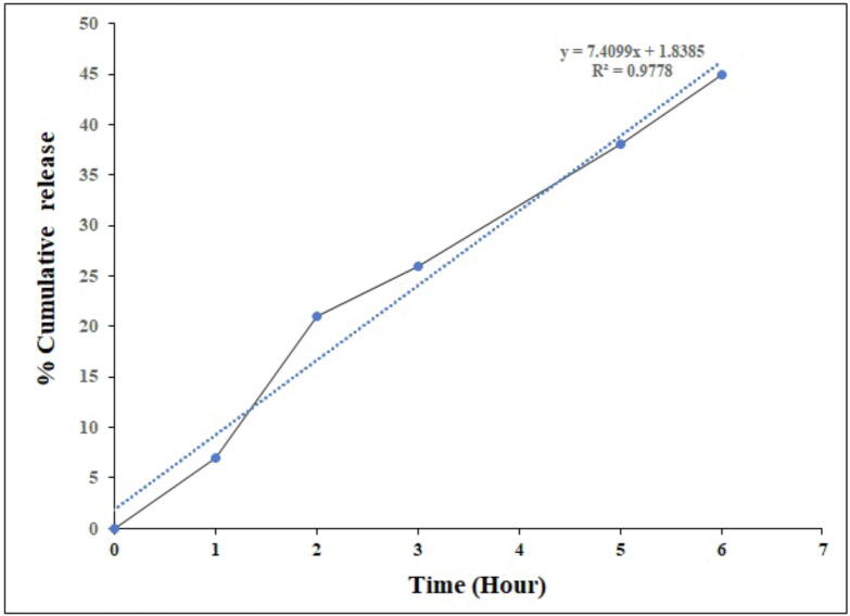
The study aimed to develop cisplatin-loaded PEGylated chitosan nanoparticles. The optimal batch of cisplatin-loaded PEGylated chitosan nanoparticles had a + 49.9 mV zeta potential, PDI of 0.347, and % PDI of 58.9. Nanoparticle zeta size was 741.4 z. d.nm, the size in diameter was 866.7 ± 470.5 nm, and nanoparticle conductivity in colloidal solution was 0.739 mS/cm. Differential scanning calorimetry (DSC) revealed that cisplatin-loaded PEGylated chitosan nanoparticles had sharp endothermic peaks at temperatures at 168.6 °C. The thermogravimetric analysis (TGA) showed the weight loss of cisplatin-loaded PEGylated chitosan nanoparticles, which was observed as 95% at 262.76 °C. XRD investigation on cisplatin-loaded PEGylated chitosan nanoparticles exhibited distinct peaks at 2θ as 9.7°, 20.4°, 22.1°, 25.3°, 36.1°, 38.1°, 39.5°, 44.3°, and 64.5°, confirming crystalline structure. The H NMR analysis showed the fingerprint region of cisplatin-loaded PEGylated chitosan nanoparticles as 0.85, 1.73, and 1.00 ppm in the proton dimension and de-shielded proton peaks appeared at 3.57, 3.58, 3.58, 3.59, 3.65, 3.67, 3,67, 3,67, 3.70, 3.71, 3.77, 3.78 and 4.71 ppm. The C NMR spectrum showed specified peaks at 63.18, 69.20, and 70.77 ppm. The FT-IR spectra of cisplatin loaded PEGylated nanoparticles show the existence of many fingerprint regions at 3186.52, 2931.68, 1453.19, 1333.98, 1253.71, 1085.19, 1019.60, 969.98, 929.53, 888.80, 706.13, and 623.67 cm. The drug release kinetics of cisplatin loaded PEGylated chitosan nanoparticles showed zero order kinetics with 48% of drug release linearity fashion which has R value of 0.9778. Studies on the MCF-7 ATCC human breast cancer cell line revealed that the IC50 value 82.08 µg /mL. Injectable nanoparticles had good physicochemical and cytotoxic properties. This method is novel since the application of the PEGylation processes leads to an increased solubility of chitosan nanoparticles at near neutral pH.
该研究旨在开发负载顺铂的聚乙二醇化壳聚糖纳米颗粒。负载顺铂的聚乙二醇化壳聚糖纳米颗粒的最佳批次具有+49.9 mV的zeta电位、0.347的PDI和58.9%的PDI。纳米颗粒的zeta尺寸为741.4 z.d.nm,直径尺寸为866.7±470.5 nm,胶体溶液中的纳米颗粒电导率为0.739 mS/cm。差示扫描量热法(DSC)显示,负载顺铂的聚乙二醇化壳聚糖纳米颗粒在168.6℃的温度下有尖锐的吸热峰。热重分析(TGA)显示了负载顺铂的聚乙二醇化壳聚糖纳米颗粒的重量损失,在262.76℃时观察到为95%。对负载顺铂的聚乙二醇化壳聚糖纳米颗粒的XRD研究在2θ为9.7°、20.4°、22.1°、25.3°、36.1°、38.1°、39.5°、44.3°和64.5°处显示出明显的峰,证实了晶体结构。1H NMR分析显示,负载顺铂的聚乙二醇化壳聚糖纳米颗粒在质子维度上的指纹区域为0.85、1.73和1.00 ppm,去屏蔽质子峰出现在3.57、3.58、3.58、3.59、3.65、3.67、3.67、3.67、3.70、3.71、3.77、3.78和4.71 ppm处。13C NMR光谱在63.18、69.20和70.77 ppm处显示出特定的峰。负载顺铂的聚乙二醇化纳米颗粒的FT-IR光谱在3186.52、2931.68、1453.19、1333.98、1253.71、1085.19、1019.60、969.98、929.53、888.80、706.13和623.67 cm处显示出许多指纹区域。负载顺铂的聚乙二醇化壳聚糖纳米颗粒的药物释放动力学显示为零级动力学,药物释放线性方式为48%,R值为0.9778。对MCF-7 ATCC人乳腺癌细胞系的研究表明,IC50值为82.08 μg/mL。可注射纳米颗粒具有良好的物理化学和细胞毒性特性。该方法是新颖的,因为聚乙二醇化过程的应用导致壳聚糖纳米颗粒在近中性pH下的溶解度增加。