Department of Pathology and Laboratory Medicine, University of Pennsylvania, Philadelphia, PA, 19104, USA; Stomatological Hospital, School of Stomatology, Southern Medical University, Guangzhou, 510280, China.
Department of Pathology and Laboratory Medicine, University of Pennsylvania, Philadelphia, PA, 19104, USA.
EBioMedicine. 2023 Jun;92:104614. doi: 10.1016/j.ebiom.2023.104614. Epub 2023 May 23.
Only a minority of melanoma patients experience durable responses to immunotherapies due to inter- and intra-tumoral heterogeneity in melanoma. As a result, there is a pressing need for suitable preclinical models to investigate resistance mechanisms and enhance treatment efficacy.
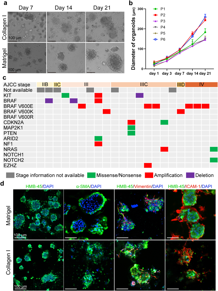
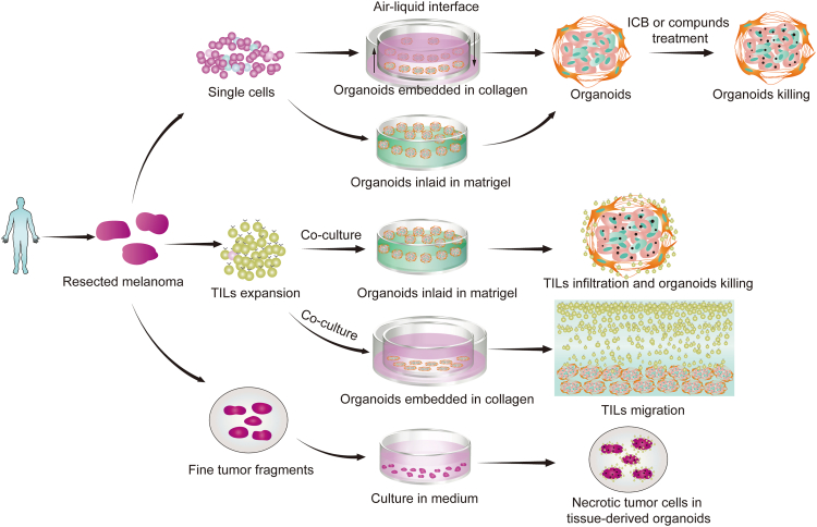
Here, we report two different methods for generating melanoma patient-derived organoids (MPDOs), one is embedded in collagen gel, and the other is inlaid in Matrigel. MPDOs in Matrigel are used for assessing the therapeutic effects of anti-PD-1 antibodies (αPD-1), autochthonous tumor infiltrating lymphocytes (TILs), and small molecule compounds. MPDOs in collagen gel are used for evaluating the chemotaxis and migratory capacity of TILs.
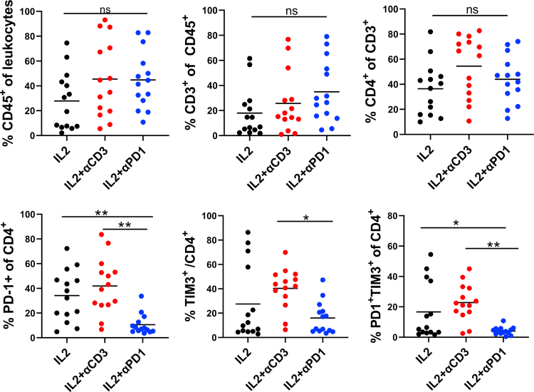
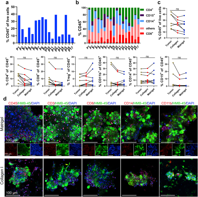
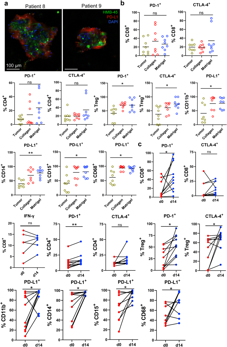
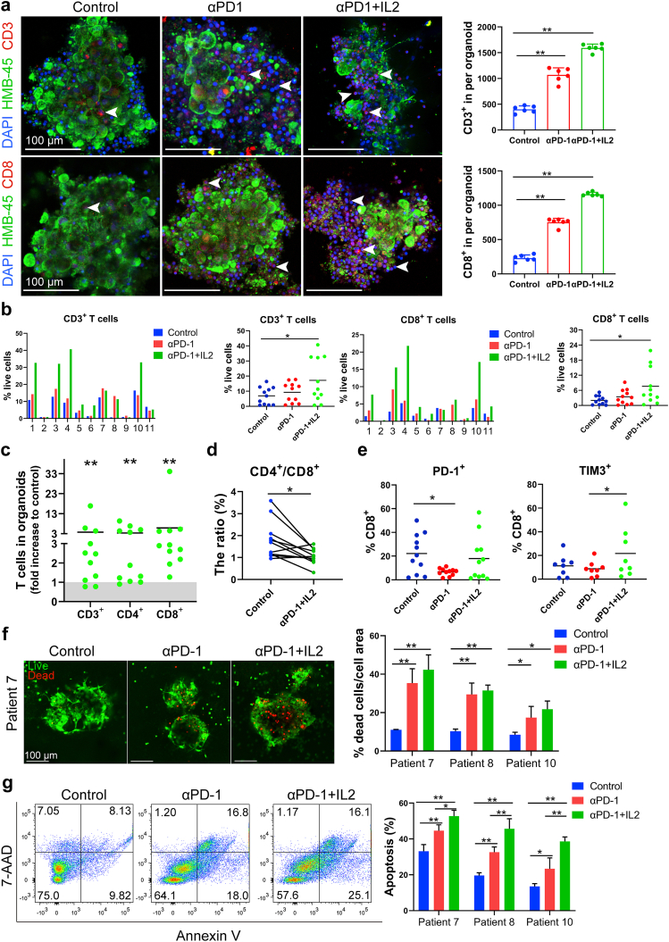
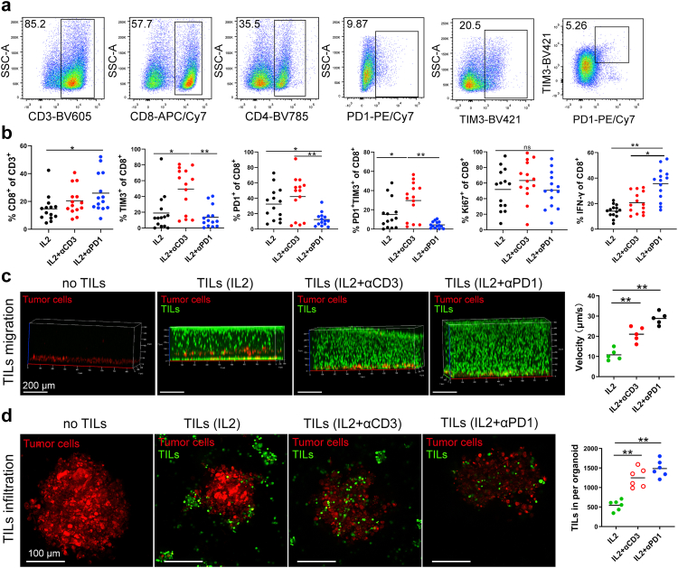
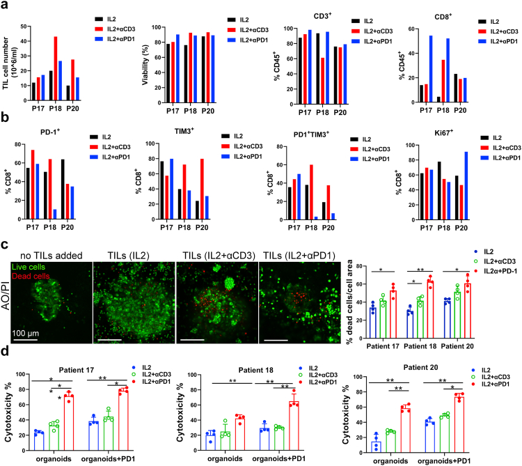
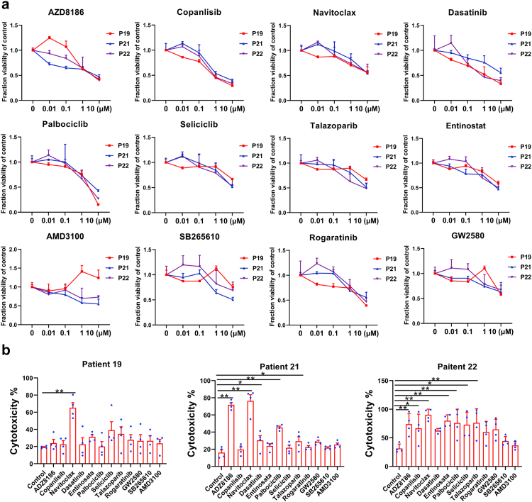
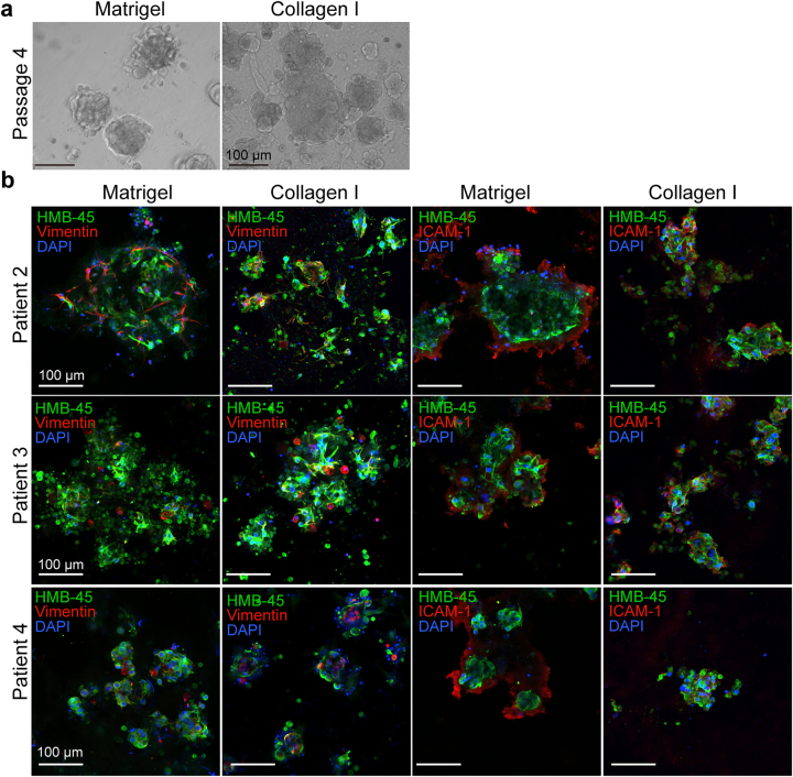
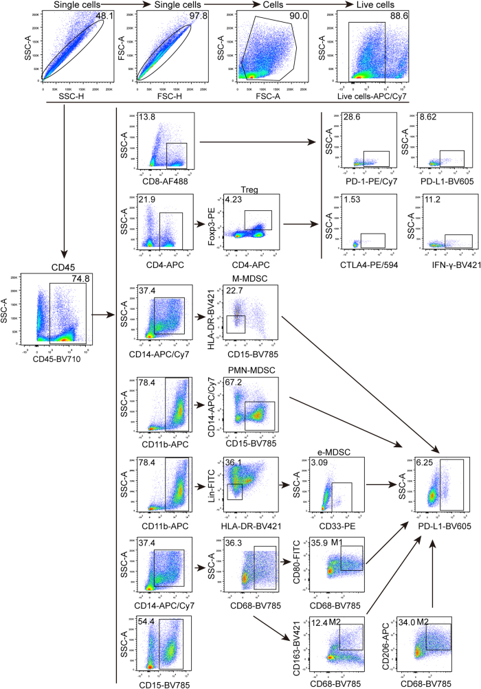
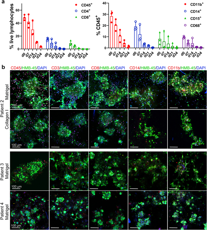
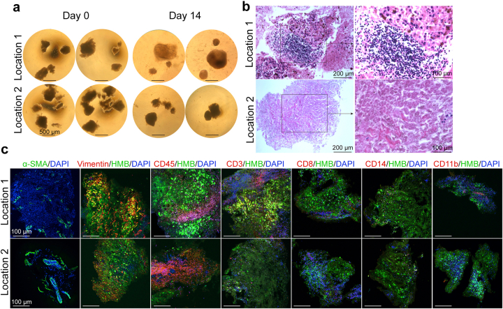
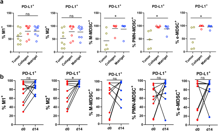
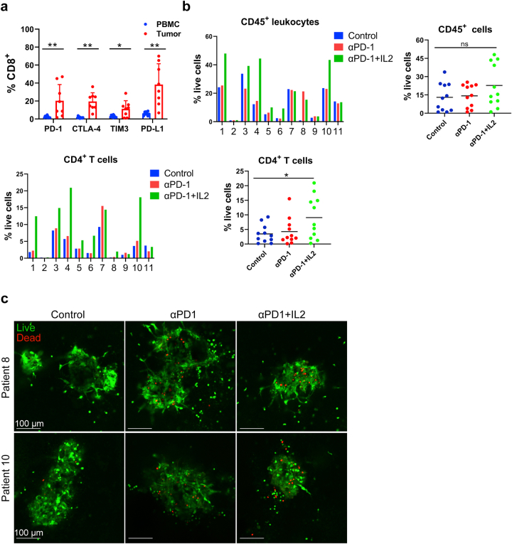
The MPDOs in collagen gel and Matrigel have similar morphology and immune cell composition to their parental melanoma tissues. MPDOs show inter- and intra-tumoral heterogeneity and contain diverse immune cells such as CD4, CD8 T, Treg, CD14 monocytic, CD15, and CD11b myeloid cells. The tumor microenvironment (TME) in MPDOs is highly immunosuppressive, and the lymphoid and myeloid lineages express similar levels of PD-1, PD-L1, and CTLA-4 as their parental melanoma tissues. Anti-PD-1 antibodies (αPD-1) reinvigorate CD8 T cells and induce melanoma cell death in the MPDOs. TILs expanded by IL-2 and αPD-1 show significantly lower expression of TIM-3, better migratory capacity and infiltration of autochthonous MPDOs, and more effective killing of melanoma cells than TILs expanded by IL-2 alone or IL-2 with αCD3. A small molecule screen discovers that Navitoclax increases the cytotoxicity of TIL therapy.
MPDOs may be used to test immune checkpoint inhibitors and cellular and targeted therapies.
This work was supported by the NIH grants CA114046, CA261608, CA258113, and the Tara Miller Melanoma Foundation.
由于黑色素瘤的肿瘤内和肿瘤间异质性,只有少数黑色素瘤患者对免疫疗法有持久的反应。因此,迫切需要合适的临床前模型来研究耐药机制并提高治疗效果。
在这里,我们报告了两种生成黑色素瘤患者来源的类器官(MPDO)的不同方法,一种是嵌入胶原凝胶中的,另一种是镶嵌在 Matrigel 中的。Matrigel 中的 MPDO 用于评估抗 PD-1 抗体(αPD-1)、自体肿瘤浸润淋巴细胞(TIL)和小分子化合物的治疗效果。胶原凝胶中的 MPDO 用于评估 TIL 的趋化性和迁移能力。
胶原凝胶和 Matrigel 中的 MPDO 具有与其亲本黑色素瘤组织相似的形态和免疫细胞组成。MPDO 显示出肿瘤内和肿瘤间的异质性,并包含多种免疫细胞,如 CD4、CD8 T、Treg、CD14 单核细胞、CD15 和 CD11b 髓样细胞。MPDO 中的肿瘤微环境(TME)高度免疫抑制,淋巴和髓样谱系表达与其亲本黑色素瘤组织相似水平的 PD-1、PD-L1 和 CTLA-4。抗 PD-1 抗体(αPD-1)使 CD8 T 细胞重新活跃,并在 MPDO 中诱导黑色素瘤细胞死亡。用 IL-2 和 αPD-1 扩增的 TIL 显示 TIM-3 的表达明显降低,更好的迁移能力和对自体 MPDO 的浸润,以及比单独用 IL-2 或 IL-2 加 αCD3 扩增的 TIL 更有效地杀死黑色素瘤细胞。小分子筛选发现 Navitoclax 增加了 TIL 治疗的细胞毒性。
MPDO 可用于测试免疫检查点抑制剂和细胞及靶向治疗。
这项工作得到了 NIH 资助 CA114046、CA261608、CA258113 和 Tara Miller 黑色素瘤基金会的支持。