Nagaraja Sridevi, Tewari Shivendra G, Reifman Jaques
Department of Defense Biotechnology High Performance Computing Software Applications Institute, Telemedicine and Advanced Technology Research Center, U.S. Army Medical Research and Development Command, Fort Detrick, MD, United States.
The Henry M. Jackson Foundation for the Advancement of Military Medicine, Inc., Bethesda, MD, United States.
Front Neurosci. 2023 May 12;17:1147437. doi: 10.3389/fnins.2023.1147437. eCollection 2023.
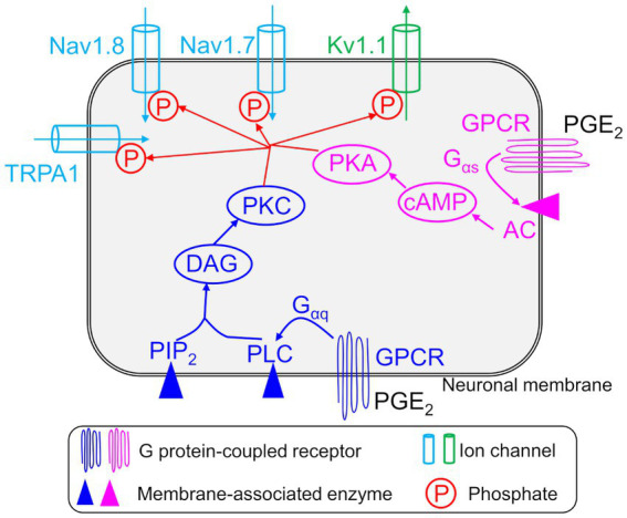
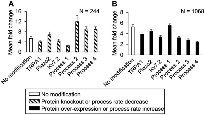
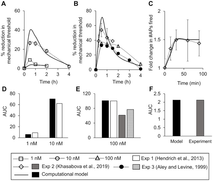
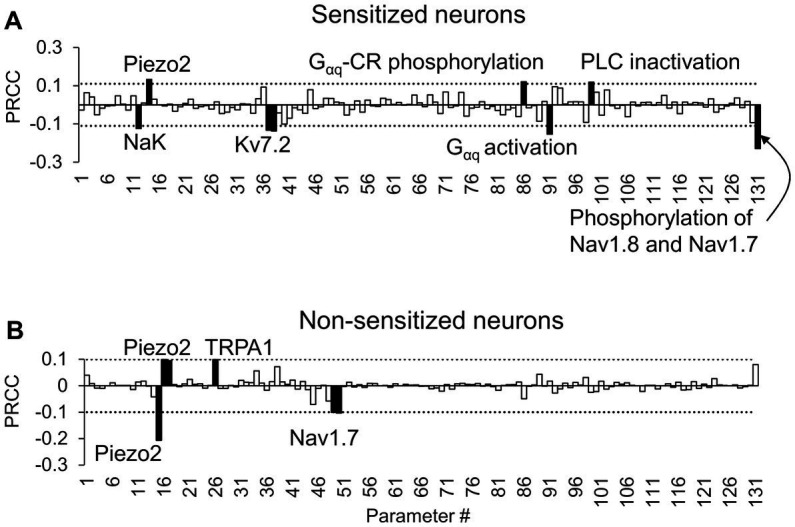
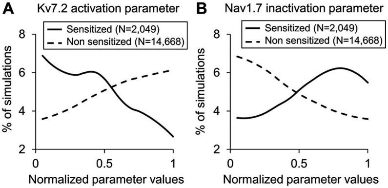
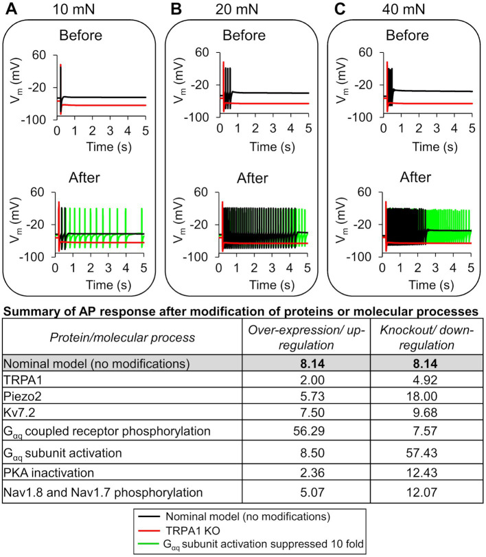
Sensory neurons embedded in muscle tissue that initiate pain sensations, i.e., nociceptors, are temporarily sensitized by inflammatory mediators during musculoskeletal trauma. These neurons transduce peripheral noxious stimuli into an electrical signal [i.e., an action potential (AP)] and, when sensitized, demonstrate lower activation thresholds and a heightened AP response. We still do not understand the relative contributions of the various transmembrane proteins and intracellular signaling processes that drive the inflammation-induced hyperexcitability of nociceptors. In this study, we used computational analysis to identify key proteins that could regulate the inflammation-induced increase in the magnitude of AP firing in mechanosensitive muscle nociceptors. First, we extended a previously validated model of a mechanosensitive mouse muscle nociceptor to incorporate two inflammation-activated G protein-coupled receptor (GPCR) signaling pathways and validated the model simulations of inflammation-induced nociceptor sensitization using literature data. Then, by performing global sensitivity analyses that simulated thousands of inflammation-induced nociceptor sensitization scenarios, we identified three ion channels and four molecular processes (from the 17 modeled transmembrane proteins and 28 intracellular signaling components) as potential regulators of the inflammation-induced increase in AP firing in response to mechanical forces. Moreover, we found that simulating single knockouts of transient receptor potential ankyrin 1 (TRPA1) and reducing the rates of G-coupled receptor phosphorylation and G subunit activation considerably altered the excitability of nociceptors (i.e., each modification increased or decreased the inflammation-induced fold change in the number of triggered APs compared to when all channels were present). These results suggest that altering the expression of TRPA1 or the concentration of intracellular G might regulate the inflammation-induced increase in AP response of mechanosensitive muscle nociceptors.
嵌入肌肉组织中引发疼痛感觉的感觉神经元,即伤害感受器,在肌肉骨骼创伤期间会被炎症介质暂时致敏。这些神经元将外周有害刺激转化为电信号[即动作电位(AP)],并且在致敏时表现出较低的激活阈值和增强的AP反应。我们仍然不了解驱动炎症诱导的伤害感受器过度兴奋的各种跨膜蛋白和细胞内信号传导过程的相对贡献。在本研究中,我们使用计算分析来识别可调节炎症诱导的机械敏感肌肉伤害感受器中AP发放幅度增加的关键蛋白。首先,我们扩展了一个先前经过验证的机械敏感小鼠肌肉伤害感受器模型,纳入两条炎症激活的G蛋白偶联受体(GPCR)信号通路,并使用文献数据验证了炎症诱导的伤害感受器致敏的模型模拟。然后,通过进行模拟数千种炎症诱导的伤害感受器致敏情况的全局敏感性分析,我们确定了三个离子通道和四个分子过程(来自17个建模的跨膜蛋白和28个细胞内信号成分)作为炎症诱导的响应机械力时AP发放增加的潜在调节因子。此外,我们发现模拟瞬时受体电位锚蛋白1(TRPA1)的单基因敲除以及降低G偶联受体磷酸化和G亚基激活的速率会显著改变伤害感受器的兴奋性(即与所有通道都存在时相比,每种修饰都会增加或减少炎症诱导的触发AP数量的倍数变化)。这些结果表明,改变TRPA1的表达或细胞内G的浓度可能调节炎症诱导的机械敏感肌肉伤害感受器的AP反应增加。