Institute of Anatomy and Cell Biology, National Yang Ming Chiao Tung University, Taipei 112304, Taiwan.
Institute of Neuroscience, National Yang Ming Chiao Tung University, Taipei 112304, Taiwan.
eNeuro. 2023 Jun 12;10(6). doi: 10.1523/ENEURO.0366-22.2023. Print 2023 Jun.
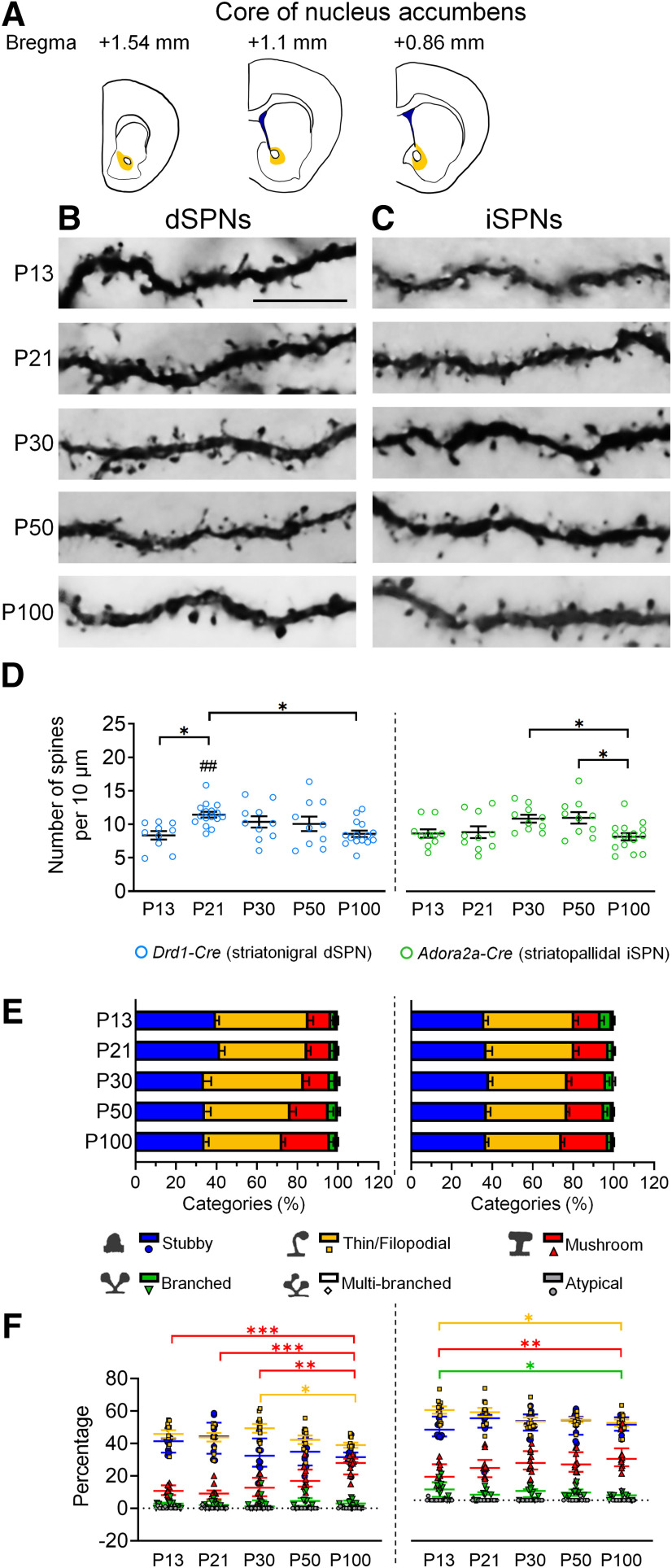
Synaptic modification in postnatal development is essential for the maturation of neural networks. Developmental maturation of excitatory synapses occurs at the loci of dendritic spines that are dynamically regulated by growth and pruning. Striatal spiny projection neurons (SPNs) receive excitatory input from the cerebral cortex and thalamus. SPNs of the striatonigral direct pathway (dSPNs) and SPNs of the striatopallidal indirect pathway (iSPNs) have different developmental roots and functions. The spatial and temporal dynamics of dendritic spine maturation of these two types of SPNs remain elusive. Here, we delineate the developmental trajectories of dendritic spines of dSPNs and iSPNs in the caudoputamen and nucleus accumbens (NAc). We labeled dendritic spines of SPNs by microinjecting Cre-dependent AAV-eYFP viruses into newborn Drd1-Cre or Adora2a-Cre mice, and analyzed spinogenesis at three levels, including different SPN cell types, subregions and postnatal times. In the dorsolateral striatum, spine pruning of dSPNs and iSPNs occurred at postnatal day (P)30-P50. In the dorsomedial striatum, the spine density of both dSPNs and iSPNs reached its peak between P30 and P50, and spine pruning occurred after P30 and P50, respectively, for dSPNs and iSPNs. In the NAc shell, spines of dSPNs and iSPNs were pruned after P21-P30, but no significant pruning was observed in iSPNs of lateral NAc shell. In the NAc core, the spine density of dSPNs and iSPNs reached its peak at P21 and P30, respectively, and subsequently declined. Collectively, the developmental maturation of dendritic spines in dSPNs and iSPNs follows distinct spatiotemporal trajectories in the dorsal and ventral striatum.
出生后发育过程中的突触修饰对于神经网络的成熟至关重要。兴奋性突触的发育成熟发生在树突棘的位置,树突棘的生长和修剪受到动态调节。纹状体棘状投射神经元(SPNs)接收来自大脑皮层和丘脑的兴奋性输入。纹状体苍白球直接通路(dSPNs)和纹状体苍白球间接通路(iSPNs)的 SPNs 具有不同的发育根源和功能。这两种类型的 SPN 树突棘成熟的时空动态仍然难以捉摸。在这里,我们描绘了尾壳核和伏隔核(NAc)中 dSPNs 和 iSPNs 的树突棘发育轨迹。我们通过将 Cre 依赖性 AAV-eYFP 病毒微注射到新生的 Drd1-Cre 或 Adora2a-Cre 小鼠中,标记 SPN 的树突棘,并在三个水平上分析 spinogenesis,包括不同的 SPN 细胞类型、亚区和出生后时间。在背外侧纹状体中,dSPNs 和 iSPNs 的树突棘修剪发生在出生后第 30-50 天。在背内侧纹状体中,dSPNs 和 iSPNs 的树突棘密度在第 30-50 天之间达到峰值,dSPNs 和 iSPNs 的树突棘修剪分别发生在第 30 天和第 50 天之后。在 NAc 壳中,dSPNs 和 iSPNs 的树突棘在第 21-30 天之间修剪,但 lateral NAc 壳中的 iSPNs 没有观察到明显的修剪。在 NAc 核中,dSPNs 和 iSPNs 的树突棘密度分别在第 21 天和第 30 天达到峰值,随后下降。总之,dSPNs 和 iSPNs 树突棘的发育成熟在背侧和腹侧纹状体中遵循不同的时空轨迹。