• 文献检索
  • 文档翻译
  • 深度研究
  • 学术资讯
  • Suppr Zotero 插件Zotero 插件
  • 邀请有礼
  • 套餐&价格
  • 历史记录
应用&插件
Suppr Zotero 插件Zotero 插件浏览器插件Mac 客户端Windows 客户端微信小程序
定价
高级版会员购买积分包购买API积分包
服务
文献检索文档翻译深度研究API 文档MCP 服务
关于我们
关于 Suppr公司介绍联系我们用户协议隐私条款
关注我们

Suppr 超能文献

核心技术专利:CN118964589B侵权必究
粤ICP备2023148730 号-1Suppr @ 2026

文献检索

告别复杂PubMed语法,用中文像聊天一样搜索,搜遍4000万医学文献。AI智能推荐,让科研检索更轻松。

立即免费搜索

文件翻译

保留排版,准确专业,支持PDF/Word/PPT等文件格式,支持 12+语言互译。

免费翻译文档

深度研究

AI帮你快速写综述,25分钟生成高质量综述,智能提取关键信息,辅助科研写作。

立即免费体验

使用全自动深度学习系统从体内共聚焦显微镜图像评估异常角膜内皮细胞。

Assessing abnormal corneal endothelial cells from in vivo confocal microscopy images using a fully automated deep learning system.

作者信息

Qu Jinghao, Qin Xiaoran, Peng Rongmei, Xiao Gege, Gu Shaofeng, Wang Haikun, Hong Jing

机构信息

Department of Ophthalmology, Peking University Third Hospital, No.49 Garden North Road, Haidian, Beijing, 100191, China.

Beijing Key Laboratory of Restoration of Damaged Ocular Nerve, Peking University Third Hospital, Beijing, China.

出版信息

Eye Vis (Lond). 2023 Jun 1;10(1):20. doi: 10.1186/s40662-023-00340-7.

DOI:10.1186/s40662-023-00340-7
PMID:37259153
原文链接:https://pmc.ncbi.nlm.nih.gov/articles/PMC10233875/
Abstract

BACKGROUND

The goal of this study is to develop a fully automated segmentation and morphometric parameter estimation system for assessing abnormal corneal endothelial cells (CECs) from LASER in vivo confocal microscopy (IVCM) images.

METHODS

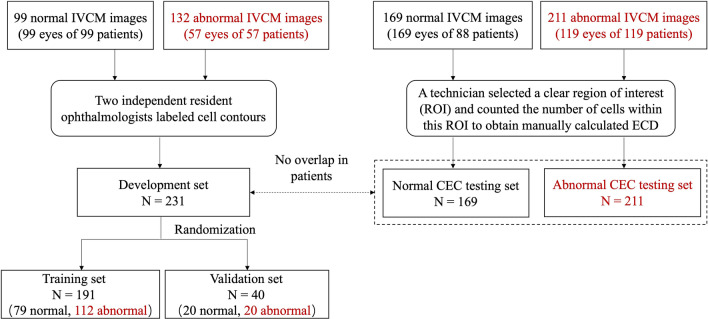
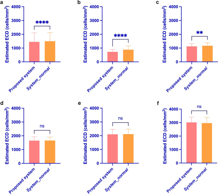
First, we developed a fully automated deep learning system for assessing abnormal CECs using a previous development set composed of normal images and a newly constructed development set composed of abnormal images. Second, two testing sets, one with 169 normal images and the other with 211 abnormal images, were used to evaluate the clinical validity and effectiveness of the proposed system on LASER IVCM images with different corneal endothelial conditions, particularly on abnormal images. Third, the automatically calculated endothelial cell density (ECD) and the manually calculated ECD were compared using both the previous and proposed systems.

RESULTS

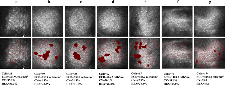
The automated morphometric parameter estimations of the average number of cells, ECD, coefficient of variation in cell area and percentage of hexagonal cells were 257 cells, 2648 ± 511 cells/mm, 32.18 ± 6.70% and 56.23 ± 8.69% for the normal CEC testing set and 83 cells, 1450 ± 656 cells/mm, 34.87 ± 10.53% and 42.55 ± 20.64% for the abnormal CEC testing set. Furthermore, for the abnormal CEC testing set, Pearson's correlation coefficient between the automatically and manually calculated ECDs was 0.9447; the 95% limits of agreement between the manually and automatically calculated ECDs were between 329.0 and - 579.5 (concordance correlation coefficient = 0.93).

CONCLUSIONS

This is the first report to count and analyze the morphology of abnormal CECs in LASER IVCM images using deep learning. Deep learning produces highly objective evaluation indicators for LASER IVCM corneal endothelium images and greatly expands the range of applications for LASER IVCM.

摘要

背景

本研究的目的是开发一种全自动分割和形态计量参数估计系统,用于从激光活体共聚焦显微镜(IVCM)图像中评估异常角膜内皮细胞(CEC)。

方法

首先,我们使用由正常图像组成的先前开发集和由异常图像组成的新构建开发集,开发了一种用于评估异常CEC的全自动深度学习系统。其次,使用两个测试集,一个包含169张正常图像,另一个包含211张异常图像,来评估所提出系统在具有不同角膜内皮状况的激光IVCM图像上的临床有效性和有效性,特别是在异常图像上。第三,使用先前和所提出的系统比较自动计算的内皮细胞密度(ECD)和手动计算的ECD。

结果

正常CEC测试集的细胞平均数、ECD、细胞面积变异系数和六边形细胞百分比的自动形态计量参数估计分别为257个细胞、2648±511个细胞/mm、32.18±6.70%和56.23±8.69%,异常CEC测试集分别为83个细胞、1450±656个细胞/mm、34.87±10.53%和42.55±20.64%。此外,对于异常CEC测试集,自动计算和手动计算的ECD之间的Pearson相关系数为0.9447;手动计算和自动计算的ECD之间的95%一致性界限在329.0和-579.5之间(一致性相关系数=0.93)。

结论

这是第一篇使用深度学习对激光IVCM图像中的异常CEC进行计数和形态分析的报告。深度学习为激光IVCM角膜内皮图像产生了高度客观的评估指标,并极大地扩展了激光IVCM的应用范围。

https://cdn.ncbi.nlm.nih.gov/pmc/blobs/890d/10233875/03d624895de3/40662_2023_340_Fig9_HTML.jpg
https://cdn.ncbi.nlm.nih.gov/pmc/blobs/890d/10233875/8e6cdacf7582/40662_2023_340_Fig1_HTML.jpg
https://cdn.ncbi.nlm.nih.gov/pmc/blobs/890d/10233875/62c4bf73f49e/40662_2023_340_Fig2_HTML.jpg
https://cdn.ncbi.nlm.nih.gov/pmc/blobs/890d/10233875/97728e416343/40662_2023_340_Fig3_HTML.jpg
https://cdn.ncbi.nlm.nih.gov/pmc/blobs/890d/10233875/4bcc05d162fb/40662_2023_340_Fig4_HTML.jpg
https://cdn.ncbi.nlm.nih.gov/pmc/blobs/890d/10233875/74ace5836ed4/40662_2023_340_Fig5_HTML.jpg
https://cdn.ncbi.nlm.nih.gov/pmc/blobs/890d/10233875/0b667e96920f/40662_2023_340_Fig6_HTML.jpg
https://cdn.ncbi.nlm.nih.gov/pmc/blobs/890d/10233875/ff857f621864/40662_2023_340_Fig7_HTML.jpg
https://cdn.ncbi.nlm.nih.gov/pmc/blobs/890d/10233875/2b51b72af483/40662_2023_340_Fig8_HTML.jpg
https://cdn.ncbi.nlm.nih.gov/pmc/blobs/890d/10233875/03d624895de3/40662_2023_340_Fig9_HTML.jpg

相似文献

1
Assessing abnormal corneal endothelial cells from in vivo confocal microscopy images using a fully automated deep learning system.使用全自动深度学习系统从体内共聚焦显微镜图像评估异常角膜内皮细胞。
Eye Vis (Lond). 2023 Jun 1;10(1):20. doi: 10.1186/s40662-023-00340-7.
2
A Fully Automated Segmentation and Morphometric Parameter Estimation System for Assessing Corneal Endothelial Cell Images.全自动角膜内皮细胞图像分割与形态参数估计算法系统
Am J Ophthalmol. 2022 Jul;239:142-153. doi: 10.1016/j.ajo.2022.02.026. Epub 2022 Mar 11.
3
A Deep Learning Model for Automated Sub-Basal Corneal Nerve Segmentation and Evaluation Using In Vivo Confocal Microscopy.一种用于使用活体共聚焦显微镜进行自动化角膜基底膜下神经分割和评估的深度学习模型。
Transl Vis Sci Technol. 2020 Jun 18;9(2):32. doi: 10.1167/tvst.9.2.32. eCollection 2020 Jun.
4
Segmentation and Evaluation of Corneal Nerves and Dendritic Cells From In Vivo Confocal Microscopy Images Using Deep Learning.基于深度学习的活体共聚焦显微镜图像中角膜神经和树突状细胞的分割和评估。
Transl Vis Sci Technol. 2022 Jun 1;11(6):24. doi: 10.1167/tvst.11.6.24.
5
A fully automated cell segmentation and morphometric parameter system for quantifying corneal endothelial cell morphology.全自动细胞分割和形态计量参数系统,用于量化角膜内皮细胞形态。
Comput Methods Programs Biomed. 2018 Jul;160:11-23. doi: 10.1016/j.cmpb.2018.03.015. Epub 2018 Mar 22.
6
Evaluation of a computer-aided diagnostic model for corneal diseases by analyzing confocal microscopy images.通过分析共焦显微镜图像评估角膜疾病的计算机辅助诊断模型
Front Med (Lausanne). 2023 Apr 20;10:1164188. doi: 10.3389/fmed.2023.1164188. eCollection 2023.
7
In vivo confocal microscopy of the corneal endothelium: comparison of three morphometry methods after corneal transplantation.角膜内皮的共聚焦显微镜活体检查:三种角膜移植术后形态计量学方法的比较。
Eye (Lond). 2011 Sep;25(9):1130-7. doi: 10.1038/eye.2011.121. Epub 2011 Jun 10.
8
Deep learning-based analysis of macaque corneal sub-basal nerve fibers in confocal microscopy images.基于深度学习的共聚焦显微镜图像中猕猴角膜基底膜下神经纤维分析
Eye Vis (Lond). 2020 May 8;7:27. doi: 10.1186/s40662-020-00192-5. eCollection 2020.
9
Comparison of Noncontact Specular and Confocal Microscopy for Evaluation of Corneal Endothelium.非接触式镜面显微镜与共焦显微镜在评估角膜内皮方面的比较
Eye Contact Lens. 2018 Sep;44 Suppl 1:S144-S150. doi: 10.1097/ICL.0000000000000362.
10
Fully automated montaging of laser scanning in vivo confocal microscopy images of the human corneal subbasal nerve plexus.全自动拼接人眼角膜基底神经丛激光共聚焦显微镜活体扫描图像。
Invest Ophthalmol Vis Sci. 2012 Apr 24;53(4):2235-42. doi: 10.1167/iovs.11-8454.

引用本文的文献

1
Assessment of Corneal Endothelial Barrier Function Based on "Y-Junctions": A Finite Element Analysis.基于“Y型连接”的角膜内皮屏障功能评估:有限元分析
Invest Ophthalmol Vis Sci. 2025 May 1;66(5):33. doi: 10.1167/iovs.66.5.33.
2
Artificial-Intelligence-Enhanced Analysis of In Vivo Confocal Microscopy in Corneal Diseases: A Review.人工智能增强的角膜疾病体内共聚焦显微镜分析:综述
Diagnostics (Basel). 2024 Mar 26;14(7):694. doi: 10.3390/diagnostics14070694.
3
Potential applications of artificial intelligence in image analysis in cornea diseases: a review.

本文引用的文献

1
A Fully Automated Segmentation and Morphometric Parameter Estimation System for Assessing Corneal Endothelial Cell Images.全自动角膜内皮细胞图像分割与形态参数估计算法系统
Am J Ophthalmol. 2022 Jul;239:142-153. doi: 10.1016/j.ajo.2022.02.026. Epub 2022 Mar 11.
2
Deep Learning for Assessing the Corneal Endothelium from Specular Microscopy Images up to 1 Year after Ultrathin-DSAEK Surgery.深度学习评估超薄角膜内皮层移植术后 1 年的角膜内皮细胞来自共焦显微镜图像。
Transl Vis Sci Technol. 2020 Aug 21;9(2):49. doi: 10.1167/tvst.9.2.49. eCollection 2020 Aug.
3
Quantitative and qualitative evaluation of deep learning automatic segmentations of corneal endothelial cell images of reduced image quality obtained following cornea transplant.
人工智能在角膜疾病图像分析中的潜在应用:综述
Eye Vis (Lond). 2024 Mar 7;11(1):10. doi: 10.1186/s40662-024-00376-3.
对角膜移植后获得的图像质量降低的角膜内皮细胞图像的深度学习自动分割进行定量和定性评估。
J Med Imaging (Bellingham). 2020 Jan;7(1):014503. doi: 10.1117/1.JMI.7.1.014503. Epub 2020 Feb 14.
4
Segmentation of corneal endothelium images using a U-Net-based convolutional neural network.基于 U-Net 的卷积神经网络的角膜内皮图像分割。
Artif Intell Med. 2018 Jun;88:1-13. doi: 10.1016/j.artmed.2018.04.004. Epub 2018 Apr 19.
5
Comparison of Noncontact Specular and Confocal Microscopy for Evaluation of Corneal Endothelium.非接触式镜面显微镜与共焦显微镜在评估角膜内皮方面的比较
Eye Contact Lens. 2018 Sep;44 Suppl 1:S144-S150. doi: 10.1097/ICL.0000000000000362.
6
Overestimation of Corneal Endothelial Cell Density in Smaller Frame Sizes in In Vivo Confocal Microscopy.体内共焦显微镜检查中小尺寸框架下角膜内皮细胞密度的高估
Cornea. 2016 Mar;35(3):363-9. doi: 10.1097/ICO.0000000000000698.
7
Comparison of 4 specular microscopes in healthy eyes and eyes with cornea guttata or corneal grafts.4种角膜内皮显微镜在健康眼及患有角膜滴状变性或角膜移植眼的比较。
Cornea. 2015 Apr;34(4):381-6. doi: 10.1097/ICO.0000000000000385.
8
Comparison of manual and automated endothelial cell density analysis in normal eyes and DSEK eyes.正常眼和 DSEK 眼中手动和自动内皮细胞密度分析的比较。
Cornea. 2013 May;32(5):567-73. doi: 10.1097/ICO.0b013e31825de8fa.
9
Behavior of corneal endothelial density over a lifetime.角膜内皮密度在一生中的变化情况。
J Cataract Refract Surg. 2001 Oct;27(10):1574-8. doi: 10.1016/s0886-3350(01)00925-7.
10
Corneal endothelium five years after transplantation.移植后五年的角膜内皮。
Am J Ophthalmol. 1994 Aug 15;118(2):185-96. doi: 10.1016/s0002-9394(14)72898-3.