• 文献检索
  • 文档翻译
  • 深度研究
  • 学术资讯
  • Suppr Zotero 插件Zotero 插件
  • 邀请有礼
  • 套餐&价格
  • 历史记录
应用&插件
Suppr Zotero 插件Zotero 插件浏览器插件Mac 客户端Windows 客户端微信小程序
定价
高级版会员购买积分包购买API积分包
服务
文献检索文档翻译深度研究API 文档MCP 服务
关于我们
关于 Suppr公司介绍联系我们用户协议隐私条款
关注我们

Suppr 超能文献

核心技术专利:CN118964589B侵权必究
粤ICP备2023148730 号-1Suppr @ 2026

文献检索

告别复杂PubMed语法,用中文像聊天一样搜索,搜遍4000万医学文献。AI智能推荐,让科研检索更轻松。

立即免费搜索

文件翻译

保留排版,准确专业,支持PDF/Word/PPT等文件格式,支持 12+语言互译。

免费翻译文档

深度研究

AI帮你快速写综述,25分钟生成高质量综述,智能提取关键信息,辅助科研写作。

立即免费体验

基于深度学习的活体共聚焦显微镜图像中角膜神经和树突状细胞的分割和评估。

Segmentation and Evaluation of Corneal Nerves and Dendritic Cells From In Vivo Confocal Microscopy Images Using Deep Learning.

机构信息

Department of Ophthalmology, Medical Faculty and University Hospital, University of Cologne, Cologne, Germany.

Division of Dry Eye and Ocular GvHD, University Hospital Cologne, University of Cologne, Cologne, Germany.

出版信息

Transl Vis Sci Technol. 2022 Jun 1;11(6):24. doi: 10.1167/tvst.11.6.24.

DOI:10.1167/tvst.11.6.24
PMID:35762938
原文链接:https://pmc.ncbi.nlm.nih.gov/articles/PMC9251793/
Abstract

PURPOSE

Segmentation and evaluation of in vivo confocal microscopy (IVCM) images requires manual intervention, which is time consuming, laborious, and non-reproducible. The aim of this research was to develop and validate deep learning-based methods that could automatically segment and evaluate corneal nerve fibers (CNFs) and dendritic cells (DCs) in IVCM images, thereby reducing processing time to analyze larger volumes of clinical images.

METHODS

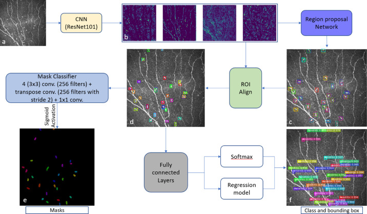
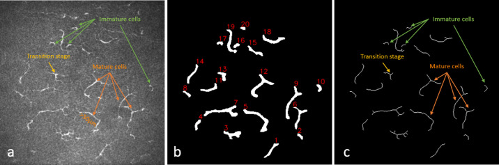
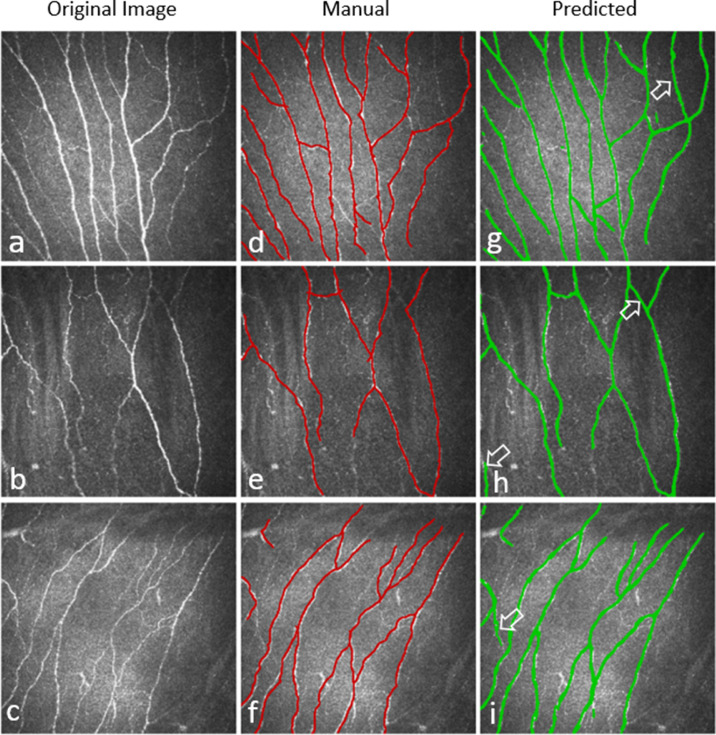
CNF and DC segmentation models were developed based on U-Net and Mask R-CNN architectures, respectively; 10-fold cross-validation was used to evaluate both models. The CNF model was trained and tested using 1097 and 122 images, and the DC model was trained and tested using 679 and 75 images, respectively, at each fold. The CNF morphology, number of nerves, number of branching points, nerve length, and tortuosity were analyzed; for DCs, number, size, and immature-mature cells were analyzed. Python-based software was written for model training, testing, and automatic morphometric parameters evaluation.

RESULTS

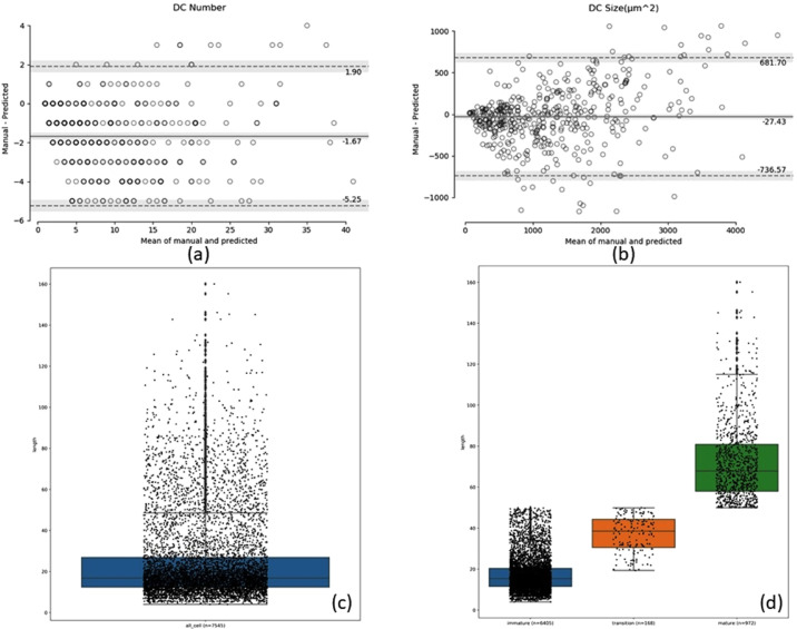
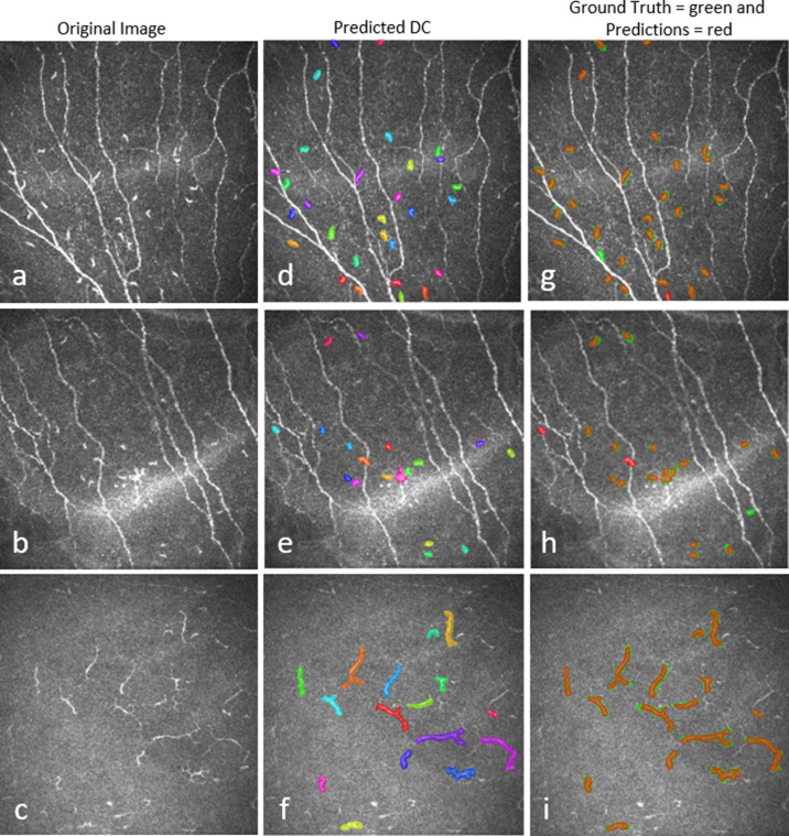
The CNF model achieved on average 86.1% sensitivity and 90.1% specificity, and the DC model achieved on average 89.37% precision, 94.43% recall, and 91.83% F1 score. The interclass correlation coefficient (ICC) between manual annotation and automatic segmentation were 0.85, 0.87, 0.95, and 0.88 for CNF number, length, branching points, and tortuosity, respectively, and the ICC for DC number and size were 0.95 and 0.92, respectively.

CONCLUSIONS

Our proposed methods demonstrated reliable consistency between manual annotation and automatic segmentation of CNF and DC with rapid speed. The results showed that these approaches have the potential to be implemented into clinical practice in IVCM images.

TRANSLATIONAL RELEVANCE

The deep learning-based automatic segmentation and quantification algorithm significantly increases the efficiency of evaluating IVCM images, thereby supporting and potentially improving the diagnosis and treatment of ocular surface disease associated with corneal nerves and dendritic cells.

摘要

目的

体内共聚焦显微镜(IVCM)图像的分割和评估需要手动干预,既耗时、费力,又不可重复。本研究旨在开发和验证基于深度学习的方法,以自动分割和评估 IVCM 图像中的角膜神经纤维(CNF)和树突状细胞(DC),从而减少分析更大体积临床图像的处理时间。

方法

基于 U-Net 和 Mask R-CNN 架构分别开发了 CNF 和 DC 分割模型;采用 10 倍交叉验证评估两个模型。CNF 模型使用 1097 个和 122 个图像进行训练和测试,DC 模型分别使用 679 个和 75 个图像进行训练和测试,每个折叠 1 次。分析了 CNF 的形态、神经数量、分支点数量、神经长度和扭曲度;对于 DCs,分析了数量、大小和幼稚成熟细胞。编写了基于 Python 的软件,用于模型训练、测试和自动形态计量参数评估。

结果

CNF 模型的平均灵敏度为 86.1%,特异性为 90.1%,DC 模型的平均精度为 89.37%,召回率为 94.43%,F1 得分为 91.83%。手动标注与自动分割之间的组内相关系数(ICC)分别为 0.85、0.87、0.95 和 0.88,用于 CNF 数量、长度、分支点和扭曲度,DC 数量和大小的 ICC 分别为 0.95 和 0.92。

结论

我们提出的方法在 CNF 和 DC 的手动标注与自动分割之间具有可靠的一致性,且速度较快。结果表明,这些方法有可能在 IVCM 图像的临床实践中得到实施。

翻译

温迪

https://cdn.ncbi.nlm.nih.gov/pmc/blobs/77ab/9251793/7dcfbdc34179/tvst-11-6-24-f010.jpg
https://cdn.ncbi.nlm.nih.gov/pmc/blobs/77ab/9251793/a713e7233325/tvst-11-6-24-f001.jpg
https://cdn.ncbi.nlm.nih.gov/pmc/blobs/77ab/9251793/55152b32a654/tvst-11-6-24-f002.jpg
https://cdn.ncbi.nlm.nih.gov/pmc/blobs/77ab/9251793/8f8ede0fd58d/tvst-11-6-24-f003.jpg
https://cdn.ncbi.nlm.nih.gov/pmc/blobs/77ab/9251793/7ff52d03d0d3/tvst-11-6-24-f004.jpg
https://cdn.ncbi.nlm.nih.gov/pmc/blobs/77ab/9251793/38bd85c559fa/tvst-11-6-24-f005.jpg
https://cdn.ncbi.nlm.nih.gov/pmc/blobs/77ab/9251793/0d64835b9b94/tvst-11-6-24-f006.jpg
https://cdn.ncbi.nlm.nih.gov/pmc/blobs/77ab/9251793/34160152d54a/tvst-11-6-24-f007.jpg
https://cdn.ncbi.nlm.nih.gov/pmc/blobs/77ab/9251793/eec79f2a704f/tvst-11-6-24-f008.jpg
https://cdn.ncbi.nlm.nih.gov/pmc/blobs/77ab/9251793/a4b2566d6b25/tvst-11-6-24-f009.jpg
https://cdn.ncbi.nlm.nih.gov/pmc/blobs/77ab/9251793/7dcfbdc34179/tvst-11-6-24-f010.jpg

相似文献

1
Segmentation and Evaluation of Corneal Nerves and Dendritic Cells From In Vivo Confocal Microscopy Images Using Deep Learning.基于深度学习的活体共聚焦显微镜图像中角膜神经和树突状细胞的分割和评估。
Transl Vis Sci Technol. 2022 Jun 1;11(6):24. doi: 10.1167/tvst.11.6.24.
2
A Deep Learning Model for Automated Sub-Basal Corneal Nerve Segmentation and Evaluation Using In Vivo Confocal Microscopy.一种用于使用活体共聚焦显微镜进行自动化角膜基底膜下神经分割和评估的深度学习模型。
Transl Vis Sci Technol. 2020 Jun 18;9(2):32. doi: 10.1167/tvst.9.2.32. eCollection 2020 Jun.
3
Generative Adversarial Network Based Automatic Segmentation of Corneal Subbasal Nerves on In Vivo Confocal Microscopy Images.基于生成对抗网络的活体共聚焦显微镜图像下角膜基底部神经的自动分割。
Transl Vis Sci Technol. 2021 May 3;10(6):33. doi: 10.1167/tvst.10.6.33.
4
An artificial intelligence-based deep learning algorithm for the diagnosis of diabetic neuropathy using corneal confocal microscopy: a development and validation study.基于人工智能的深度学习算法利用角膜共聚焦显微镜诊断糖尿病周围神经病变:一项开发和验证研究。
Diabetologia. 2020 Feb;63(2):419-430. doi: 10.1007/s00125-019-05023-4. Epub 2019 Nov 12.
5
Combining In Vivo Corneal Confocal Microscopy With Deep Learning-Based Analysis Reveals Sensory Nerve Fiber Loss in Acute Simian Immunodeficiency Virus Infection.结合体内角膜共焦显微镜与基于深度学习的分析揭示急性猴免疫缺陷病毒感染中的感觉神经纤维损失。
Cornea. 2021 May 1;40(5):635-642. doi: 10.1097/ICO.0000000000002661.
6
Deep learning-based analysis of macaque corneal sub-basal nerve fibers in confocal microscopy images.基于深度学习的共聚焦显微镜图像中猕猴角膜基底膜下神经纤维分析
Eye Vis (Lond). 2020 May 8;7:27. doi: 10.1186/s40662-020-00192-5. eCollection 2020.
7
Characteristics of Toxic Keratopathy, an In Vivo Confocal Microscopy Study.毒蕈样角膜病变的特征:一项活体共聚焦显微镜研究。
Transl Vis Sci Technol. 2021 Sep 1;10(11):11. doi: 10.1167/tvst.10.11.11.
8
Segmentation and multiparametric evaluation of corneal whorl-like nerves for in vivo confocal microscopy images in dry eye disease.干眼病活体共聚焦显微镜图像中环状神经的分割和多参数评估。
BMJ Open Ophthalmol. 2024 Oct 7;9(1):e001861. doi: 10.1136/bmjophth-2024-001861.
9
In vivo confocal microscopy detects bilateral changes of corneal immune cells and nerves in unilateral herpes zoster ophthalmicus.在体共聚焦显微镜检测单侧带状疱疹性眼病双侧角膜免疫细胞和神经的变化。
Ocul Surf. 2018 Jan;16(1):101-111. doi: 10.1016/j.jtos.2017.09.004. Epub 2017 Sep 18.
10
Inflammation and the nervous system: the connection in the cornea in patients with infectious keratitis.炎症与神经系统:感染性角膜炎患者角膜中的联系。
Invest Ophthalmol Vis Sci. 2011 Jul 11;52(8):5136-43. doi: 10.1167/iovs.10-7048.

引用本文的文献

1
Advances in ocular aging: combining deep learning, imaging, and liquid biopsy biomarkers.眼部衰老研究进展:深度学习、成像技术与液体活检生物标志物的结合
Front Med (Lausanne). 2025 Jul 23;12:1591936. doi: 10.3389/fmed.2025.1591936. eCollection 2025.
2
AxonFinder: Automated segmentation of tumor innervating neuronal fibers.轴突定位器:肿瘤支配神经纤维的自动分割
Heliyon. 2024 Dec 15;11(1):e41209. doi: 10.1016/j.heliyon.2024.e41209. eCollection 2025 Jan 15.
3
Segmentation and multiparametric evaluation of corneal whorl-like nerves for in vivo confocal microscopy images in dry eye disease.

本文引用的文献

1
Comparing the results of manual and automated quantitative corneal neuroanalysing modules for beginners.比较手动和自动定量角膜神经分析模块在初学者中的结果。
Sci Rep. 2021 Sep 14;11(1):18208. doi: 10.1038/s41598-021-97567-y.
2
Generative Adversarial Network Based Automatic Segmentation of Corneal Subbasal Nerves on In Vivo Confocal Microscopy Images.基于生成对抗网络的活体共聚焦显微镜图像下角膜基底部神经的自动分割。
Transl Vis Sci Technol. 2021 May 3;10(6):33. doi: 10.1167/tvst.10.6.33.
3
Corneal Immune Cells Are Increased in Patients With Multiple Sclerosis.
干眼病活体共聚焦显微镜图像中环状神经的分割和多参数评估。
BMJ Open Ophthalmol. 2024 Oct 7;9(1):e001861. doi: 10.1136/bmjophth-2024-001861.
4
Diabetic Peripheral Neuropathy: Emerging Treatments of Neuropathic Pain and Novel Diagnostic Methods.糖尿病性周围神经病变:神经性疼痛的新兴治疗方法及新型诊断方法
J Diabetes Sci Technol. 2024 Sep 16:19322968241279553. doi: 10.1177/19322968241279553.
5
Multimodal Approach in Dry Eye Disease Combining In Vivo Confocal Microscopy and HLA-DR Expression.多模态方法在干眼症中的应用,结合了活体共聚焦显微镜和 HLA-DR 表达。
Transl Vis Sci Technol. 2024 Aug 1;13(8):39. doi: 10.1167/tvst.13.8.39.
6
Three-Dimensional Reconstruction of Subbasal Nerve Density in Eyes With Limbal Stem Cell Deficiency: A Pilot Study.角膜缘干细胞缺陷眼中的基底神经丛密度的三维重建:一项初步研究。
Cornea. 2024 Oct 1;43(10):1278-1284. doi: 10.1097/ICO.0000000000003571. Epub 2024 Jun 26.
7
Artificial-Intelligence-Enhanced Analysis of In Vivo Confocal Microscopy in Corneal Diseases: A Review.人工智能增强的角膜疾病体内共聚焦显微镜分析:综述
Diagnostics (Basel). 2024 Mar 26;14(7):694. doi: 10.3390/diagnostics14070694.
8
Potential applications of artificial intelligence in image analysis in cornea diseases: a review.人工智能在角膜疾病图像分析中的潜在应用:综述
Eye Vis (Lond). 2024 Mar 7;11(1):10. doi: 10.1186/s40662-024-00376-3.
9
Open-Source Automated Segmentation of Neuronal Structures in Corneal Confocal Microscopy Images of the Subbasal Nerve Plexus With Accuracy on Par With Human Segmentation.开源自动化分割角膜共聚焦显微镜图像下的神经结构亚基底神经丛,其准确性与人工分割相当。
Cornea. 2023 Oct 1;42(10):1309-1319. doi: 10.1097/ICO.0000000000003319. Epub 2023 Jul 7.
10
In-vivo corneal confocal microscopy: Imaging analysis, biological insights and future directions.体内角膜共聚焦显微镜:成像分析、生物学见解和未来方向。
Commun Biol. 2023 Jun 19;6(1):652. doi: 10.1038/s42003-023-05005-8.
多发性硬化症患者的角膜免疫细胞增加。
Transl Vis Sci Technol. 2021 Apr 1;10(4):19. doi: 10.1167/tvst.10.4.19.
4
Deep learning-based automatic meibomian gland segmentation and morphology assessment in infrared meibography.基于深度学习的红外睑板腺图像中睑板腺自动分割与形态评估。
Sci Rep. 2021 Apr 7;11(1):7649. doi: 10.1038/s41598-021-87314-8.
5
A Deep Learning Model for Automated Sub-Basal Corneal Nerve Segmentation and Evaluation Using In Vivo Confocal Microscopy.一种用于使用活体共聚焦显微镜进行自动化角膜基底膜下神经分割和评估的深度学习模型。
Transl Vis Sci Technol. 2020 Jun 18;9(2):32. doi: 10.1167/tvst.9.2.32. eCollection 2020 Jun.
6
Validation of the Use of Automated and Manual Quantitative Analysis of Corneal Nerve Plexus Following Refractive Surgery.屈光手术后角膜神经丛自动和手动定量分析应用的验证
Diagnostics (Basel). 2020 Jul 18;10(7):493. doi: 10.3390/diagnostics10070493.
7
Visualization of microneuromas by using in vivo confocal microscopy: An objective biomarker for the diagnosis of neuropathic corneal pain?应用活体共聚焦显微镜观察微神经瘤:诊断神经病理性角膜痛的客观生物标志物?
Ocul Surf. 2020 Oct;18(4):651-656. doi: 10.1016/j.jtos.2020.07.004. Epub 2020 Jul 11.
8
Automated Retinal Layer Segmentation Using Graph-based Algorithm Incorporating Deep-learning-derived Information.基于图算法结合深度学习信息的自动视网膜层分割。
Sci Rep. 2020 Jun 12;10(1):9541. doi: 10.1038/s41598-020-66355-5.
9
Deep learning-based analysis of macaque corneal sub-basal nerve fibers in confocal microscopy images.基于深度学习的共聚焦显微镜图像中猕猴角膜基底膜下神经纤维分析
Eye Vis (Lond). 2020 May 8;7:27. doi: 10.1186/s40662-020-00192-5. eCollection 2020.
10
Automated Tortuosity Analysis of Nerve Fibers in Corneal Confocal Microscopy.角膜共焦显微镜下神经纤维自动迂曲度分析。
IEEE Trans Med Imaging. 2020 Sep;39(9):2725-2737. doi: 10.1109/TMI.2020.2974499. Epub 2020 Feb 17.