Ma Qianqing, Shen Chunyun, Gao Yankun, Duan Yayang, Li Wanyan, Lu Gensheng, Qin Xiachuan, Zhang Chaoxue, Wang Junli
Department of Ultrasound, the First Affiliated Hospital of Anhui Medical University, Hefei, People's Republic of China.
Department of Ultrasound, Wuhu No. 2 People's Hospital, Wuhu, People's Republic of China.
Breast Cancer (Dove Med Press). 2023 May 26;15:381-390. doi: 10.2147/BCTT.S410356. eCollection 2023.
Breast cancer is the most common tumor globally. Automated Breast Volume Scanner (ABVS) and strain elastography (SE) can provide more useful breast information. The use of radiomics combined with ABVS and SE images to predict breast cancer has become a new focus. Therefore, this study developed and validated a radiomics analysis of breast lesions in combination with coronal plane of ABVS and SE to improve the differential diagnosis of benign and malignant breast diseases.
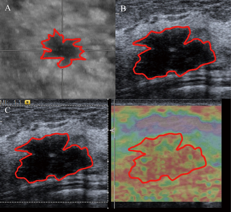
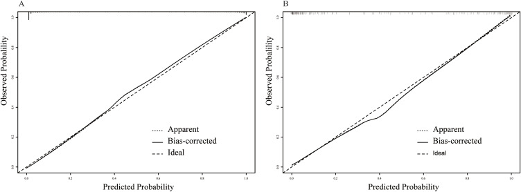
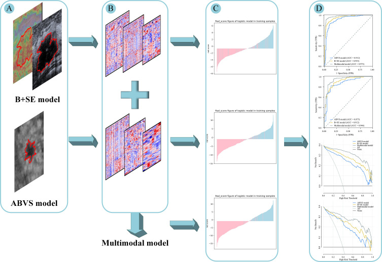
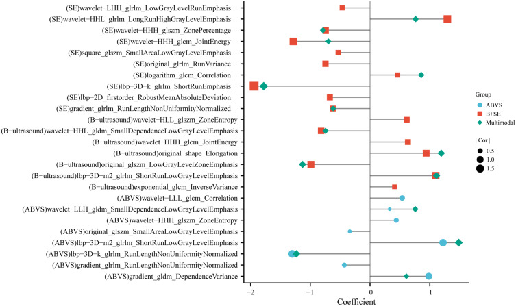
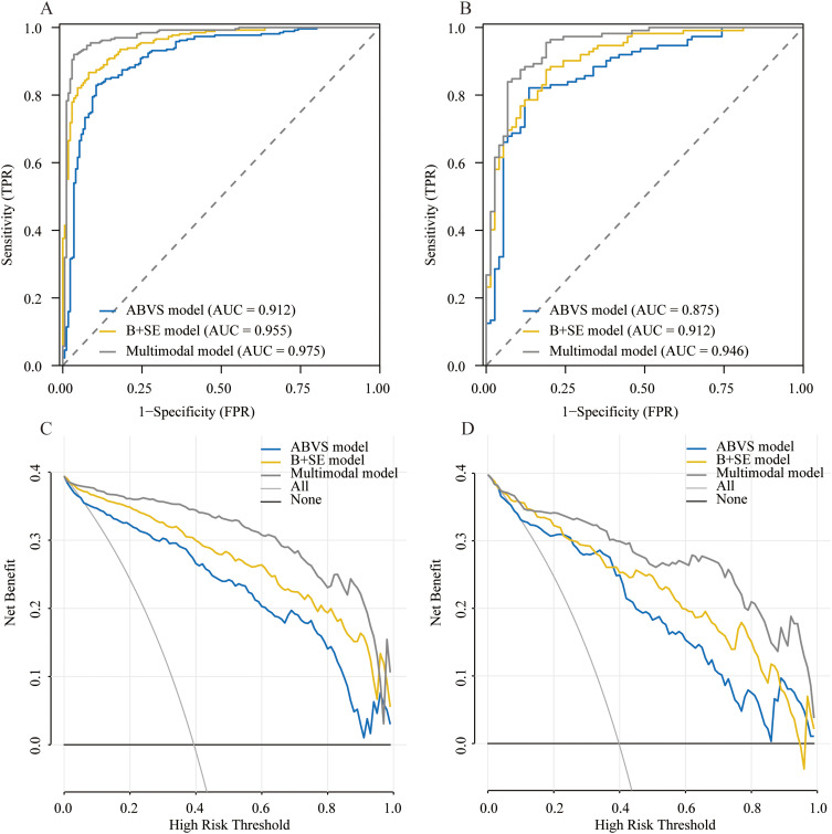
620 pathologically confirmed breast lesions from January 2017 to August 2021 were retrospectively analyzed and randomly divided into a training set (n=434) and a validation set (n=186). Radiomic features of the lesions were extracted from ABVS, B-ultrasound, and strain elastography (SE) images, respectively. These were then filtered by Gradient Boosted Decision Tree (GBDT) and multiple logistic regression. The ABVS model is based on coronal plane features for the breast, B+SE model is based on features of B-ultrasound and SE, and the multimodal model is based on features of three examinations. The evaluation of the predicted performance of the three models used the receiver operating characteristic (ROC) and decision curve analysis (DCA).
The area under the curve, accuracy, specificity, and sensitivity of the multimodal model in the training set are 0.975 (95% CI:0.959-0.991),93.78%, 92.02%, and 96.49%, respectively, and 0.946 (95% CI:0.913 -0.978), 87.63%, 83.93%, and 93.24% in the validation set, respectively. The multimodal model outperformed the ABVS model and B+SE model in both the training ( < 0.001, = 0.002, respectively) and validation sets ( < 0.001, = 0.034, respectively).
Radiomics from the coronal plane of the breast lesion provide valuable information for identification. A multimodal model combination with radiomics from ABVS, B-ultrasound, and SE could improve the diagnostic efficacy of breast masses.
乳腺癌是全球最常见的肿瘤。自动乳腺容积扫描仪(ABVS)和应变弹性成像(SE)能够提供更多有用的乳腺信息。将放射组学与ABVS和SE图像相结合用于预测乳腺癌已成为一个新的研究热点。因此,本研究开发并验证了一种结合ABVS冠状面和SE的乳腺病变放射组学分析方法,以改善乳腺良恶性疾病的鉴别诊断。
回顾性分析2017年1月至2021年8月间620例经病理证实的乳腺病变,并将其随机分为训练集(n = 434)和验证集(n = 186)。分别从ABVS、B超和应变弹性成像(SE)图像中提取病变的放射组学特征。然后通过梯度提升决策树(GBDT)和多元逻辑回归进行筛选。ABVS模型基于乳腺冠状面特征,B + SE模型基于B超和SE特征,多模态模型基于三种检查的特征。使用受试者操作特征(ROC)和决策曲线分析(DCA)对三种模型的预测性能进行评估。
多模态模型在训练集中的曲线下面积、准确率、特异性和敏感性分别为0.975(95%CI:0.959 - 0.991)、93.78%、92.02%和96.49%,在验证集中分别为0.946(95%CI:0.913 - 0.978)、87.63%、83.93%和93.24%。多模态模型在训练集(分别为P < 0.001,P = 0.002)和验证集(分别为P < 0.001,P = 0.034)中均优于ABVS模型和B + SE模型。
乳腺病变冠状面的放射组学为鉴别诊断提供了有价值的信息。结合ABVS、B超和SE的放射组学的多模态模型可提高乳腺肿块的诊断效能。