Wang Lei, Li Pengfei, Zhou Yu, Gu Renjun, Lu Ge, Zhang Chunbing
College of First Clinical Medicine, Nanjing University of Chinese Medicine, Nanjing, Jiangsu, 210023, People's Republic of China.
Department of Clinical Laboratory, Jiangsu Province Hospital of Chinese Medicine, The Affiliated Hospital of Nanjing University of Chinese Medicine, Nanjing, Jiangsu, 210029, People's Republic of China.
J Inflamm Res. 2023 May 27;16:2271-2296. doi: 10.2147/JIR.S406298. eCollection 2023.
Magnoflorine (Mag) has been reported to have anxiolytics, anti-cancer, and anti-inflammatory properties. In this study, we aim to investigate the effects of Mag on the rheumatoid arthritis (RA) and explore the underlying mechanism using a collagen-induced arthritis (CIA) mouse model and a lipopolysaccharide (LPS)-stimulated macrophage inflammation model.
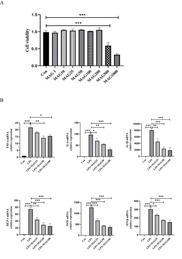
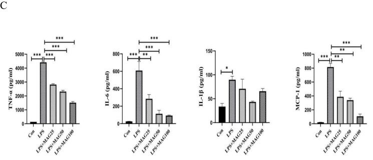
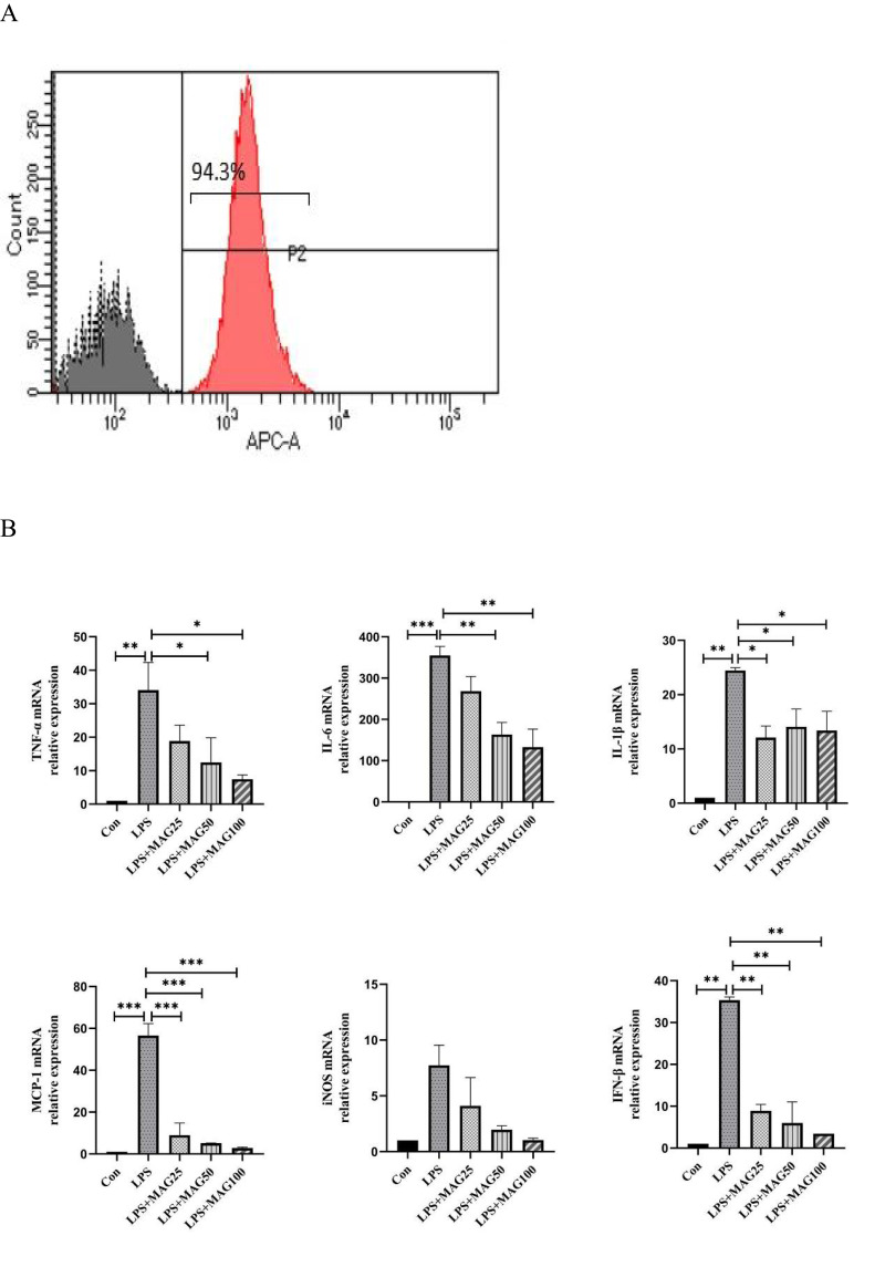
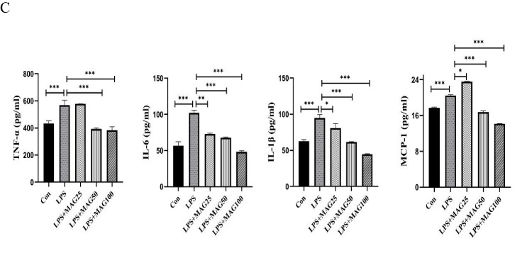
The in vivo effects of Mag on CIA were studied by inducing CIA in a mouse model using DBA/1J mice followed by treatment with vehicle, methotrexate (MTX, 1 mg/kg/d), and Mag (5 mg/kg/d, 10 mg/kg/d, and 20 mg/kg/d), and the in vitro effects of Mag on macrophages were examined by stimulation of RAW264.7 cells line and peritoneal macrophages (PMs) by LPS in the presence of different concentrations of Mag. Network pharmacology and molecular docking was then performed to predict the the binding ability between Mag and its targets. Inflammatory mediators were assayed by quantitative real-time PCR and enzyme linked immunosorbent assay (ELISA). Signaling pathway changes were subsequently determined by Western blotting and immunohistochemistry (IHC).
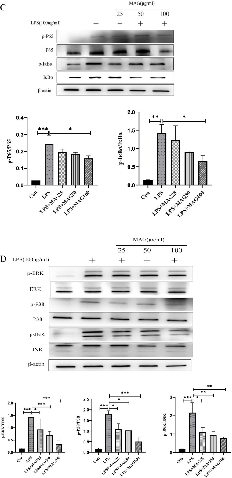
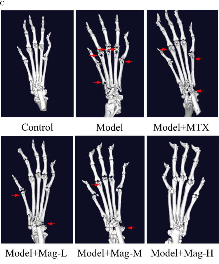
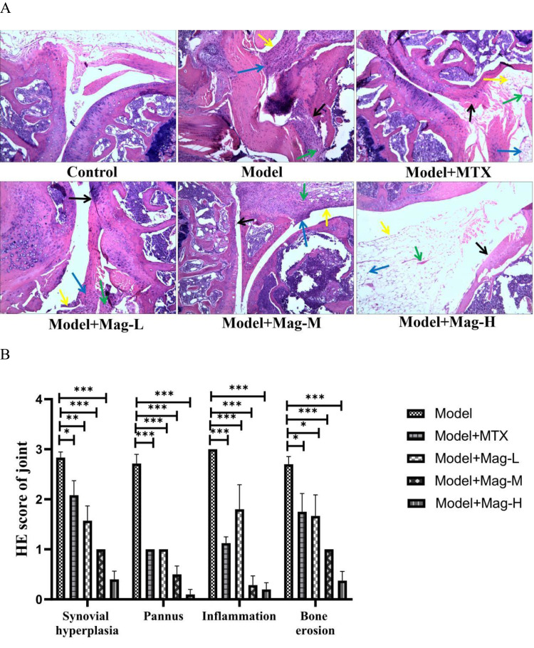
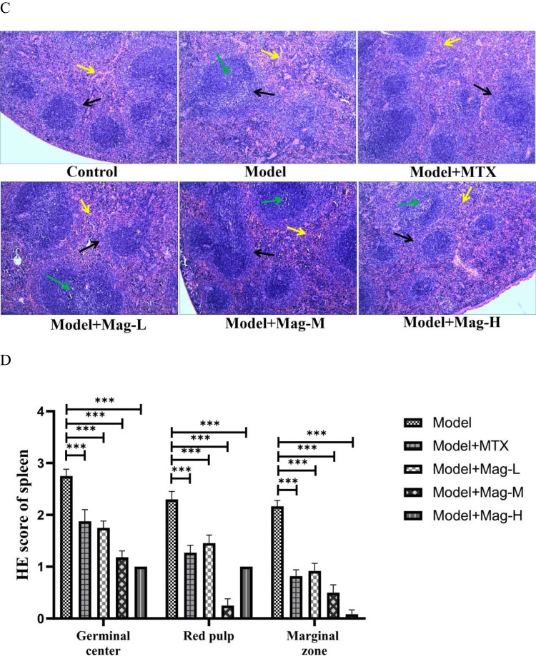
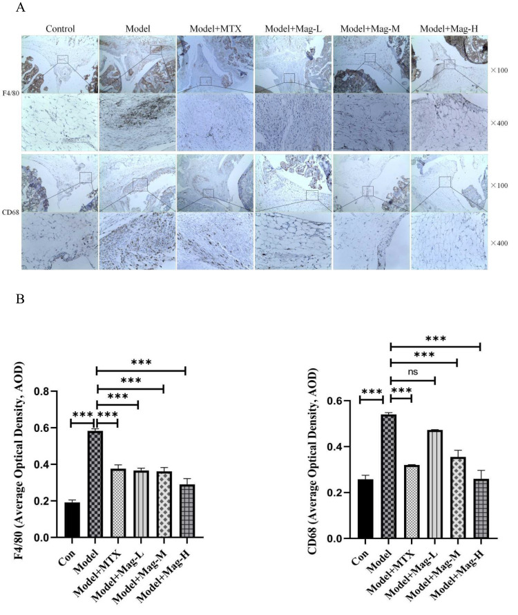
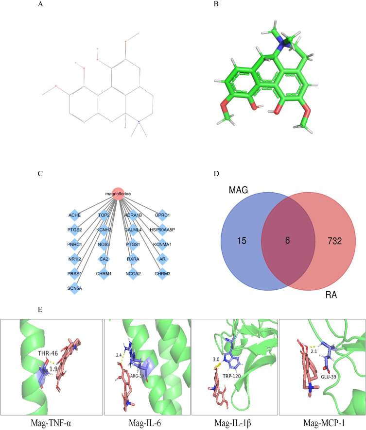
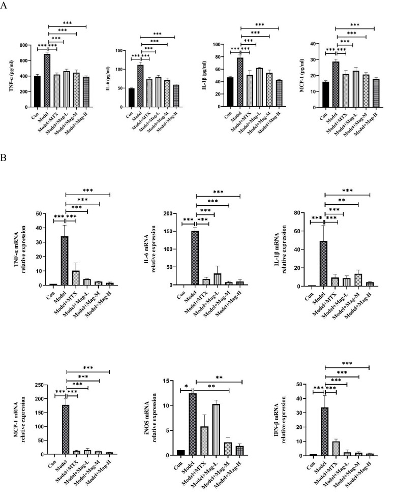
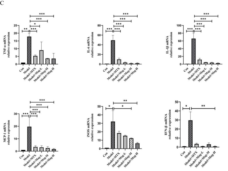
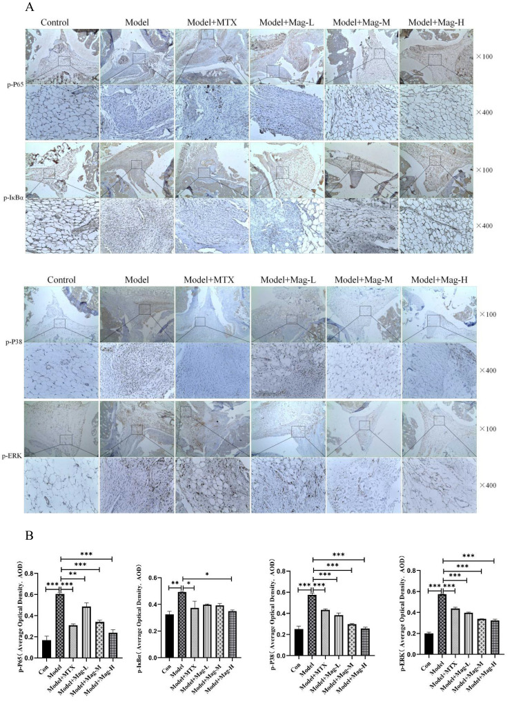
In vivo experiments demonstrated that Mag decreased arthritis severity scores, joints destruction, and macrophages infiltration into the synovial tissues of the CIA mice. Network pharmacology analysis revealed that Mag interacted with TNF-α, IL-6, IL-1β, and MCP-1. Consistent with this, analysis of the serum, synovial tissue of the CIA mice, and the supernatant of the cultured RAW264.7 cells and PMs showed that Mag suppressed the expression of TNF-α, IL-6, IL-1β, MCP-1, iNOS, and IFN-β. Furthermore, Mag attenuated the phosphorylation of p65, IκBα, ERK, JNK, and p38 MAPKs in the synovial tissues of the CIA mice and LPS-stimulated RAW 264.7 cells.
Mag may exert anti-arthritic and anti-inflammatory effects by inhibiting the activation of NF-κB and MAPK signaling pathways.
已报道小檗碱(Mag)具有抗焦虑、抗癌和抗炎特性。在本研究中,我们旨在研究小檗碱对类风湿性关节炎(RA)的影响,并使用胶原诱导性关节炎(CIA)小鼠模型和脂多糖(LPS)刺激的巨噬细胞炎症模型探索其潜在机制。
通过用DBA/1J小鼠诱导CIA小鼠模型,然后分别用赋形剂、甲氨蝶呤(MTX,1mg/kg/d)和小檗碱(5mg/kg/d、10mg/kg/d和20mg/kg/d)进行处理,研究小檗碱对CIA的体内作用;在不同浓度小檗碱存在的情况下,通过LPS刺激RAW264.7细胞系和腹腔巨噬细胞(PMs),研究小檗碱对巨噬细胞的体外作用。随后进行网络药理学和分子对接以预测小檗碱与其靶点之间的结合能力。通过定量实时PCR和酶联免疫吸附测定(ELISA)检测炎症介质。随后通过蛋白质印迹法和免疫组织化学(IHC)确定信号通路变化。
体内实验表明,小檗碱降低了CIA小鼠的关节炎严重程度评分、关节破坏以及巨噬细胞向滑膜组织的浸润。网络药理学分析显示,小檗碱与肿瘤坏死因子-α(TNF-α)、白细胞介素-6(IL-6)、白细胞介素-1β(IL-1β)和单核细胞趋化蛋白-1(MCP-1)相互作用。与此一致的是,对CIA小鼠的血清、滑膜组织以及培养的RAW264.7细胞和PMs的上清液分析表明,小檗碱抑制了TNF-α、IL-6、IL-1β、MCP-1、诱导型一氧化氮合酶(iNOS)和干扰素-β(IFN-β)的表达。此外,小檗碱减弱了CIA小鼠滑膜组织和LPS刺激的RAW 264.7细胞中p65、IκBα、细胞外调节蛋白激酶(ERK)、应激活化蛋白激酶(JNK)和p38丝裂原活化蛋白激酶(MAPKs)的磷酸化。
小檗碱可能通过抑制核因子-κB(NF-κB)和丝裂原活化蛋白激酶(MAPK)信号通路的激活发挥抗关节炎和抗炎作用。