Department of Radiology and Imaging Sciences, Emory University, 1364 Clifton Rd, Atlanta, GA, 30322, USA.
Division of Nuclear Medicine and Molecular Imaging, Massachusetts General Hospital & Department of Radiology, Harvard Medical School, Boston, MA, 02114, USA.
Nat Commun. 2023 Jun 5;14(1):3257. doi: 10.1038/s41467-023-36377-4.
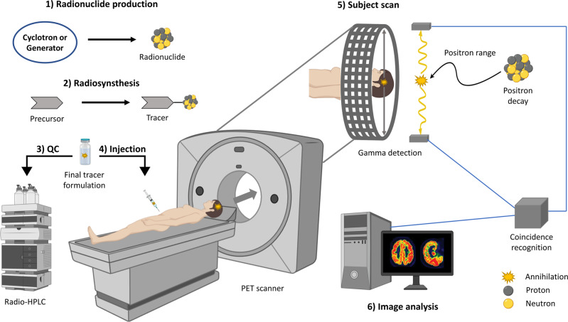
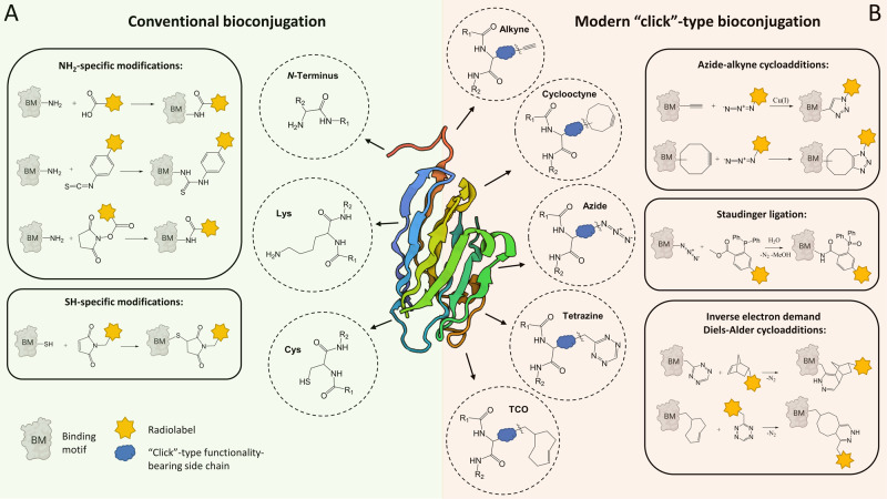
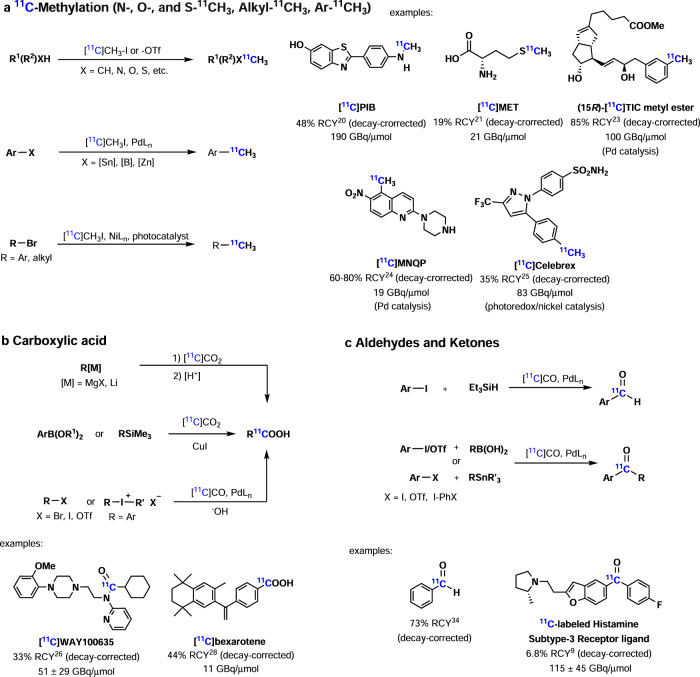
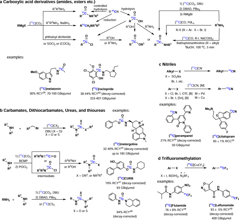
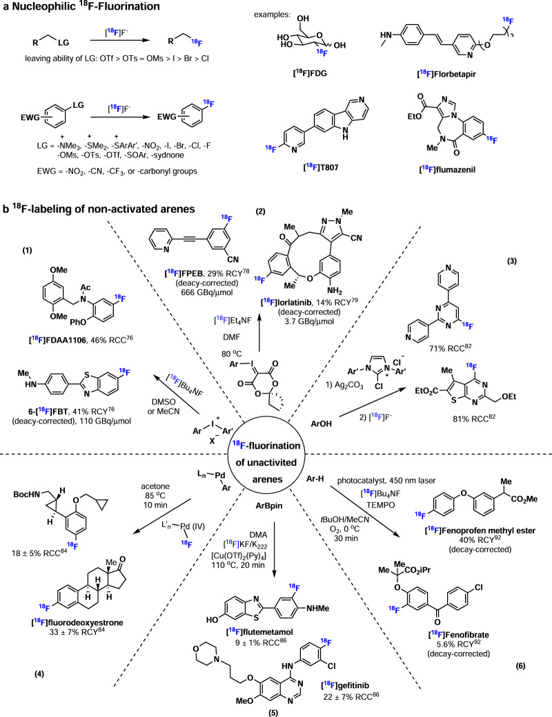
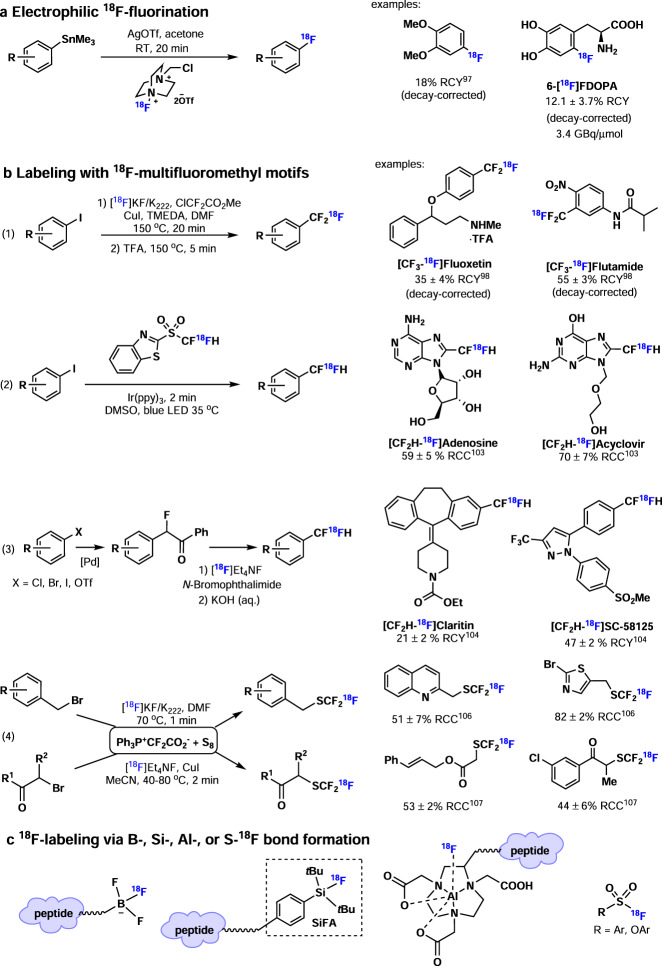
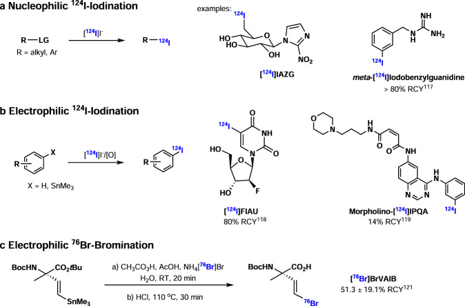
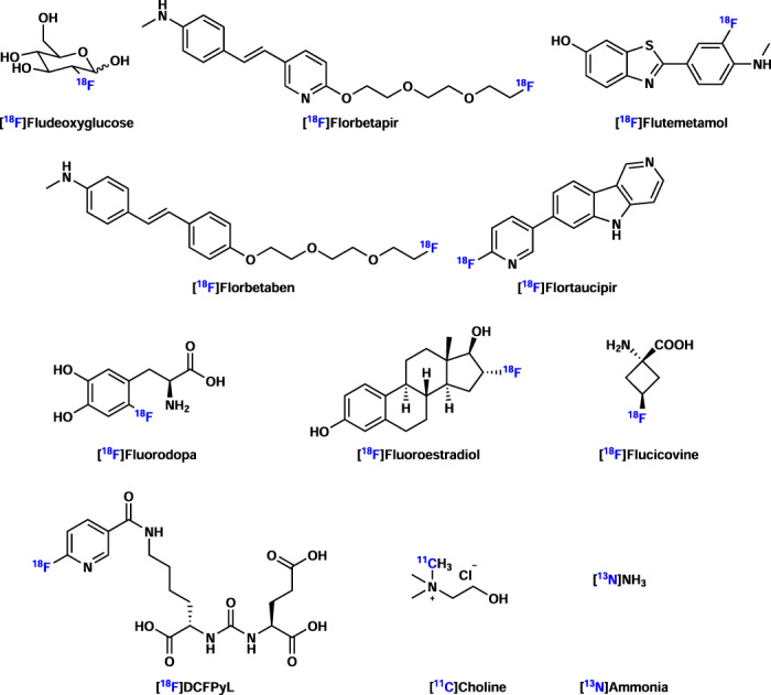
Positron emission tomography (PET) constitutes a functional imaging technique that is harnessed to probe biological processes in vivo. PET imaging has been used to diagnose and monitor the progression of diseases, as well as to facilitate drug development efforts at both preclinical and clinical stages. The wide applications and rapid development of PET have ultimately led to an increasing demand for new methods in radiochemistry, with the aim to expand the scope of synthons amenable for radiolabeling. In this work, we provide an overview of commonly used chemical transformations for the syntheses of PET tracers in all aspects of radiochemistry, thereby highlighting recent breakthrough discoveries and contemporary challenges in the field. We discuss the use of biologicals for PET imaging and highlight general examples of successful probe discoveries for molecular imaging with PET - with a particular focus on translational and scalable radiochemistry concepts that have been entered to clinical use.
正电子发射断层扫描(PET)是一种功能成像技术,用于在体探测生物过程。PET 成像已被用于诊断和监测疾病的进展,以及促进临床前和临床阶段的药物开发工作。PET 的广泛应用和快速发展最终导致对放射化学中新技术的需求不断增加,目的是扩大可用于放射性标记的缩合剂的范围。在这项工作中,我们全面概述了放射化学各个方面用于 PET 示踪剂合成的常用化学转化,从而突出了该领域的最新突破性发现和当代挑战。我们讨论了用于 PET 成像的生物制剂,并强调了使用 PET 进行分子成像的成功探针发现的一般示例 - 特别关注已进入临床使用的转化和可扩展放射化学概念。