Department of Biomedical Imaging and Image-Guided Therapy, Division of Computational Imaging Research (CIR), Medical University of Vienna, Währinger Gürtel 18-20, 1090, Vienna, Austria.
Department of Biomedical Imaging and Image-Guided Therapy, Division of General and Pediatric Radiology, Medical University of Vienna, Vienna, Austria.
Eur Radiol Exp. 2023 Jun 7;7(1):32. doi: 10.1186/s41747-023-00343-y.
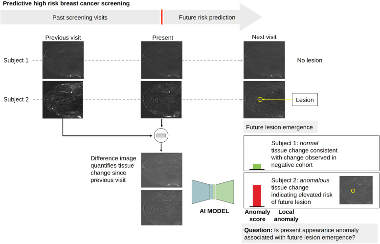
International societies have issued guidelines for high-risk breast cancer (BC) screening, recommending contrast-enhanced magnetic resonance imaging (CE-MRI) of the breast as a supplemental diagnostic tool. In our study, we tested the applicability of deep learning-based anomaly detection to identify anomalous changes in negative breast CE-MRI screens associated with future lesion emergence.
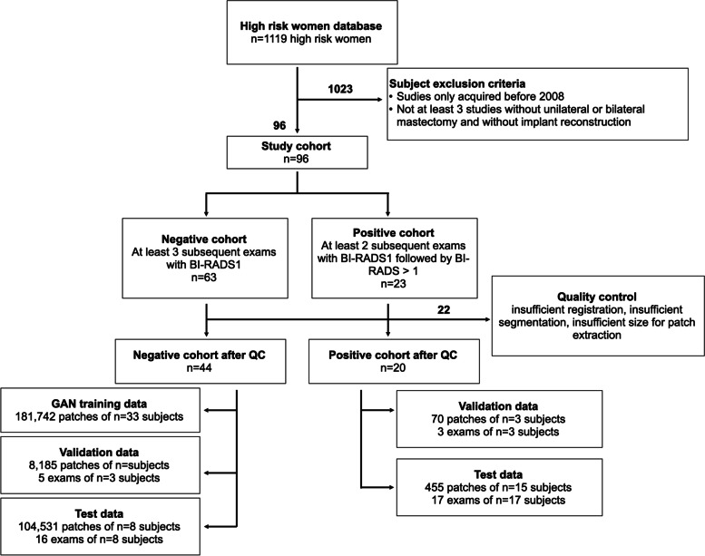
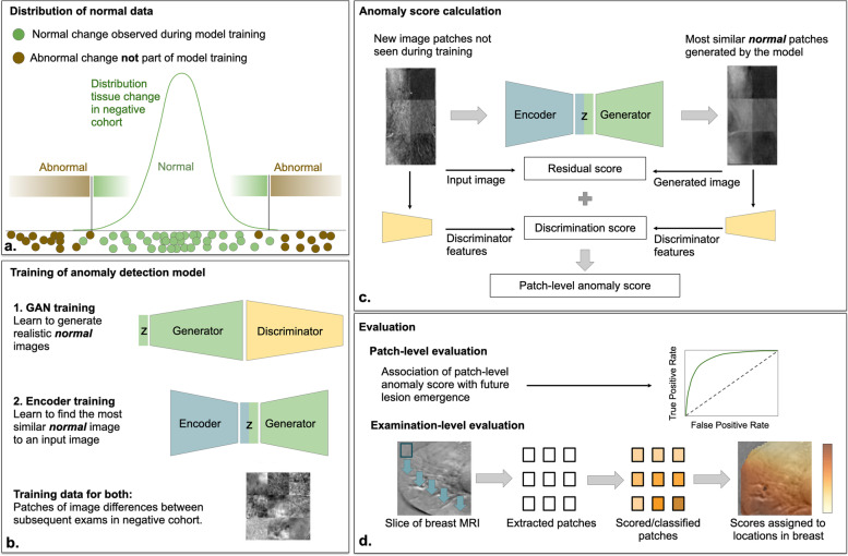
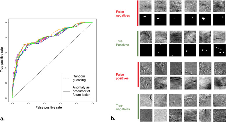
In this prospective study, we trained a generative adversarial network on dynamic CE-MRI of 33 high-risk women who participated in a screening program but did not develop BC. We defined an anomaly score as the deviation of an observed CE-MRI scan from the model of normal breast tissue variability. We evaluated the anomaly score's association with future lesion emergence on the level of local image patches (104,531 normal patches, 455 patches of future lesion location) and entire CE-MRI exams (21 normal, 20 with future lesion). Associations were analyzed by receiver operating characteristic (ROC) curves on the patch level and logistic regression on the examination level.
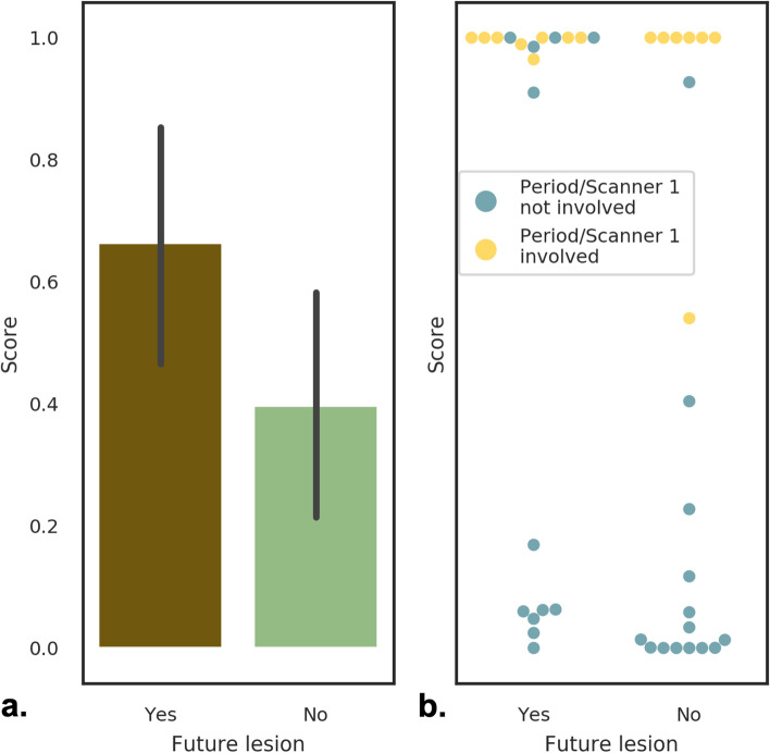
The local anomaly score on image patches was a good predictor for future lesion emergence (area under the ROC curve 0.804). An exam-level summary score was significantly associated with the emergence of lesions at any location at a later time point (p = 0.045).
Breast cancer lesions are associated with anomalous appearance changes in breast CE-MRI occurring before the lesion emerges in high-risk women. These early image signatures are detectable and may be a basis for adjusting individual BC risk and personalized screening.
Anomalies in screening MRI preceding lesion emergence in women at high-risk of breast cancer may inform individualized screening and intervention strategies.
• Breast lesions are associated with preceding anomalies in CE-MRI of high-risk women. • Deep learning-based anomaly detection can help to adjust risk assessment for future lesions. • An appearance anomaly score may be used for adjusting screening interval times.
国际社会已经发布了高危乳腺癌(BC)筛查指南,建议将乳腺对比增强磁共振成像(CE-MRI)作为补充诊断工具。在我们的研究中,我们测试了基于深度学习的异常检测在识别与未来病变出现相关的阴性乳腺 CE-MRI 筛查中异常变化的适用性。
在这项前瞻性研究中,我们在 33 名参加筛查计划但未患乳腺癌的高危女性的动态 CE-MRI 上训练了一个生成对抗网络。我们将异常评分定义为观察到的 CE-MRI 扫描与正常乳腺组织变异性模型的偏差。我们评估了局部图像斑块(104,531 个正常斑块,455 个未来病变位置斑块)和整个 CE-MRI 检查(21 个正常,20 个有未来病变)上异常评分与未来病变出现的相关性。在斑块水平上通过接收者操作特征(ROC)曲线分析关联,在检查水平上通过逻辑回归分析关联。
图像斑块上的局部异常评分是未来病变出现的良好预测指标(ROC 曲线下面积为 0.804)。在任何位置出现病变的检查水平汇总评分与之后的病变出现显著相关(p = 0.045)。
在高危女性中,乳腺癌病变与 CE-MRI 筛查前病变出现之前的异常外观变化相关。这些早期的图像特征是可检测的,可能为调整个体乳腺癌风险和个性化筛查提供依据。
在乳腺癌高危女性中,CE-MRI 筛查前病变出现之前的异常可能为个体化筛查和干预策略提供信息。
• 高危女性的 CE-MRI 中,病变前存在异常。• 基于深度学习的异常检测有助于调整未来病变的风险评估。• 外观异常评分可用于调整筛查间隔时间。