Leibniz Institute of Photonic Technology (Member of Leibniz Health Technologies, Member of the Leibniz Centre for Photonics in Infection Research, LPI), 07745 Jena, Germany.
Center for Sepsis Control and Care, Jena University Hospital, 07747 Jena, Germany.
Int J Mol Sci. 2023 Jun 5;24(11):9762. doi: 10.3390/ijms24119762.
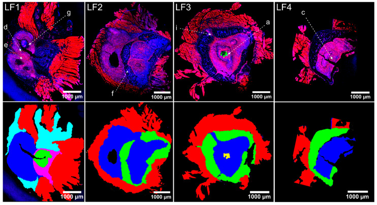
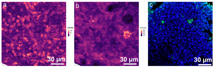
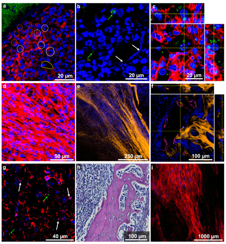
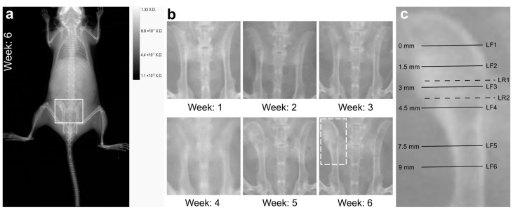
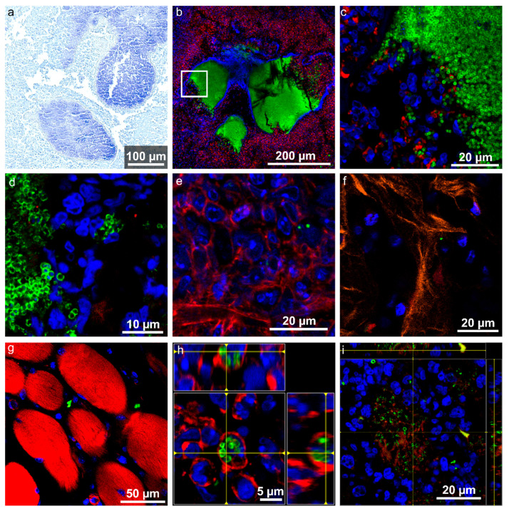
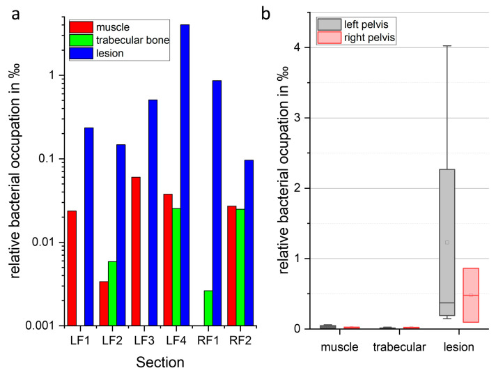
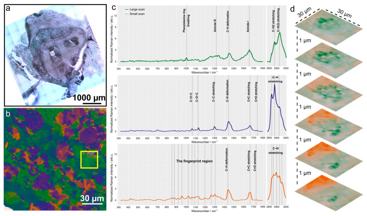
Osteomyelitis is an infection of the bone that is often difficult to treat and causes a significant healthcare burden. is the most common pathogen causing osteomyelitis. Osteomyelitis mouse models have been established to gain further insights into the pathogenesis and host response. Here, we use an established hematogenous osteomyelitis mouse model to investigate morphological tissue changes and bacterial localization in chronic osteomyelitis with a focus on the pelvis. X-ray imaging was performed to follow the disease progression. Six weeks post infection, when osteomyelitis had manifested itself with a macroscopically visible bone deformation in the pelvis, we used two orthogonal methods, namely fluorescence imaging and label-free Raman spectroscopy, to characterise tissue changes on a microscopic scale and to localise bacteria in different tissue regions. Hematoxylin and eosin as well as Gram staining were performed as a reference method. We could detect all signs of a chronically florid tissue infection with osseous and soft tissue changes as well as with different inflammatory infiltrate patterns. Large lesions dominated in the investigated tissue samples. Bacteria were found to form abscesses and were distributed in high numbers in the lesion, where they could occasionally also be detected intracellularly. In addition, bacteria were found in lower numbers in surrounding muscle tissue and even in lower numbers in trabecular bone tissue. The Raman spectroscopic imaging revealed a metabolic state of the bacteria with reduced activity in agreement with small cell variants found in other studies. In conclusion, we present novel optical methods to characterise bone infections, including inflammatory host tissue reactions and bacterial adaptation.
骨髓炎是一种骨感染,通常难以治疗,给医疗保健带来巨大负担。 是引起骨髓炎最常见的病原体。已建立骨髓炎小鼠模型以深入了解发病机制和宿主反应。在这里,我们使用已建立的血源性骨髓炎小鼠模型来研究慢性骨髓炎的形态组织变化和细菌定位,重点是骨盆。进行 X 射线成像以跟踪疾病进展。感染后 6 周,当骨髓炎在骨盆中表现出肉眼可见的骨变形时,我们使用两种正交方法,即荧光成像和无标记拉曼光谱,在微观尺度上描述组织变化,并在不同组织区域定位细菌。进行苏木精和伊红以及革兰氏染色作为参考方法。我们可以检测到慢性炎症组织感染的所有迹象,包括骨和软组织变化以及不同的炎症浸润模式。在研究的组织样本中,大病灶占主导地位。细菌形成脓肿,并在病变部位大量分布,偶尔也可以在细胞内检测到。此外,在周围肌肉组织中的细菌数量较少,在小梁骨组织中的细菌数量甚至更少。拉曼光谱成像显示细菌的代谢状态与其他研究中发现的小细胞变体一致,活性降低。总之,我们提出了新的光学方法来描述骨感染,包括炎症宿主组织反应和细菌适应。