• 文献检索
  • 文档翻译
  • 深度研究
  • 学术资讯
  • Suppr Zotero 插件Zotero 插件
  • 邀请有礼
  • 套餐&价格
  • 历史记录
应用&插件
Suppr Zotero 插件Zotero 插件浏览器插件Mac 客户端Windows 客户端微信小程序
定价
高级版会员购买积分包购买API积分包
服务
文献检索文档翻译深度研究API 文档MCP 服务
关于我们
关于 Suppr公司介绍联系我们用户协议隐私条款
关注我们

Suppr 超能文献

核心技术专利:CN118964589B侵权必究
粤ICP备2023148730 号-1Suppr @ 2026

文献检索

告别复杂PubMed语法,用中文像聊天一样搜索,搜遍4000万医学文献。AI智能推荐,让科研检索更轻松。

立即免费搜索

文件翻译

保留排版,准确专业,支持PDF/Word/PPT等文件格式,支持 12+语言互译。

免费翻译文档

深度研究

AI帮你快速写综述,25分钟生成高质量综述,智能提取关键信息,辅助科研写作。

立即免费体验

胸部计算机断层扫描图像、肿瘤标志物和代谢组学在鉴别肺结节良恶性中的预测价值。

The predictive value of chest computed tomography images, tumor markers, and metabolomics in the identification of benign and malignant pulmonary nodules.

作者信息

Xu Shuhua, Ge Jingjun, Liu Xiaoli, He Qiyu, Xu Dongjin, Cao Weikang, Ding Junning, Kai Xinghua, Zhou Guoping

机构信息

Department of Cardiothoracic Surgery, Dongtai Hospital of Traditional Chinese Medicine, Dongtai, China.

Department of Radiology Imaging, Dongtai Hospital of Traditional Chinese Medicine, Dongtai, China.

出版信息

J Thorac Dis. 2023 May 30;15(5):2668-2679. doi: 10.21037/jtd-23-250. Epub 2023 May 24.

DOI:10.21037/jtd-23-250
PMID:37324101
原文链接:https://pmc.ncbi.nlm.nih.gov/articles/PMC10267948/
Abstract

BACKGROUND

Invasive puncture biopsy is currently the main method of identifying benign and malignant pulmonary nodules (PNs). This study aimed to investigate the application effect of chest computed tomography (CT) images, tumor markers (TMs), and metabolomics in the identification of benign and malignant PNs (MPNs).

METHODS

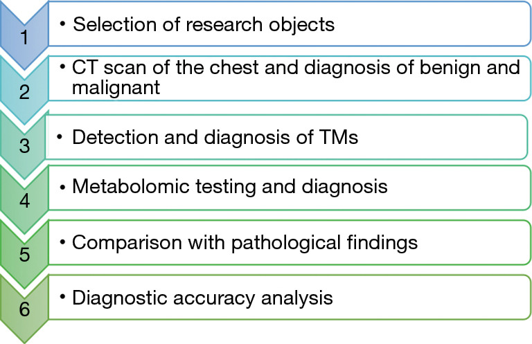
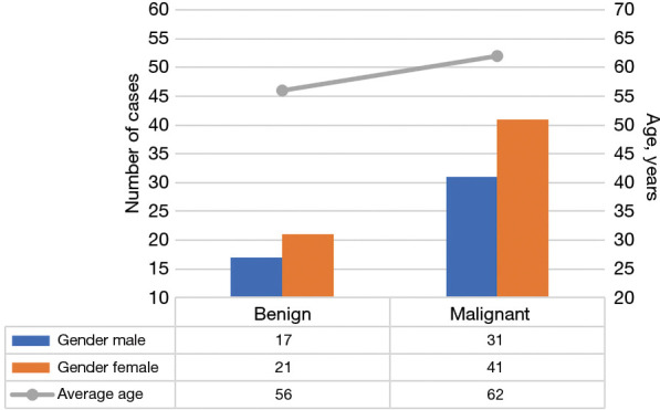
A total of 110 patients with PNs who were hospitalized in Dongtai Hospital of Traditional Chinese Medicine from March 2021 to March 2022 were selected as the study cohort. A retrospective analysis study of chest CT imaging, serum TMs testing, and plasma fatty acid (FA) metabolomics was performed on all participants.

RESULTS

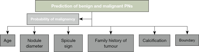
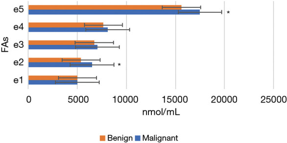
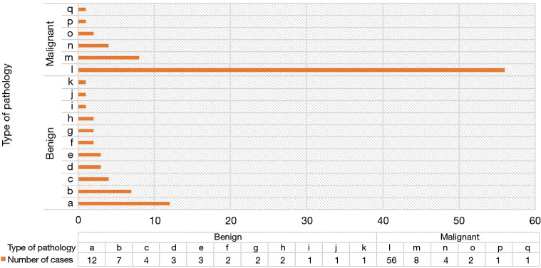
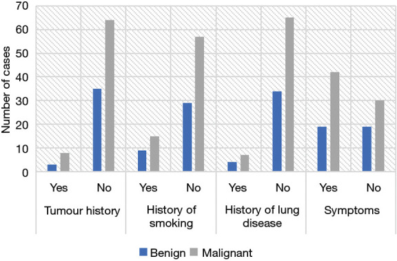
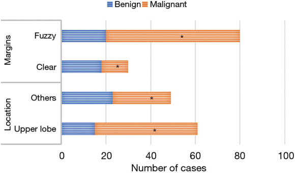
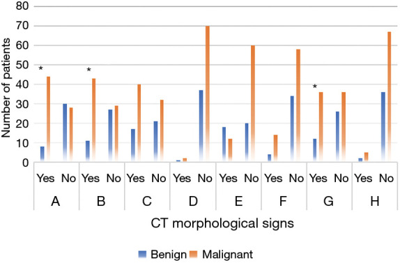
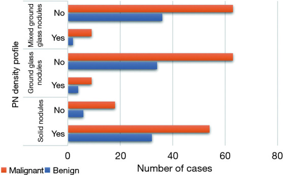
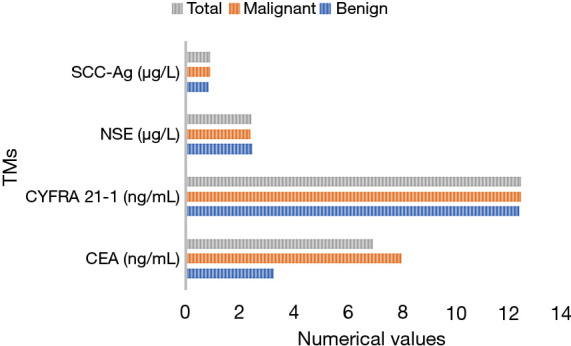
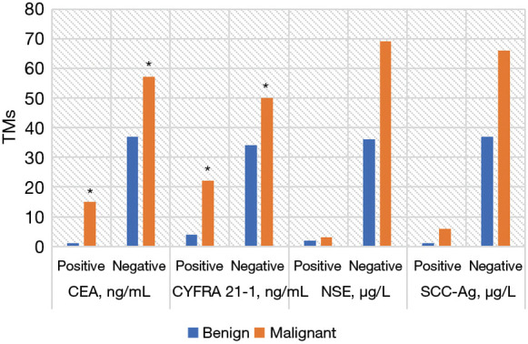
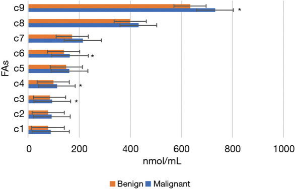
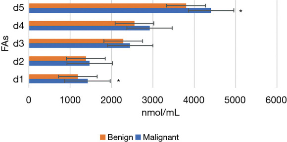
According to the pathological results, participants were divided into a MPN group (n=72) and a benign PN (BPN) group (n=38). The morphological signs of CT images, the levels and positive rate of serum TMs, and the plasma FA indicator were compared between groups. There were significant differences between the MPN group and the BPN group in the CT morphological signs, including location of PN and the number of patients with or without lobulation sign, spicule sign, and vessel convergence sign (P<0.05). Serum carcinoembryonic antigen (CEA), cytokeratin-19 fragment (CYFRA 21-1), neuron-specific enolase (NSE), and squamous cell carcinoma antigen (SCC-Ag) were not significantly different between the 2 groups. The serum contents of CEA and CYFRA 21-1 in the MPN group were remarkably higher than those in the BPN group (P<0.05). The plasma levels of palmitic acid, total omega-3 polyunsaturated FA (W3), nervonic acid, stearic acid, docosatetraenoic acid, linolenic acid, eicosapentaenoic acid, total saturated FA, and total FA were much higher in the MPN group than the BPN group (P<0.05).

CONCLUSIONS

In conclusion, chest CT images and TMs, combined with metabolomics, has a good application effect in the diagnosis of BPNs and MPNs, and is worthy of further promotion.

摘要

背景

侵入性穿刺活检是目前鉴别肺结节(PNs)良恶性的主要方法。本研究旨在探讨胸部计算机断层扫描(CT)图像、肿瘤标志物(TMs)和代谢组学在鉴别良恶性PNs(MPNs)中的应用效果。

方法

选取2021年3月至2022年3月在东台市中医院住院的110例PNs患者作为研究队列。对所有参与者进行胸部CT成像、血清TMs检测和血浆脂肪酸(FA)代谢组学的回顾性分析研究。

结果

根据病理结果,参与者分为MPN组(n = 72)和良性PN(BPN)组(n = 38)。比较两组间CT图像的形态学征象、血清TMs水平及阳性率和血浆FA指标。MPN组和BPN组在CT形态学征象方面存在显著差异,包括PN的位置以及有或无分叶征、毛刺征和血管集束征的患者数量(P < 0.05)。两组间血清癌胚抗原(CEA)、细胞角蛋白19片段(CYFRA 21 - 1)、神经元特异性烯醇化酶(NSE)和鳞状细胞癌抗原(SCC - Ag)无显著差异。MPN组血清CEA和CYFRA 21 - 1含量明显高于BPN组(P < 0.05)。MPN组血浆棕榈酸、总ω - 3多不饱和脂肪酸(W3)、神经酸、硬脂酸、二十二碳四烯酸、亚麻酸、二十碳五烯酸、总饱和脂肪酸和总脂肪酸水平均显著高于BPN组(P < 0.05)。

结论

总之,胸部CT图像和TMs,结合代谢组学,在BPNs和MPNs的诊断中具有良好的应用效果,值得进一步推广。

https://cdn.ncbi.nlm.nih.gov/pmc/blobs/2ef6/10267948/2f562c23144a/jtd-15-05-2668-f15.jpg
https://cdn.ncbi.nlm.nih.gov/pmc/blobs/2ef6/10267948/1c297b318d1d/jtd-15-05-2668-f1.jpg
https://cdn.ncbi.nlm.nih.gov/pmc/blobs/2ef6/10267948/23f83836e2c3/jtd-15-05-2668-f2.jpg
https://cdn.ncbi.nlm.nih.gov/pmc/blobs/2ef6/10267948/7cdc7960fcd6/jtd-15-05-2668-f3.jpg
https://cdn.ncbi.nlm.nih.gov/pmc/blobs/2ef6/10267948/3e0ffb132d55/jtd-15-05-2668-f4.jpg
https://cdn.ncbi.nlm.nih.gov/pmc/blobs/2ef6/10267948/4ce66ed39537/jtd-15-05-2668-f5.jpg
https://cdn.ncbi.nlm.nih.gov/pmc/blobs/2ef6/10267948/a96512622d1e/jtd-15-05-2668-f6.jpg
https://cdn.ncbi.nlm.nih.gov/pmc/blobs/2ef6/10267948/c2aa771db7ea/jtd-15-05-2668-f7.jpg
https://cdn.ncbi.nlm.nih.gov/pmc/blobs/2ef6/10267948/b3723f9af67d/jtd-15-05-2668-f8.jpg
https://cdn.ncbi.nlm.nih.gov/pmc/blobs/2ef6/10267948/620be436418d/jtd-15-05-2668-f9.jpg
https://cdn.ncbi.nlm.nih.gov/pmc/blobs/2ef6/10267948/047b1bc97202/jtd-15-05-2668-f10.jpg
https://cdn.ncbi.nlm.nih.gov/pmc/blobs/2ef6/10267948/486910cddb9c/jtd-15-05-2668-f11.jpg
https://cdn.ncbi.nlm.nih.gov/pmc/blobs/2ef6/10267948/f13cbb98025b/jtd-15-05-2668-f12.jpg
https://cdn.ncbi.nlm.nih.gov/pmc/blobs/2ef6/10267948/133964a43ee2/jtd-15-05-2668-f13.jpg
https://cdn.ncbi.nlm.nih.gov/pmc/blobs/2ef6/10267948/3d6cb0a69298/jtd-15-05-2668-f14.jpg
https://cdn.ncbi.nlm.nih.gov/pmc/blobs/2ef6/10267948/2f562c23144a/jtd-15-05-2668-f15.jpg

相似文献

1
The predictive value of chest computed tomography images, tumor markers, and metabolomics in the identification of benign and malignant pulmonary nodules.胸部计算机断层扫描图像、肿瘤标志物和代谢组学在鉴别肺结节良恶性中的预测价值。
J Thorac Dis. 2023 May 30;15(5):2668-2679. doi: 10.21037/jtd-23-250. Epub 2023 May 24.
2
Combining serum miRNAs, CEA, and CYFRA21-1 with imaging and clinical features to distinguish benign and malignant pulmonary nodules: a pilot study : Xianfeng Li et al.: Combining biomarker, imaging, and clinical features to distinguish pulmonary nodules.联合血清微小RNA、癌胚抗原和细胞角蛋白19片段与影像学及临床特征鉴别肺结节的良恶性:一项初步研究:李先锋等人:联合生物标志物、影像学和临床特征鉴别肺结节
World J Surg Oncol. 2017 May 25;15(1):107. doi: 10.1186/s12957-017-1171-y.
3
Combination of serum ACSL4 levels and low-dose 256-slice spiral CT exhibits the potential in the early screening of lung cancer.血清 ACSL4 水平与低剂量 256 层螺旋 CT 的联合应用具有早期筛查肺癌的潜力。
Medicine (Baltimore). 2023 Feb 3;102(5):e32733. doi: 10.1097/MD.0000000000032733.
4
Utility of squamous cell carcinoma antigen, carcinoembryonic antigen, Cyfra 21-1 and neuron specific enolase in lung cancer diagnosis: a prospective study from China.鳞状细胞癌抗原、癌胚抗原、细胞角蛋白 19 片段 21-1 和神经元特异性烯醇化酶在肺癌诊断中的应用:来自中国的一项前瞻性研究。
Chin Med J (Engl). 2011 Oct;124(20):3244-8.
5
Diagnostic value of chest radiography, computed tomography and tumour markers in the differentiation of malignant from benign solitary pulmonary lesions.胸部X线摄影、计算机断层扫描及肿瘤标志物在鉴别孤立性肺良性与恶性病变中的诊断价值
Eur J Med Res. 1999 Aug 25;4(8):313-27.
6
Combination of positron emission tomography/computed tomography and chest thin-layer high-resolution computed tomography for evaluation of pulmonary nodules: Correlation with imaging features, maximum standardized uptake value, and pathology.正电子发射断层扫描/计算机断层扫描与胸部薄层高分辨率计算机断层扫描联合用于评估肺结节:与影像学特征、最大标准化摄取值及病理的相关性
Medicine (Baltimore). 2018 Aug;97(31):e11640. doi: 10.1097/MD.0000000000011640.
7
Cytokeratin 19 fragment CYFRA 21-1 compared with carcinoembryonic antigen, squamous cell carcinoma antigen and neuron-specific enolase in lung cancer. Results of an international multicentre study.细胞角蛋白19片段CYFRA 21-1与癌胚抗原、鳞状细胞癌抗原及神经元特异性烯醇化酶在肺癌中的比较:一项国际多中心研究结果
Eur J Clin Chem Clin Biochem. 1994 Mar;32(3):189-99.
8
An evaluation of the tumour markers, carcinoembryonic antigen (CEA), cytokeratin marker (CYFRA 21-1) and neuron-specific enolase (NSE) in the differentiation of malignant from benign solitary pulmonary lesions.肿瘤标志物癌胚抗原(CEA)、细胞角蛋白标志物(CYFRA 21-1)和神经元特异性烯醇化酶(NSE)在鉴别孤立性肺良性病变与恶性病变中的评估。
Lung Cancer. 1999 Dec;26(3):149-55. doi: 10.1016/s0169-5002(99)00084-7.
9
Classification of solid pulmonary nodules using a machine-learning nomogram based on F-FDG PET/CT radiomics integrated clinicobiological features.基于F-FDG PET/CT影像组学整合临床生物学特征的机器学习列线图对实性肺结节的分类
Ann Transl Med. 2022 Dec;10(23):1265. doi: 10.21037/atm-22-2647.
10
[Diagnostic value of serum tumor markers in differentiating malignant from benign solitary pulmonary nodules].[血清肿瘤标志物在鉴别孤立性肺结节良恶性中的诊断价值]
Beijing Da Xue Xue Bao Yi Xue Ban. 2014 Oct 18;46(5):707-10.

引用本文的文献

1
Development and validation of CT radiomics diagnostic models: differentiating benign from malignant pulmonary nodules and evaluating malignancy degree.CT影像组学诊断模型的开发与验证:鉴别肺结节的良恶性及评估恶性程度
J Thorac Dis. 2025 Mar 31;17(3):1645-1672. doi: 10.21037/jtd-2025-152. Epub 2025 Mar 20.
2
Minimally invasive biomarkers for triaging lung nodules-challenges and future perspectives.用于肺结节分类的微创生物标志物——挑战与未来展望
Cancer Metastasis Rev. 2025 Jan 31;44(1):29. doi: 10.1007/s10555-025-10247-5.

本文引用的文献

1
Integrative Serum Metabolic Fingerprints Based Multi-Modal Platforms for Lung Adenocarcinoma Early Detection and Pulmonary Nodule Classification.基于整合血清代谢指纹的多模态平台用于肺腺癌早期检测和肺结节分类。
Adv Sci (Weinh). 2022 Dec;9(34):e2203786. doi: 10.1002/advs.202203786. Epub 2022 Oct 18.
2
Plasm Metabolomics Study in Pulmonary Metastatic Carcinoma.肺转移性癌的血浆代谢组学研究
J Oncol. 2022 Aug 21;2022:9460019. doi: 10.1155/2022/9460019. eCollection 2022.
3
Comprehensive plasma metabolomics and lipidomics of benign and malignant solitary pulmonary nodules.
良性和恶性孤立性肺结节的综合血浆代谢组学和脂质组学研究。
Metabolomics. 2022 Aug 29;18(9):71. doi: 10.1007/s11306-022-01929-0.
4
Computer-aided Classification of Lung Nodules on CT Images with Expert Knowledge.基于专家知识的CT图像上肺结节的计算机辅助分类
Proc SPIE Int Soc Opt Eng. 2021 Feb;11598. doi: 10.1117/12.2581888. Epub 2021 Feb 15.
5
Malignant Nodules Detected on Lung Cancer Screening CT: Yield of Short-Term Follow-Up CT in Showing Nodule Growth.肺癌筛查 CT 发现的恶性结节:短期随访 CT 在显示结节生长方面的收益。
AJR Am J Roentgenol. 2022 Nov;219(5):735-741. doi: 10.2214/AJR.22.27869. Epub 2022 Jun 8.
6
FLUORO-CT GUIDED BIOPSY OF LUNG NODULES: A STEP BY STEP REVISION.氟代 CT 引导下的肺结节活检:步步修订。
Port J Card Thorac Vasc Surg. 2022 Jan 4;28(4):43-46. doi: 10.48729/pjctvs.223.
7
Clinical impact of a deep learning system for automated detection of missed pulmonary nodules on routine body computed tomography including the chest region.一种深度学习系统对常规胸部计算机断层扫描中漏诊肺结节进行自动检测的临床影响。
Eur Radiol. 2022 May;32(5):2891-2900. doi: 10.1007/s00330-021-08412-9. Epub 2022 Jan 9.
8
Epigenetic imprinting alterations as effective diagnostic biomarkers for early-stage lung cancer and small pulmonary nodules.表观遗传印迹改变作为早期肺癌和小肺结节的有效诊断生物标志物。
Clin Epigenetics. 2021 Dec 14;13(1):220. doi: 10.1186/s13148-021-01203-5.
9
Cancer Risk in Nodules Detected at Follow-Up Lung Cancer Screening CT.随访肺癌筛查 CT 检测到的结节中的癌症风险。
AJR Am J Roentgenol. 2022 Apr;218(4):634-641. doi: 10.2214/AJR.21.26927. Epub 2021 Nov 10.
10
Accurate segmentation for different types of lung nodules on CT images using improved U-Net convolutional network.利用改进的 U-Net 卷积网络对 CT 图像上的不同类型肺结节进行精确分割。
Medicine (Baltimore). 2021 Oct 8;100(40):e27491. doi: 10.1097/MD.0000000000027491.