• 文献检索
  • 文档翻译
  • 深度研究
  • 学术资讯
  • Suppr Zotero 插件Zotero 插件
  • 邀请有礼
  • 套餐&价格
  • 历史记录
应用&插件
Suppr Zotero 插件Zotero 插件浏览器插件Mac 客户端Windows 客户端微信小程序
定价
高级版会员购买积分包购买API积分包
服务
文献检索文档翻译深度研究API 文档MCP 服务
关于我们
关于 Suppr公司介绍联系我们用户协议隐私条款
关注我们

Suppr 超能文献

核心技术专利:CN118964589B侵权必究
粤ICP备2023148730 号-1Suppr @ 2026

文献检索

告别复杂PubMed语法,用中文像聊天一样搜索,搜遍4000万医学文献。AI智能推荐,让科研检索更轻松。

立即免费搜索

文件翻译

保留排版,准确专业,支持PDF/Word/PPT等文件格式,支持 12+语言互译。

免费翻译文档

深度研究

AI帮你快速写综述,25分钟生成高质量综述,智能提取关键信息,辅助科研写作。

立即免费体验

[肛周瘘管:概述]

[Perianal Fistula: An Overview].

作者信息

Kim Min Ju

出版信息

J Korean Soc Radiol. 2023 May;84(3):565-585. doi: 10.3348/jksr.2023.0018. Epub 2023 May 30.

DOI:10.3348/jksr.2023.0018
PMID:37325009
原文链接:https://pmc.ncbi.nlm.nih.gov/articles/PMC10265243/
Abstract

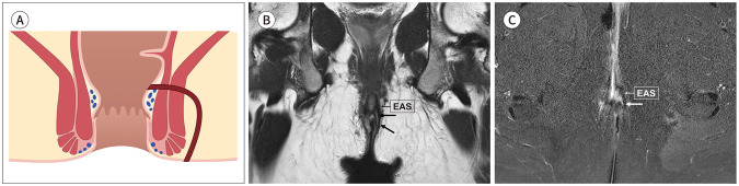
Perianal fistula is a common inflammatory condition in the general population and affects the area around the anal canal. Although most cases are benign, they cause serious morbidity and require surgical treatment due to a high risk of recurrence. MR imaging is a gold standard technique for the evaluation of perianal fistulas and provides accurate information on the anatomy of the anal canal, its relationship to the anal sphincter complex, accurate identification of secondary tracts or abscesses, and reporting associated complications. MR imaging can help monitor treatment effects and determine treatment methods. Crohn's disease-related fistulas often require medical rather than surgical treatment. The radiologist is required to know the anatomy and MR imaging findings of the perianal fistula to present an accurate diagnosis to the clinician.

摘要

肛周瘘是普通人群中一种常见的炎症性疾病,累及肛管周围区域。尽管大多数病例为良性,但由于复发风险高,它们会导致严重的发病率且需要手术治疗。磁共振成像(MR)是评估肛周瘘的金标准技术,可提供有关肛管解剖结构、其与肛门括约肌复合体的关系、准确识别继发管道或脓肿以及报告相关并发症的准确信息。MR成像有助于监测治疗效果并确定治疗方法。克罗恩病相关的瘘通常需要药物治疗而非手术治疗。放射科医生需要了解肛周瘘的解剖结构和MR成像表现,以便向临床医生提供准确的诊断。

https://cdn.ncbi.nlm.nih.gov/pmc/blobs/d165/10265243/0542a4719e65/jksr-84-565-g013.jpg
https://cdn.ncbi.nlm.nih.gov/pmc/blobs/d165/10265243/36b06a255424/jksr-84-565-g001.jpg
https://cdn.ncbi.nlm.nih.gov/pmc/blobs/d165/10265243/2b6656b20086/jksr-84-565-g002.jpg
https://cdn.ncbi.nlm.nih.gov/pmc/blobs/d165/10265243/e7199bdbef72/jksr-84-565-g003.jpg
https://cdn.ncbi.nlm.nih.gov/pmc/blobs/d165/10265243/92ec1ce555af/jksr-84-565-g004.jpg
https://cdn.ncbi.nlm.nih.gov/pmc/blobs/d165/10265243/840642450679/jksr-84-565-g005.jpg
https://cdn.ncbi.nlm.nih.gov/pmc/blobs/d165/10265243/de65307729e4/jksr-84-565-g006.jpg
https://cdn.ncbi.nlm.nih.gov/pmc/blobs/d165/10265243/9d69012ded38/jksr-84-565-g007.jpg
https://cdn.ncbi.nlm.nih.gov/pmc/blobs/d165/10265243/041fbecbf5fd/jksr-84-565-g008.jpg
https://cdn.ncbi.nlm.nih.gov/pmc/blobs/d165/10265243/47a05845e3bc/jksr-84-565-g009.jpg
https://cdn.ncbi.nlm.nih.gov/pmc/blobs/d165/10265243/1ca831965a3a/jksr-84-565-g010.jpg
https://cdn.ncbi.nlm.nih.gov/pmc/blobs/d165/10265243/02c196808b49/jksr-84-565-g011.jpg
https://cdn.ncbi.nlm.nih.gov/pmc/blobs/d165/10265243/c3f29e18ad6c/jksr-84-565-g012.jpg
https://cdn.ncbi.nlm.nih.gov/pmc/blobs/d165/10265243/0542a4719e65/jksr-84-565-g013.jpg

相似文献

1
[Perianal Fistula: An Overview].[肛周瘘管:概述]
J Korean Soc Radiol. 2023 May;84(3):565-585. doi: 10.3348/jksr.2023.0018. Epub 2023 May 30.
2
MR imaging evaluation of perianal fistulas: spectrum of imaging features.磁共振成像评估肛周瘘管:影像学特征谱。
Radiographics. 2012 Jan-Feb;32(1):175-94. doi: 10.1148/rg.321115040.
3
Treatment of Perianal Fistula and Abscess: Crohn's and Non-Crohn's.肛周瘘管和脓肿的治疗:克罗恩病型与非克罗恩病型。
Curr Treat Options Gastroenterol. 2007 Jun;10(3):211-20. doi: 10.1007/s11938-007-0014-2.
4
Anovaginal fistulas: evaluation with endoanal MR imaging.肛门阴道瘘:经肛门磁共振成像评估
Radiology. 2004 Apr;231(1):123-8. doi: 10.1148/radiol.2311021190.
5
Supplementary Anal Imaging by Magnetic Resonance Enterography in Patients with Crohn's Disease Not Suspected of Having Perianal Fistulas.疑似患有克罗恩病但无肛周瘘的患者的磁共振肠造影术补充分析成像。
Clin Gastroenterol Hepatol. 2020 Feb;18(2):415-423.e4. doi: 10.1016/j.cgh.2019.07.038. Epub 2019 Jul 25.
6
[Perianal Crohn's disease].[肛周克罗恩病]
Ther Umsch. 2013 Jul;70(7):417-28. doi: 10.1024/0040-5930/a000427.
7
The contribution of preoperative MRI to the surgical management of anal fistulas.术前磁共振成像在肛瘘手术治疗中的作用。
Diagn Interv Radiol. 2018 Nov;24(6):321-327. doi: 10.5152/dir.2018.18340.
8
MRI of perianal fistulas in Crohn's disease.MRI 对克罗恩病肛周瘘管的诊断。
Acta Biomed. 2020 Jul 13;91(8-S):27-33. doi: 10.23750/abm.v91i8-S.9970.
9
Diagnosis and treatment of fistulising Crohn's disease.瘘管性克罗恩病的诊断与治疗
Dan Med Bull. 2011 Oct;58(10):C4338.
10
Perianal fistulas in Crohn's disease: MRI diagnosis and surgical planning: MRI in fistulazing perianal Crohn's disease.克罗恩病中的肛周瘘管:MRI诊断与手术规划:肛周克罗恩病瘘管形成的MRI表现
Abdom Imaging. 2007 Nov;32(6):705-18. doi: 10.1007/s00261-007-9188-2. Epub 2007 Mar 3.

本文引用的文献

1
The development of a minimum dataset for MRI reporting of anorectal fistula: a multi-disciplinary, expert consensus process.肛门直肠瘘磁共振报告最小数据集的制定:多学科专家共识过程。
Eur Radiol. 2022 Dec;32(12):8306-8316. doi: 10.1007/s00330-022-08931-z. Epub 2022 Jun 23.
2
Perianal Imaging in Crohn Disease: Current Status With a Focus on MRI, From the Special Series on Imaging of Inflammation.克罗恩病的肛周影像学:以磁共振成像为重点的现状,来自炎症成像特刊
AJR Am J Roentgenol. 2022 May;218(5):781-792. doi: 10.2214/AJR.21.26615. Epub 2021 Sep 22.
3
AGA Clinical Practice Guidelines on the Medical Management of Moderate to Severe Luminal and Perianal Fistulizing Crohn's Disease.
美国胃肠病学会中重度管腔及肛周瘘管型克罗恩病医学管理临床实践指南
Gastroenterology. 2021 Jun;160(7):2496-2508. doi: 10.1053/j.gastro.2021.04.022.
4
Current concepts in the pathogenesis of cryptoglandular perianal fistula.肛门腺源性肛隐窝瘘发病机制的现代概念。
J Int Med Res. 2021 Feb;49(2):300060520986669. doi: 10.1177/0300060520986669.
5
ESGAR consensus statement on the imaging of fistula-in-ano and other causes of anal sepsis.ESGAR 共识声明:肛门瘘管和其他肛门感染原因的影像学诊断。
Eur Radiol. 2020 Sep;30(9):4734-4740. doi: 10.1007/s00330-020-06826-5. Epub 2020 Apr 19.
6
The role of magnetic resonance imaging in the preoperative evaluation of anal fistulas.磁共振成像在肛瘘术前评估中的作用。
Sci Rep. 2019 Nov 29;9(1):17947. doi: 10.1038/s41598-019-54441-2.
7
Imaging features of Benign Perianal lesions.肛周良性病变的影像学特征。
J Med Imaging Radiat Oncol. 2019 Oct;63(5):617-623. doi: 10.1111/1754-9485.12934. Epub 2019 Aug 1.
8
Development and Validation of a Magnetic Resonance Index for Assessing Fistulas in Patients With Crohn's Disease.发展和验证一种磁共振指数,用于评估克罗恩病患者的瘘管。
Gastroenterology. 2019 Nov;157(5):1233-1244.e5. doi: 10.1053/j.gastro.2019.07.027. Epub 2019 Jul 20.
9
MR Imaging of Perianal Fistulas.肛周瘘管的磁共振成像
Radiol Clin North Am. 2018 Sep;56(5):775-789. doi: 10.1016/j.rcl.2018.04.005. Epub 2018 Jul 11.
10
MRI of anal canal: normal anatomy, imaging protocol, and perianal fistulas: Part 1.MRI 检查肛门管:正常解剖、成像方案和肛周瘘:第 1 部分。
Abdom Radiol (NY). 2018 Jun;43(6):1334-1352. doi: 10.1007/s00261-017-1305-2.