Yang Dongming, Li Jie, Li Zhiping, Zhao Mengyang, Wang Dongdong, Sun Zhixin, Wen Pei, Gou Fengting, Dai Yuexin, Ji Yilan, Li Wen, Zhao Deming, Yang Lifeng
National Animal Transmissible Spongiform Encephalopathy Laboratory, College of Veterinary Medicine, State Key Laboratories for Agrobiotechnology, Key Laboratory of Animal Epidemiology of Ministry of Agriculture and Rural Affairs, China Agricultural University, Beijing, China.
Front Mol Neurosci. 2023 Jun 2;16:1163981. doi: 10.3389/fnmol.2023.1163981. eCollection 2023.
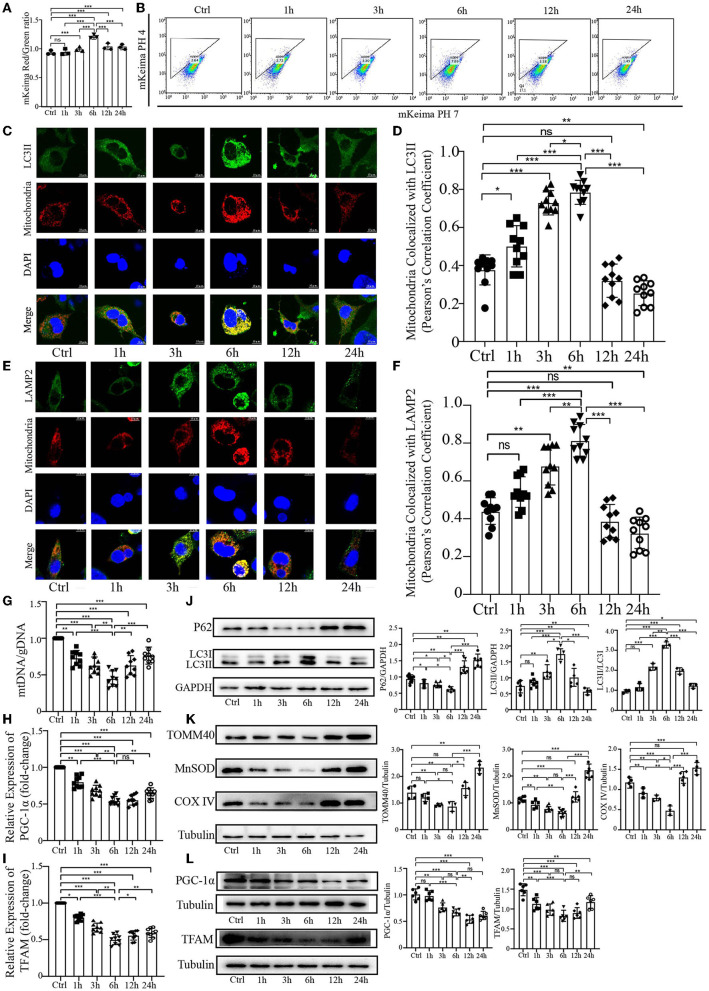
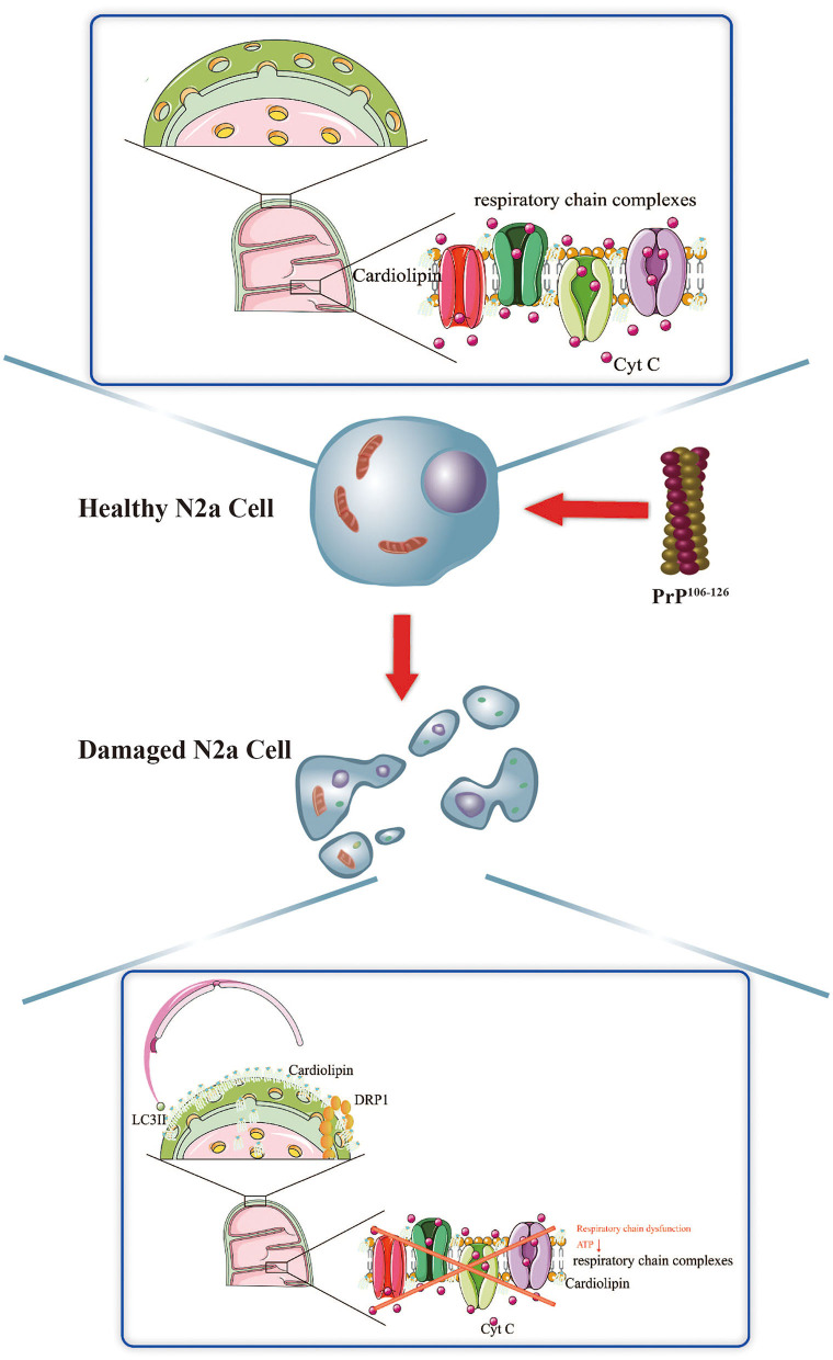
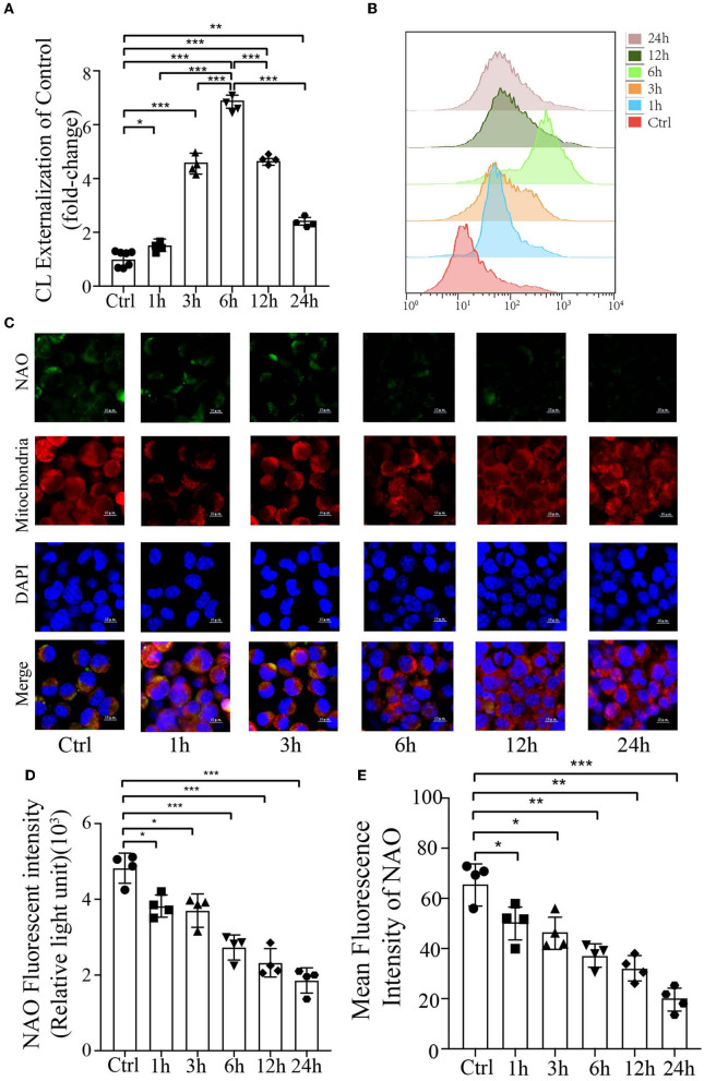
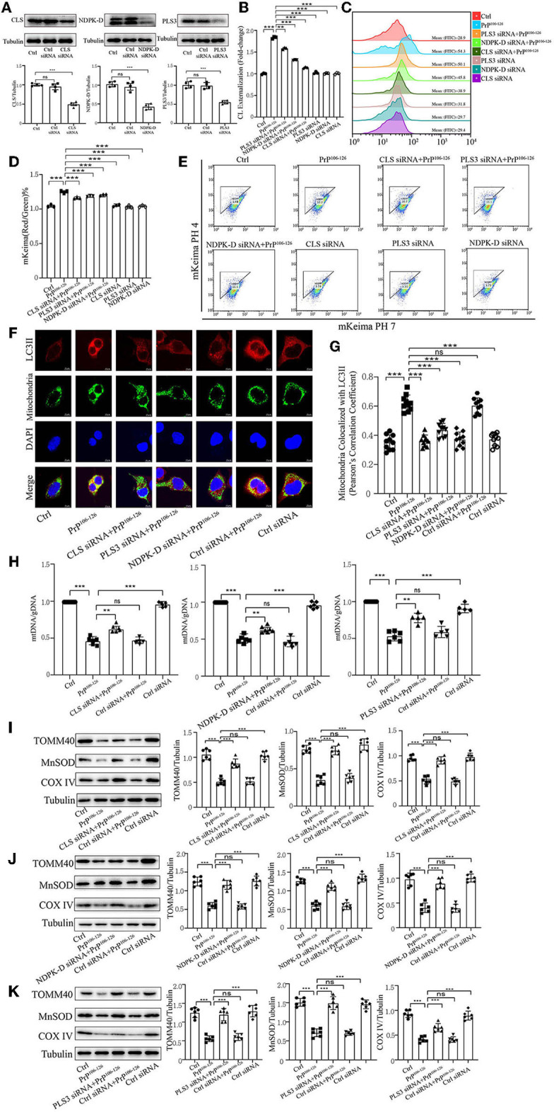
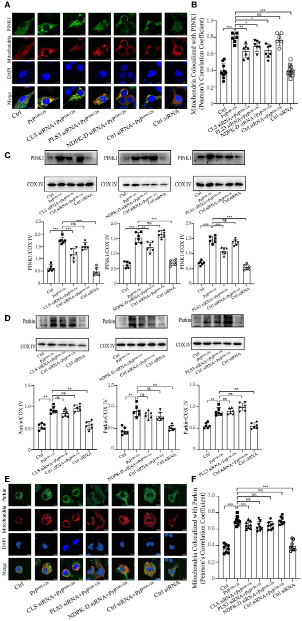
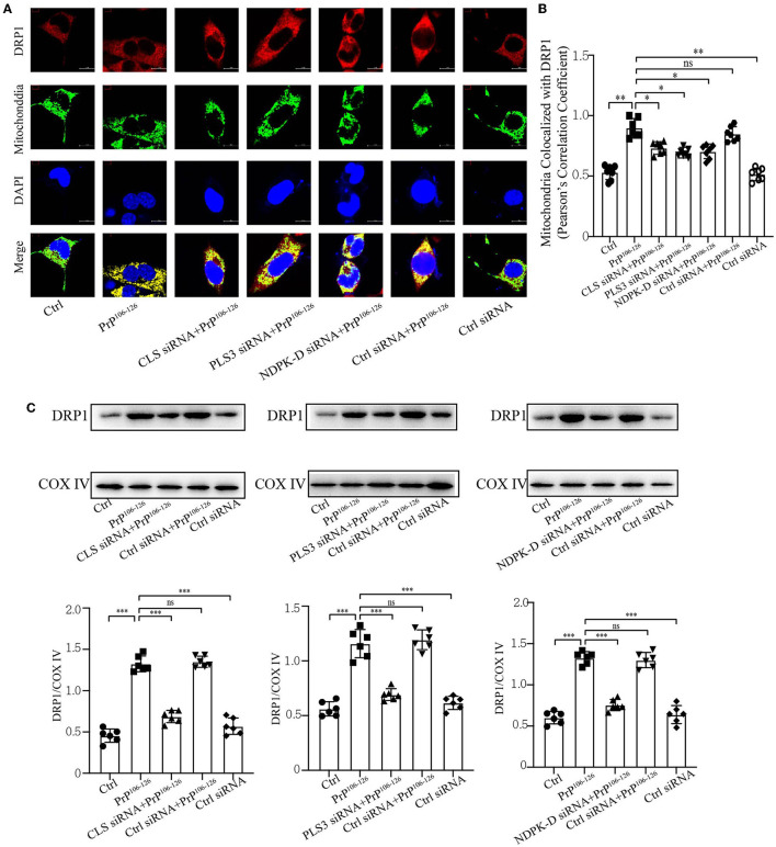
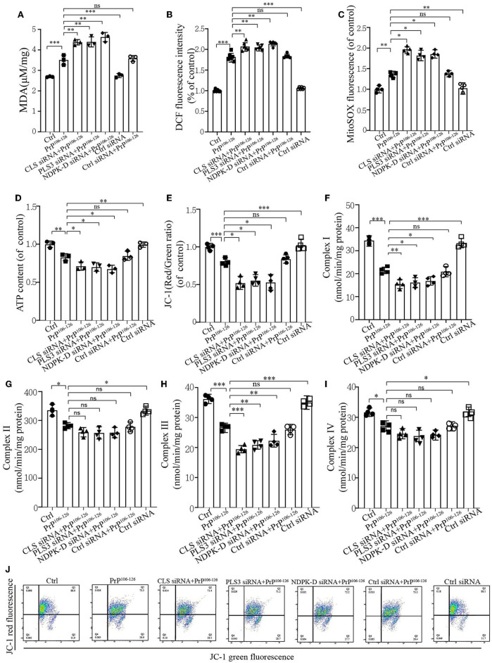
Proper mitochondrial performance is imperative for the maintenance of normal neuronal function to prevent the development of neurodegenerative diseases. Persistent accumulation of damaged mitochondria plays a role in prion disease pathogenesis, which involves a chain of events that culminate in the generation of reactive oxygen species and neuronal death. Our previous studies have demonstrated that PINK1/Parkin-mediated mitophagy induced by PrP is defective and leads to an accumulation of damaged mitochondria after PrP treatment. Externalized cardiolipin (CL), a mitochondria-specific phospholipid, has been reported to play a role in mitophagy by directly interacting with LC3II at the outer mitochondrial membrane. The involvement of CL externalization in PrP-induced mitophagy and its significance in other physiological processes of N2a cells treated with PrP remain unknown. We demonstrate that the PrP peptide caused a temporal course of mitophagy in N2a cells, which gradually increased and subsequently decreased. A similar trend in CL externalization to the mitochondrial surface was seen, resulting in a gradual decrease in CL content at the cellular level. Inhibition of CL externalization by knockdown of CL synthase, responsible for synthesis of CL, or phospholipid scramblase-3 and NDPK-D, responsible for CL translocation to the mitochondrial surface, significantly decreased PrP-induced mitophagy in N2a cells. Meanwhile, the inhibition of CL redistribution significantly decreased PINK1 and DRP1 recruitment in PrP treatment but had no significant decrease in Parkin recruitment. Furthermore, the inhibition of CL externalization resulted in impaired oxidative phosphorylation and severe oxidative stress, which led to mitochondrial dysfunction. Our results indicate that CL externalization induced by PrP on N2a cells plays a positive role in the initiation of mitophagy, leading to the stabilization of mitochondrial function.
正常的线粒体功能对于维持正常神经元功能以预防神经退行性疾病的发生至关重要。受损线粒体的持续积累在朊病毒病发病机制中起作用,这涉及一系列最终导致活性氧生成和神经元死亡的事件。我们之前的研究表明,由朊蛋白(PrP)诱导的PINK1/Parkin介导的线粒体自噬存在缺陷,并导致PrP处理后受损线粒体的积累。线粒体外膜磷脂酰甘油(CL),一种线粒体特异性磷脂,已被报道通过在外膜与LC3II直接相互作用而在线粒体自噬中发挥作用。CL外化在PrP诱导的线粒体自噬中的作用及其在PrP处理的N2a细胞其他生理过程中的意义尚不清楚。我们证明,PrP肽在N2a细胞中引起了线粒体自噬的时间进程,其逐渐增加随后减少。观察到CL向线粒体表面外化的类似趋势,导致细胞水平CL含量逐渐降低。通过敲低负责CL合成的CL合酶或负责CL转运到线粒体表面的磷脂翻转酶-3和NDPK-D来抑制CL外化,显著降低了PrP诱导的N2a细胞线粒体自噬。同时,CL重新分布的抑制显著降低了PrP处理中PINK1和动力相关蛋白1(DRP1)的募集,但对Parkin的募集没有显著降低。此外,CL外化的抑制导致氧化磷酸化受损和严重的氧化应激,从而导致线粒体功能障碍。我们的结果表明,PrP在N2a细胞上诱导的CL外化在启动线粒体自噬中起积极作用,从而导致线粒体功能的稳定。