Department of Radiology, Neuroradiology and Nuclear Medicine , Johannes Wesling University Hospital, Ruhr-University, Bochum, Germany.
Institute of Pathology, University of Magdeburg, Leipziger Str. 44, 39112, Magdeburg, Germany.
Eur Radiol. 2023 Sep;33(9):5955-5964. doi: 10.1007/s00330-023-09788-6. Epub 2023 Jun 22.
To investigate associations between apparent diffusion coefficient (ADC) and cell count, Ki 67, tumor-stroma ratio (TSR), and tumoral lymphocytes in different hepatic malignancies.
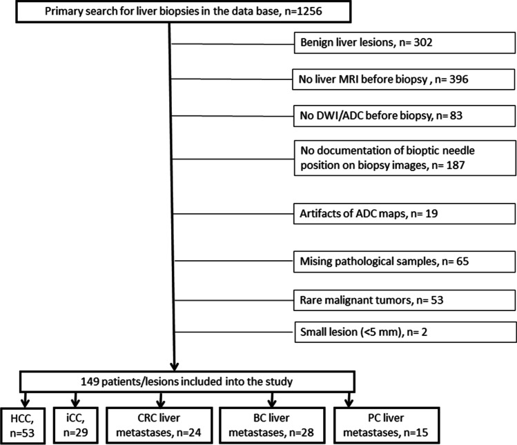
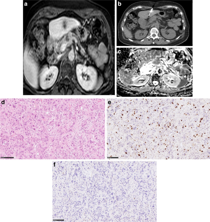
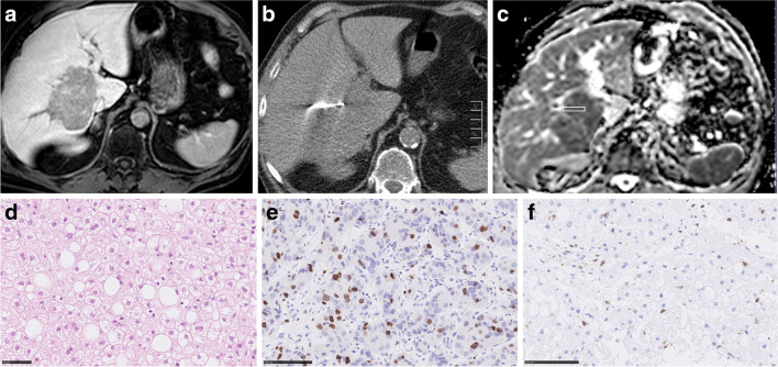
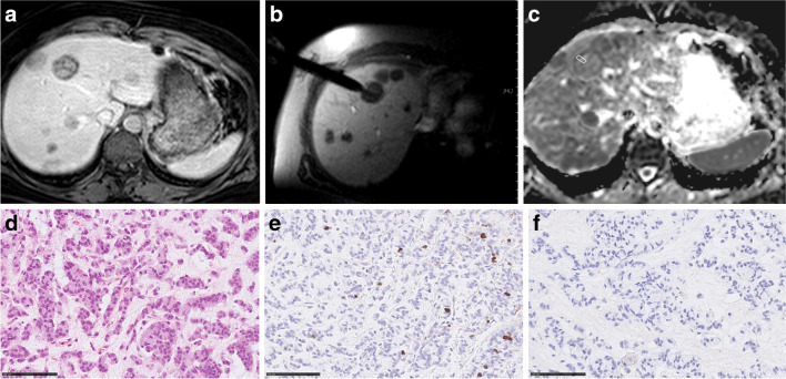
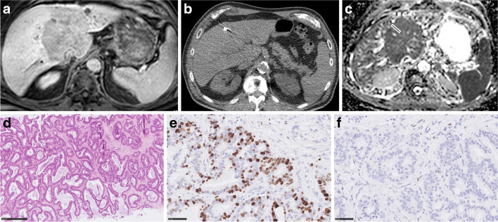
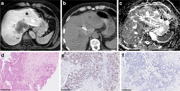
We identified 149 cases with performed liver biopsies: hepatocellular cancer (HCC, n = 53), intrahepatic cholangiocarcinoma (iCC, n = 29), metastases of colorectal cancer (CRC, n = 24), metastases of breast cancer (BC, n = 28), and metastases of pancreatic cancer (PC, n = 15). MRI was performed on a 1.5-T scanner with an axial echo-planar sequence. MRI was done before biopsy. Biopsy images of target lesions were selected. The cylindrical region of interest was placed on the ADC map of target lesions in accordance with the needle position on the biopsy images. Mean ADC values were estimated. TSR, cell counts, proliferation index Ki 67, and number of tumor-infiltrating lymphocytes were estimated. Spearman's rank correlation coefficients and intraclass correlation coefficients were calculated.
Inter-reader agreement was excellent regarding the ADC measurements. In HCC, ADC correlated with cell count (r = - 0.68, p < 0.001) and with TSR (r = 0.31, p = 0.024). In iCC, ADC correlated with TSR (r = 0.60, p < 0.001) and with cell count (r = - 0.54, p = 0.002). In CRC metastases, ADC correlated with cell count (r = - 0.54 p = 0.006) and with Ki 67 (r = - 0.46, p = 0.024). In BC liver metastases, ADC correlated with TSR (r = 0.55, p < 0.002) and with Ki 67 (r = - 0.51, p = 0.006). In PC metastases, no significant correlations were found.
ADC correlated with tumor cellularity in HCC, iCC, and CRC liver metastases. ADC reflects TSR in BC liver metastases, HCC, and iCC. ADC cannot reflect intratumoral lymphocytes.
The present study shows that the apparent diffusion coefficient can be used as a surrogate imaging marker for different histopathological features in several malignant hepatic lesions.
• ADC reflects different histopathological features in several hepatic tumors. • ADC correlates with tumor cellularity in HCC, iCC, and CRC metastases. • ADC strongly correlates with tumor-stroma ratio in BC metastases and iCC.
研究表观扩散系数(ADC)与细胞计数、Ki 67、肿瘤间质比(TSR)和不同肝恶性肿瘤中肿瘤浸润淋巴细胞之间的关系。
我们共纳入 149 例接受肝脏活检的患者:肝细胞癌(HCC,n=53)、肝内胆管细胞癌(iCC,n=29)、结直肠癌转移(CRC,n=24)、乳腺癌转移(BC,n=28)和胰腺癌转移(PC,n=15)。使用 1.5-T 扫描仪进行 MRI 检查,采用轴位回波平面序列。在活检前进行 MRI 检查。选择目标病变的活检图像。根据活检图像上的针位,将圆柱形感兴趣区放置在目标病变的 ADC 图上。估计平均 ADC 值。估计 TSR、细胞计数、增殖指数 Ki 67 和肿瘤浸润淋巴细胞数量。计算 Spearman 秩相关系数和组内相关系数。
ADC 测量的两位观察者之间的一致性很好。在 HCC 中,ADC 与细胞计数(r=-0.68,p<0.001)和 TSR(r=0.31,p=0.024)呈负相关。在 iCC 中,ADC 与 TSR(r=0.60,p<0.001)和细胞计数(r=-0.54,p=0.002)呈负相关。在 CRC 转移中,ADC 与细胞计数(r=-0.54,p=0.006)和 Ki 67(r=-0.46,p=0.024)呈负相关。在 BC 肝转移中,ADC 与 TSR(r=0.55,p<0.002)和 Ki 67(r=-0.51,p=0.006)呈负相关。在 PC 转移中,未发现显著相关性。
ADC 与 HCC、iCC 和 CRC 肝转移中的肿瘤细胞密度相关。ADC 反映 BC 肝转移、HCC 和 iCC 中的 TSR。ADC 不能反映肿瘤内淋巴细胞。
本研究表明,表观扩散系数可作为几种肝恶性病变中不同组织病理学特征的替代成像标志物。
• ADC 反映几种肝肿瘤的不同组织病理学特征。• ADC 与 HCC、iCC 和 CRC 转移中的肿瘤细胞密度相关。• ADC 与 BC 转移和 iCC 中的肿瘤间质比呈强相关性。