Hara Takeshi, Mizuno Masaki, Hida Kazutoshi, Sasamori Toru, Miyoshi Yasuyuki, Uchikado Hisaaki, Ohashi Hiroki, Sugawara Taku, Takeshima Yasuhiro, Ohara Yukoh, Kondo Akihide, Endo Toshiki
Spine and Spinal Cord center, Juntendo University, Tokyo, Japan.
Department of Neurosurgery, Juntendo University, Tokyo, Japan.
Neurospine. 2023 Sep;20(3):747-755. doi: 10.14245/ns.2346376.188. Epub 2023 Jun 20.
This study was aimed to report the clinical characteristics of intramedullary schwannomas and discuss imaging findings and treatment strategies.
The inclusion criterion was consecutive patients with intramedullary schwannomas who were surgically treated at 8 centers between 2009 and 2020. Clinical characteristics included age, sex, clinical presentation, disease duration, and follow-up period. The modified McCormick scale was used to compare the preoperative and postoperative conditions. Pre- and postoperative magnetic resonance images (MRI) of each case were analyzed.
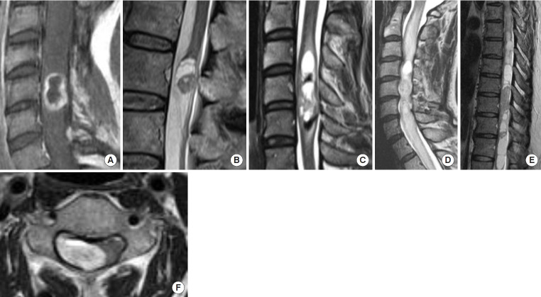
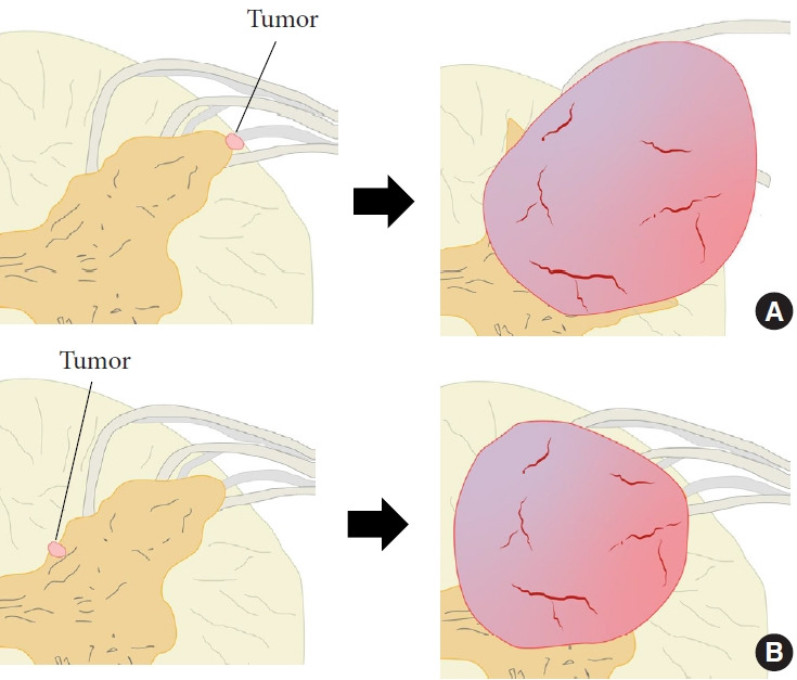
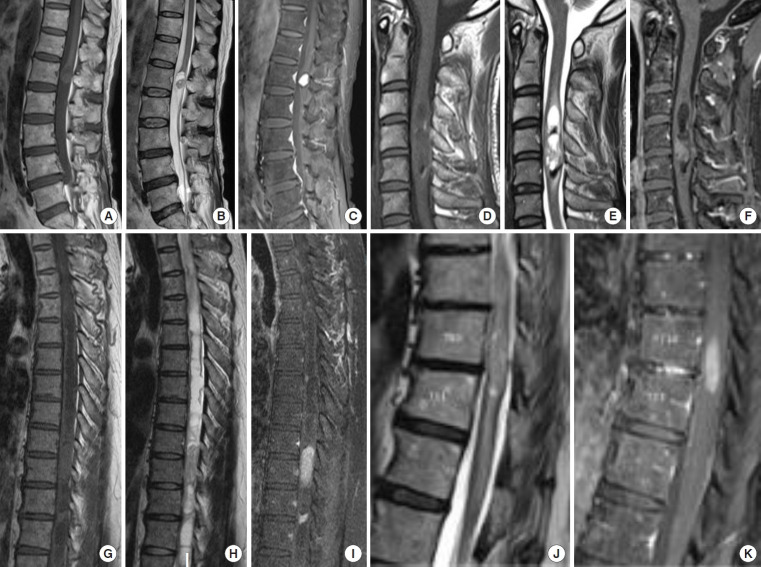
The mean age of the total 11 patients at the operation was 50.2 years. The mean duration of the symptoms was 23 months, with limb paresthesia being the most common clinical presentation. The cervical spine was the most common localization level of the tumor in 6 cases. The mean follow-up duration was 49.4 months. Gross total resection (GTR) and subtotal resection (STR) was achieved in 9 and 2 cases, respectively. According to the modified McCormick scale at 6 months postoperatively, 7 cases (63.6%) had improved and 4 cases (36.3%) had unchanged grades. Typical MRI findings of the intramedullary schwannoma included ring-like enhancement, syringomyelia, cystic formation, intramedullary edema, and hemosiderin deposition. Gadolinium enhancement was homogenous in 8 cases (72.7%). The tumor margins were well demarcated in all cases.
Intramedullary schwannoma should be considered when sharp margins and well-enhanced tumors are present at the cervical spine level and the initial symptoms are relatively mild, such as dysesthesia. When GTR cannot be achieved, STR for tumor decompression is recommended.
本研究旨在报告髓内神经鞘瘤的临床特征,并探讨影像学表现及治疗策略。
纳入标准为2009年至2020年间在8个中心接受手术治疗的连续性髓内神经鞘瘤患者。临床特征包括年龄、性别、临床表现、病程及随访时间。采用改良的 McCormick 量表比较术前和术后情况。分析每个病例术前和术后的磁共振成像(MRI)。
11例患者手术时的平均年龄为50.2岁。症状的平均持续时间为23个月,肢体感觉异常是最常见的临床表现。颈椎是6例肿瘤最常见的定位水平。平均随访时间为49.4个月。分别有9例和2例实现了全切除(GTR)和次全切除(STR)。根据术后6个月的改良 McCormick 量表,7例(63.6%)病情改善,4例(36.3%)分级未变。髓内神经鞘瘤的典型MRI表现包括环状强化、脊髓空洞症、囊性形成、髓内水肿和含铁血黄素沉积。8例(72.7%)钆增强均匀。所有病例肿瘤边界均清晰。
当颈椎水平出现边界清晰且强化良好的肿瘤,且初始症状相对较轻(如感觉异常)时,应考虑髓内神经鞘瘤。当无法实现GTR时,建议行STR以进行肿瘤减压。