Department of Physical and Rehabilitation Medicine, Korea University Guro Hospital, 148 Gurodong-ro, Guro-gu, Seoul, 08308, Republic of Korea.
Department of Computer Science and Engineering, Korea University, 145, Anam-ro, Seoul, 02841, Republic of Korea.
BMC Musculoskelet Disord. 2023 Jun 27;24(1):524. doi: 10.1186/s12891-023-06623-3.
In case of focal neuropathy, the muscle fibers innervated by the corresponding nerves are replaced with fat or fibrous tissue due to denervation, which results in increased echo intensity (EI) on ultrasonography. EI analysis can be conducted quantitatively using gray scale analysis. Mean value of pixel brightness of muscle image defined as EI. However, the accuracy achieved by using this parameter alone to differentiate between normal and abnormal muscles is limited. Recently, attempts have been made to increase the accuracy using artificial intelligence (AI) in the analysis of muscle ultrasound images. CTS is the most common disease among focal neuropathy. In this study, we aimed to verify the utility of AI assisted quantitative analysis of muscle ultrasound in CTS.
This is retrospective study that used data from adult who underwent ultrasonographic examination of hand muscles. The patient with CTS confirmed by electromyography and subjects without CTS were included. Ultrasound images of the unaffected hands of patients or subjects without CTS were used as controls. Ultrasonography was performed by one physician in same sonographic settings. Both conventional quantitative grayscale analysis and machine learning (ML) analysis were performed for comparison.
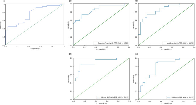
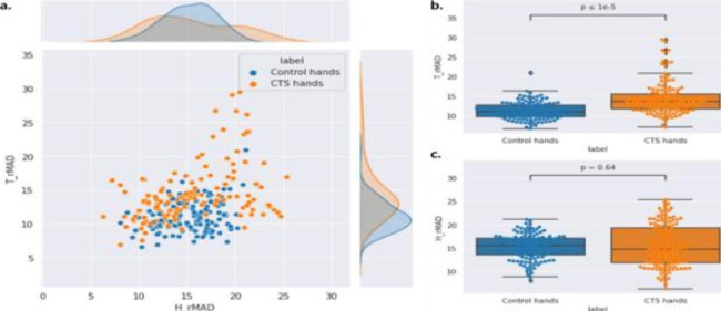
A total of 47 hands with CTS and 27 control hands were analyzed. On conventional quantitative analysis, mean EI ratio (i.e. mean thenar EI/mean hypothenar EI ratio) were significantly higher in the patient group than in the control group, and the AUC was 0.76 in ROC analysis. In the analysis using machine learning, the AUC was the highest for the linear support vector classifier (AUC = 0.86). When recursive feature elimination was applied to the classifier, the AUC value improved to 0.89.
This study showed a significant increase in diagnostic accuracy when AI was used for quantitative analysis of muscle ultrasonography. If an analysis protocol using machine learning can be established and mounted on an ultrasound machine, a noninvasive and non-time-consuming muscle ultrasound examination can be conducted as an ancillary tool for diagnosis.
在局灶性神经病的情况下,由于失神经支配,相应神经支配的肌纤维被脂肪或纤维组织取代,这导致超声检查的回声强度(EI)增加。EI 分析可以通过灰度分析进行定量分析。定义为 EI 的肌肉图像像素亮度的平均值。然而,仅使用此参数来区分正常和异常肌肉的准确性是有限的。最近,有人试图通过人工智能(AI)在肌肉超声图像分析中增加准确性。肌萎缩性侧索硬化症是局灶性神经病中最常见的疾病。在这项研究中,我们旨在验证 AI 辅助肌肉超声定量分析在肌萎缩性侧索硬化症中的实用性。
这是一项回顾性研究,使用接受手部肌肉超声检查的成人数据。包括通过肌电图证实的肌萎缩性侧索硬化症患者和无肌萎缩性侧索硬化症患者。患者或无肌萎缩性侧索硬化症患者的未受影响手部的超声图像被用作对照。超声检查由一名医生在相同的超声设置下进行。同时进行常规定量灰度分析和机器学习(ML)分析进行比较。
共分析了 47 只患有肌萎缩性侧索硬化症的手和 27 只对照组的手。在常规定量分析中,患者组的平均 EI 比值(即鱼际 EI/小鱼际 EI 比值的平均值)显著高于对照组,ROC 分析的 AUC 为 0.76。在使用机器学习的分析中,线性支持向量分类器的 AUC 最高(AUC=0.86)。当将递归特征消除应用于分类器时,AUC 值提高到 0.89。
这项研究表明,当 AI 用于肌肉超声的定量分析时,诊断准确性显著提高。如果可以建立并安装在超声机上的机器学习分析协议,那么可以作为辅助诊断工具,进行非侵入性和非耗时的肌肉超声检查。