- Faculdade de Ciências da Saúde de Barretos Dr. Paulo Prata - FACISB, Medicina - Barretos - SP - Brasil.
- IRCAD América Latina - Unidade de Barretos - Barretos - SP - Brasil.
Rev Col Bras Cir. 2024 Nov 25;51:e20243789. doi: 10.1590/0100-6991e-20243789-en. eCollection 2024.
All forms of access to the peritoneal cavity in laparoscopy could damage intra-abdominal structures. Currently, ultrasound (USG) is being used in several procedures to guide needles: breast biopsy, central venous access puncture, anesthetic nerve blocks, etc. Therefore, this research seeks to verify the feasibility and viability of performing pneumoperitoneum using USG-guided puncture in a pilot study using a porcine model.
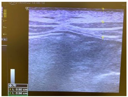
The cross-sectional study was carried out with a sample of 10 anesthetized sows in the IRCAD-América Latina Barretos Unit laboratory. The experiment consisted of an abdominal puncture guided by USG with a linear transducer to create the pneumoperitoneum. After the puncture, the drop test was performed, and CO2 was insufflated into the cavity. Subsequently, a 10mm trocar was introduced to insert the optic. The parameters from the USG were the thickness of the abdominal wall layers, intraperitoneal needle measurement, drop test, and the presence of complications.
The average measurement of the layers was 0.45 centimeters of subcutaneous tissue, 0.67 centimeters of muscle, and 0.15 centimeters of peritoneum. The mean measurement of the intraperitoneal needle was 1.17cm. Furthermore, the drop test was positive in 100% of cases, and there was no bleeding or lesions on any attempt.
Ultrasound-guided pneumoperitoneum is feasible and safe in the porcine model. The subcutaneous, muscular, and peritoneum layers are identifiable and measurable in this model. Subsequent studies are necessary to verify the importance of this new procedure.
腹腔镜下进入腹腔的所有方式都可能损伤腹腔内结构。目前,超声(USG)已被用于指导多种操作中的针头:乳腺活检、中心静脉穿刺、麻醉神经阻滞等。因此,本研究旨在通过使用猪模型进行的初步研究来验证使用 USG 引导穿刺进行气腹的可行性和实用性。
该横断面研究在 IRCAD-América Latina Barretos 单位实验室对 10 头麻醉母猪进行了样本研究。实验包括使用线性换能器进行 USG 引导的腹部穿刺以建立气腹。穿刺后进行滴注试验,并向腔内注入 CO2。随后,引入 10mm trocar 插入光学元件。USG 的参数包括腹壁各层的厚度、腹腔内针的测量、滴注试验以及并发症的发生情况。
各层的平均测量值为 0.45 厘米的皮下组织、0.67 厘米的肌肉和 0.15 厘米的腹膜。腹腔内针的平均测量值为 1.17cm。此外,100%的病例滴注试验呈阳性,且在所有尝试中均未出现出血或损伤。
超声引导气腹在猪模型中是可行且安全的。在该模型中可识别和测量皮下、肌肉和腹膜各层。需要进一步的研究来验证这一新程序的重要性。