Doan Khanh-Hung, Liu Tai-Li, Yun Won-Sik, Kim Yi-Sik, Yun Kyeong Ho, Oh Seok Kyu, Park Jong-Pil, Rhew Jay Young, Lee Sang-Rok
Division of Cardiology, Jeonbuk National University Hospital, Jeonju 54907, Republic of Korea.
Division of Cardiology, Wonkwang University Hospital, Iksan 54538, Republic of Korea.
J Clin Med. 2023 Jun 15;12(12):4073. doi: 10.3390/jcm12124073.
Calcified coronary lesions can cause stent under-expansion, malapposition, and polymer degradation, hence increasing the risk of adverse clinical outcomes. Percutaneous coronary intervention (PCI) guided by intravascular ultrasound (IVUS) has been used regularly to improve outcomes. Our primary aim was to evaluate the clinical efficacy of IVUS-guided PCI in calcified coronary lesions.
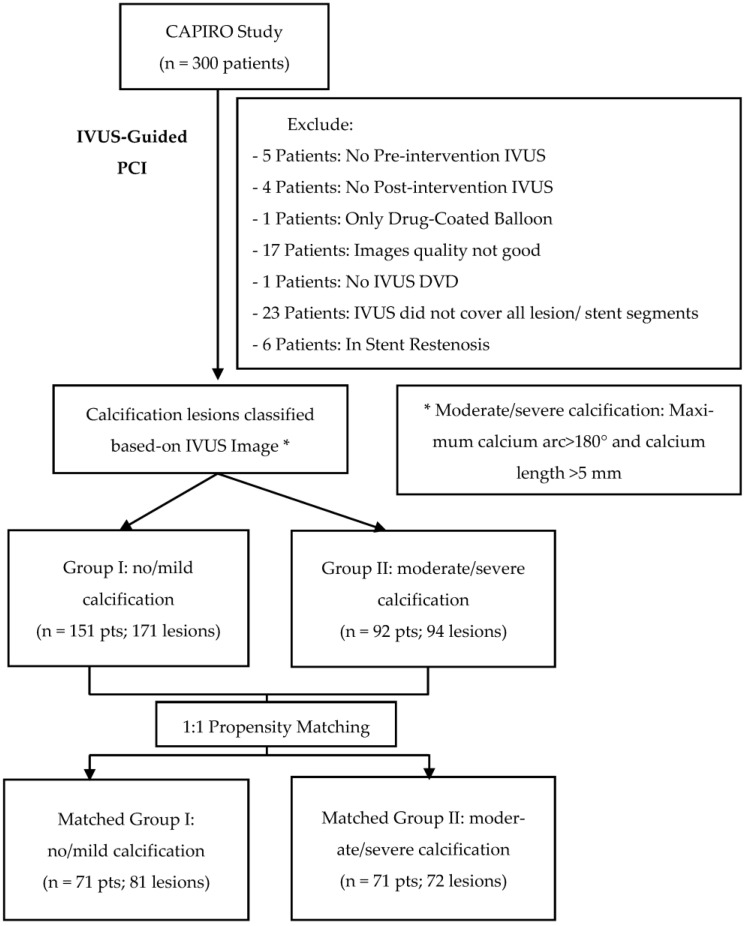
From August 2018 to December 2021, we prospectively included 300 patients in the CAPIRO study (CAlcified plaque in patients receiving Resolute Onyx) at three educational hospitals in Jeonbuk Province. We studied 243 patients (265 lesions) who were followed up for over a year. Based on coronary calcification by IVUS analysis, the patient population was categorized into two groups (Group I: non/mild calcification; Group II: moderate/severe calcification (maximum calcium arc >180° and calcium length > 5 mm)). One-to-one Propensity Score Matching was used to match the baseline characteristics. The stent expansion rate was analyzed by recent criteria. The primary clinical outcome was Major Adverse Cardiac Events (MACE), which included Cardiac death, Myocardial Infarction (MI), and Target Lesion Revascularization (TLR).
After follow-up time, the MACE rate in Group I was 1.99%, comparable to Group II's 1.09% ( = 0.594). The components of MACE did not significantly differ between the two groups. Based on absolute MSA or MSA/MVA at MSA site criteria, the stent expansion rate in Group II was lower than that of Group I. Nevertheless, based on recent relative criteria, the stent expansion rate in both groups was comparable.
After more than a year of follow-up, IVUS-guided PCI in moderate/severe calcification lesions was associated with good clinical outcomes, which was comparable with non/mild calcification lesions. Future studies with a larger sample size and a more extended follow-up period are required to clarify our findings.
冠状动脉钙化病变可导致支架扩张不全、贴壁不良及聚合物降解,从而增加不良临床结局的风险。血管内超声(IVUS)引导下的经皮冠状动脉介入治疗(PCI)已被常规用于改善治疗效果。我们的主要目的是评估IVUS引导下PCI治疗冠状动脉钙化病变的临床疗效。
2018年8月至2021年12月,我们在全北道的三家教学医院前瞻性纳入了CAPIRO研究(接受Resolute Onyx治疗患者的钙化斑块)中的300例患者。我们研究了243例患者(265个病变),这些患者接受了超过一年的随访。根据IVUS分析的冠状动脉钙化情况,将患者人群分为两组(I组:无/轻度钙化;II组:中度/重度钙化(最大钙化弧>180°且钙化长度>5mm))。采用一对一倾向评分匹配法来匹配基线特征。根据最新标准分析支架扩张率。主要临床结局为主要不良心脏事件(MACE),包括心源性死亡、心肌梗死(MI)和靶病变血运重建(TLR)。
随访期后,I组的MACE发生率为1.99%,与II组的1.09%相当(P = 0.594)。两组间MACE的组成部分无显著差异。根据MSA部位的绝对MSA或MSA/MVA标准,II组的支架扩张率低于I组。然而,根据最新的相对标准,两组的支架扩张率相当。
经过一年多的随访,IVUS引导下对中度/重度钙化病变进行PCI治疗具有良好的临床疗效,与无/轻度钙化病变相当。需要开展样本量更大、随访期更长的未来研究来阐明我们的研究结果。