Horgos Bianca, Mecea Miruna, Boer Armand, Buruiana Andrei, Ciortea Razvan, Mihu Carmen-Mihaela, Florian Ioan Stefan, Florian Alexandru Ioan, Stamatian Florin, Szabo Bianca, Albu Camelia, Susman Sergiu, Pascalau Raluca
Faculty of Medicine, "Iuliu Haţieganu" University of Medicine and Pharmacy, Cluj-Napoca, Romania.
Department of Oncology, "Ion Chiricuţă" Institute of Oncology, Cluj-Napoca, Romania.
Front Neuroanat. 2023 Jun 14;17:1160742. doi: 10.3389/fnana.2023.1160742. eCollection 2023.
Ventriculomegaly (VM) is a fetal brain malformation which may present independently (isolated form) or in association with different cerebral malformations, genetic syndromes or other pathologies (non-isolated form).
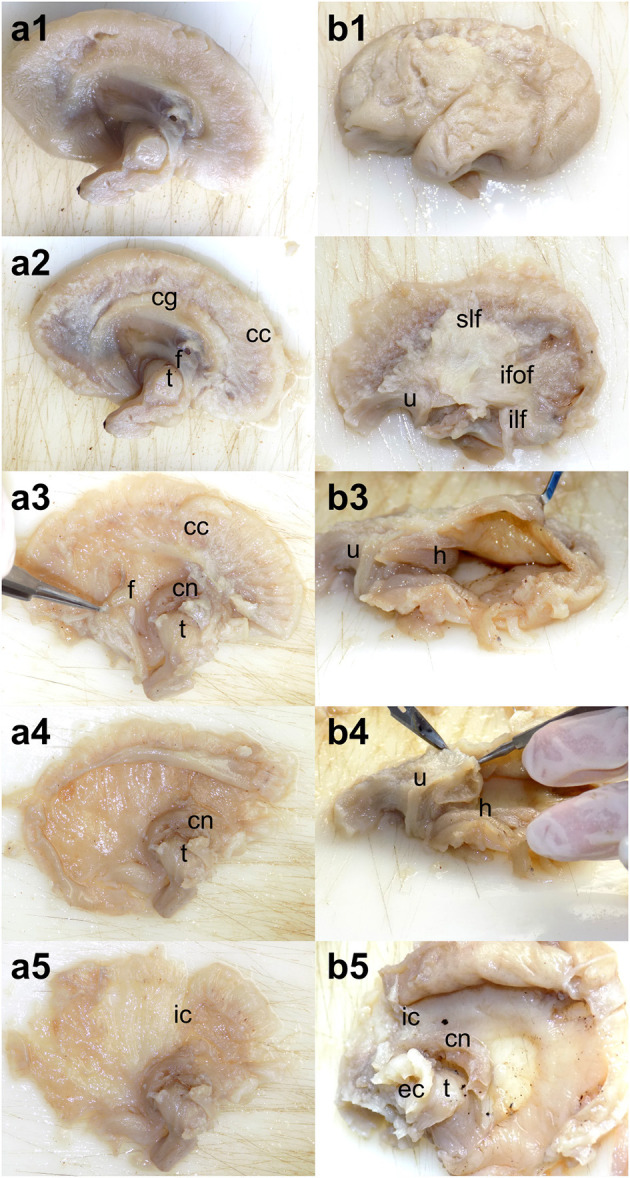
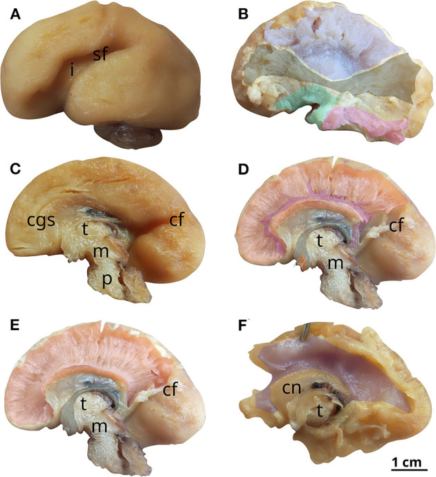
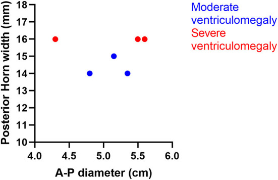
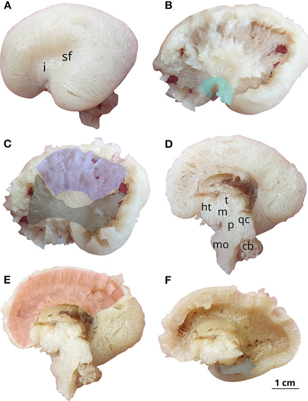
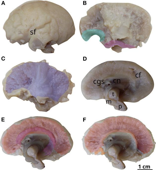
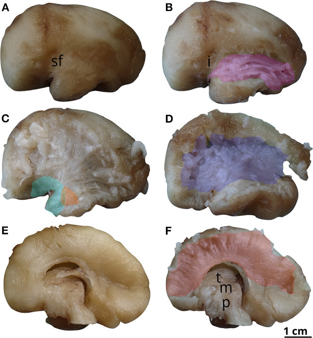
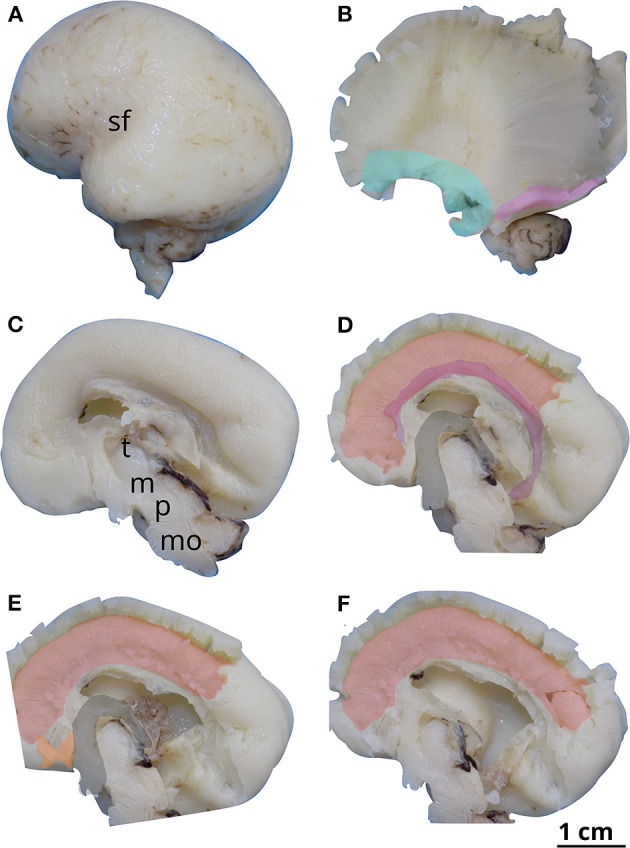
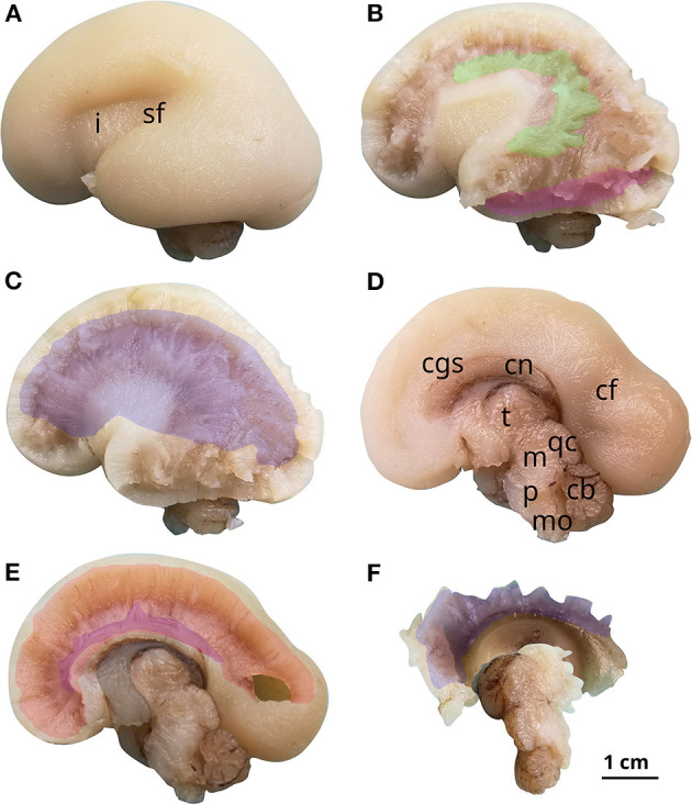
This paper aims to study the effect of ventriculomegaly on the internal tridimensional architecture of fetal brains by way of Klingler's dissection. Ventriculomegaly was diagnosed using fetal ultrasonography during pregnancy and subsequently confirmed by necropsy. Taking into consideration the diameter of the lateral ventricle (measured at the level of the atrium), the brains were divided into two groups: moderate ventriculomegaly (with atrial diameter between 13 and 15 mm) and severe ventriculomegaly (with atrial diameter above 15 mm).
The results of each dissection were described and illustrated, then compared with age-matched reference brains. In the pathological brains, fascicles in direct contact with the enlarged ventricles were found to be thinner and displaced inferiorly, the opening of the uncinate fasciculus was wider, the fornix was no longer in contact with the corpus callosum and the convexity of the corpus callosum was inverted. We have studied the prevalence of neurodevelopmental delay in children born with ventriculomegaly in the literature and discovered that a normal developmental outcome was found in over 90% of the mild VM cases, approximately 75% of the moderate and 60% in severe VM, with the correlated neurological impairments ranging from attention deficits to psychiatric disorders.
脑室扩大(VM)是一种胎儿脑部畸形,可单独出现(孤立型)或与不同的脑部畸形、遗传综合征或其他病理情况相关(非孤立型)。
本文旨在通过克林格勒解剖法研究脑室扩大对胎儿脑部内部三维结构的影响。在孕期通过胎儿超声诊断脑室扩大,随后经尸检证实。根据侧脑室直径(在心房水平测量),将脑部分为两组:中度脑室扩大(心房直径在13至15毫米之间)和重度脑室扩大(心房直径大于15毫米)。
描述并展示了每次解剖的结果,然后与年龄匹配的对照脑进行比较。在病理脑中,发现与扩大脑室直接接触的纤维束更细且向下移位,钩束开口更宽,穹窿不再与胼胝体接触,胼胝体凸面反转。我们研究了文献中患有脑室扩大的儿童神经发育迟缓的患病率,发现超过90%的轻度VM病例发育结果正常,中度VM约为75%,重度VM为60%,相关神经功能障碍范围从注意力缺陷到精神疾病。