Indiana Center for Regenerative Medicine & Engineering, Indiana University Health Comprehensive Wound Center, Department of Surgery, Indiana University School of Medicine.
Indiana Center for Regenerative Medicine & Engineering, Indiana University Health Comprehensive Wound Center, Department of Surgery, Indiana University School of Medicine;
J Vis Exp. 2023 Jun 16(196). doi: 10.3791/65301.
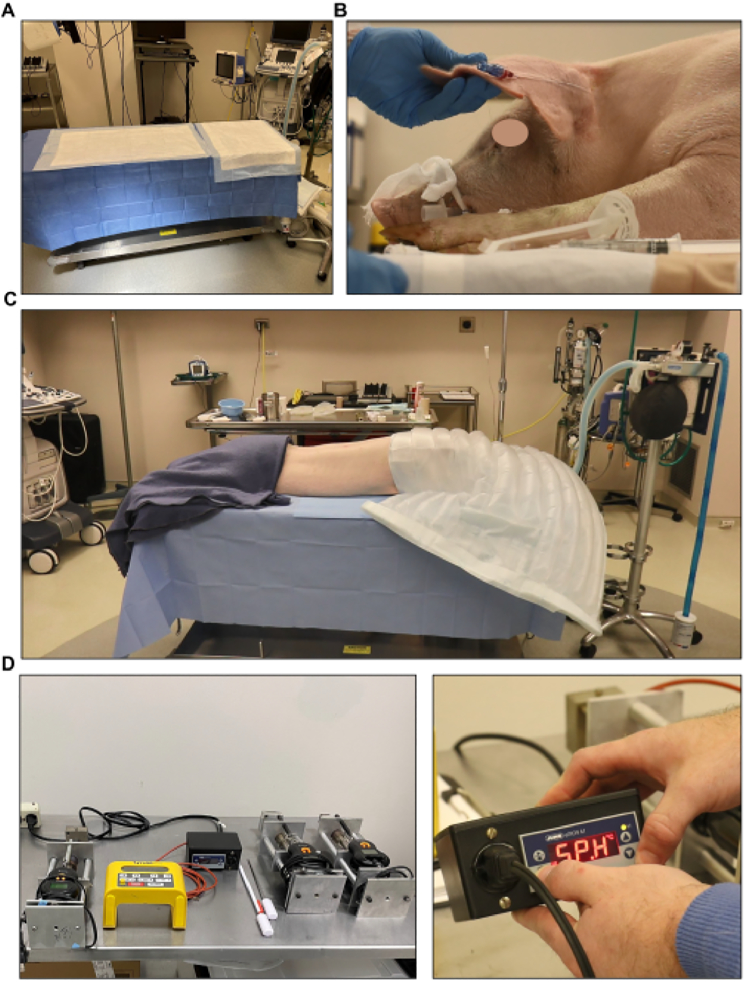
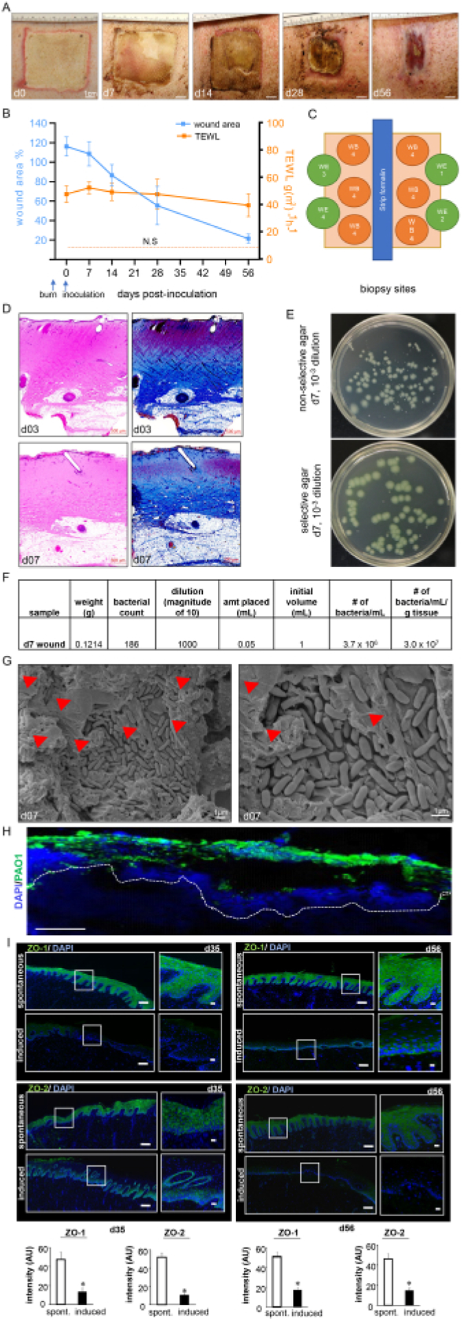
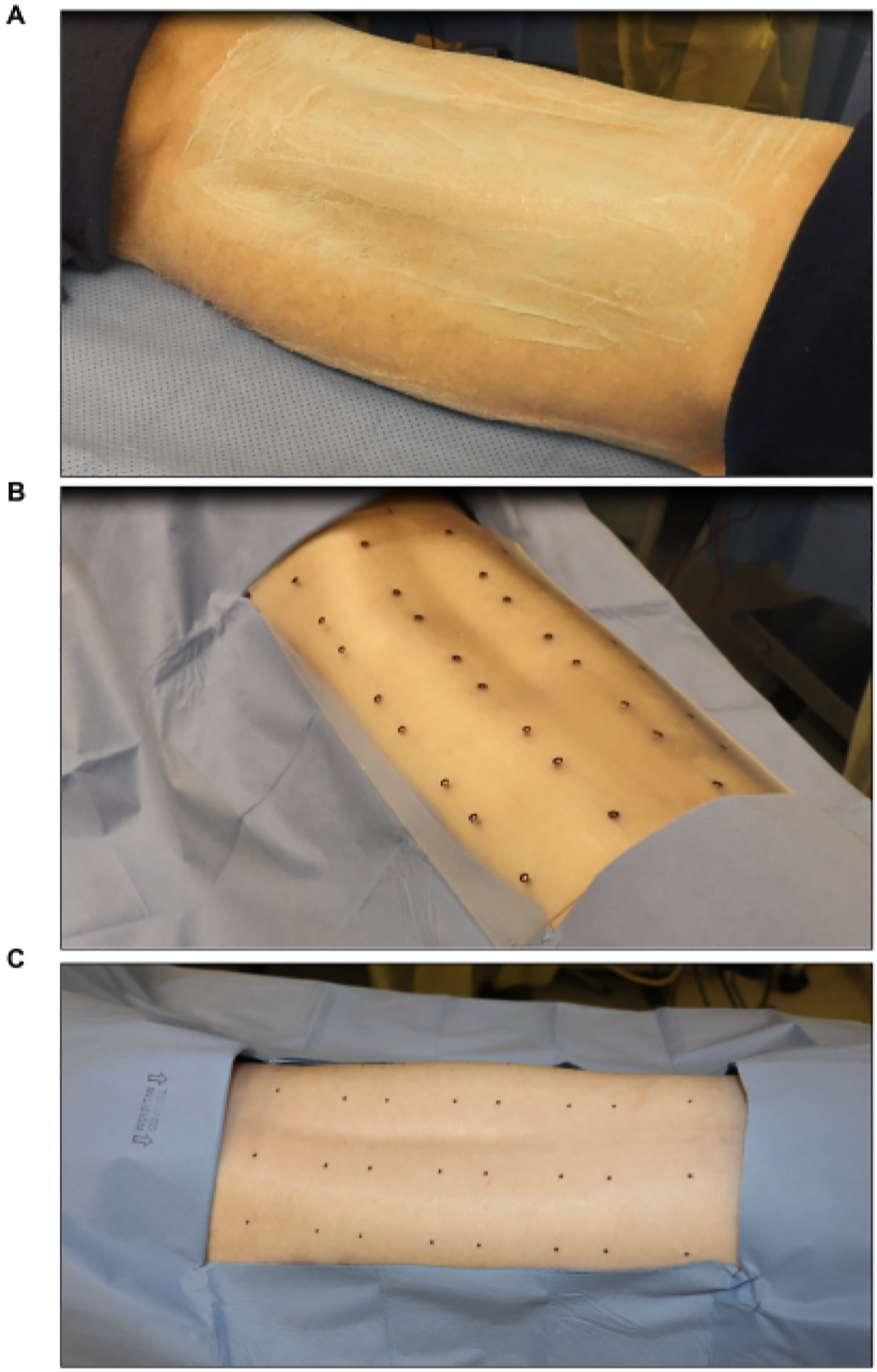
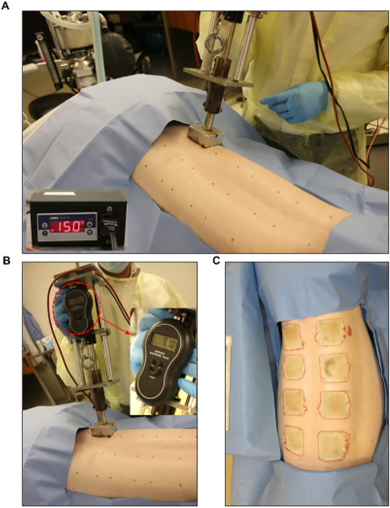
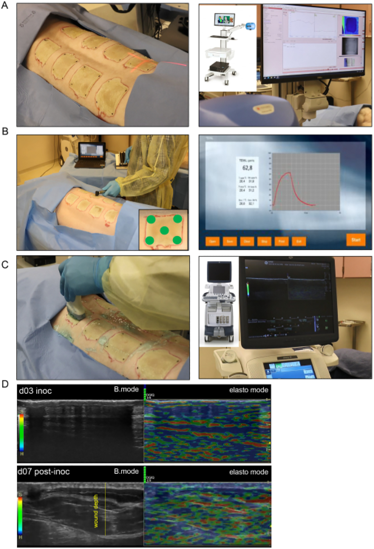
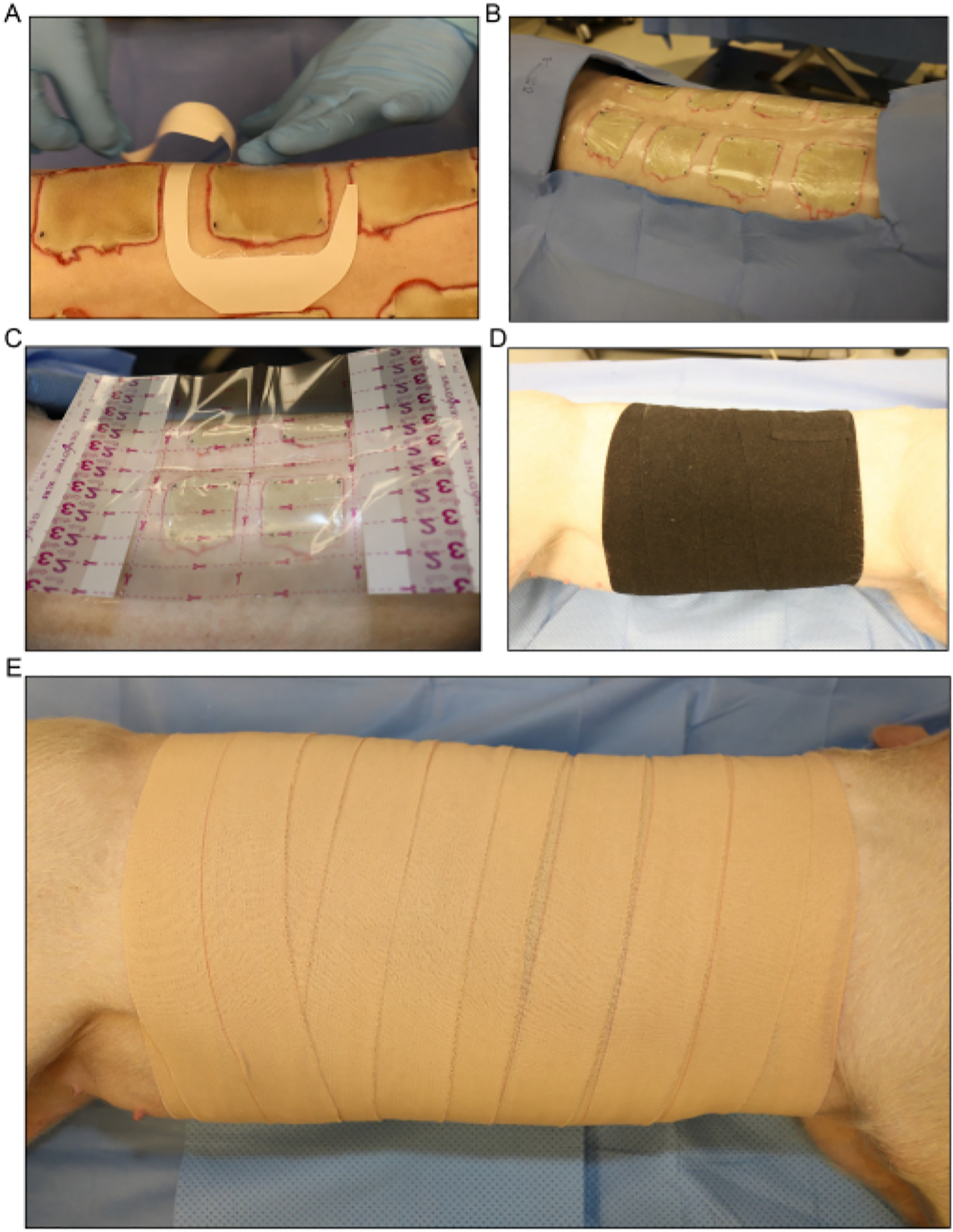
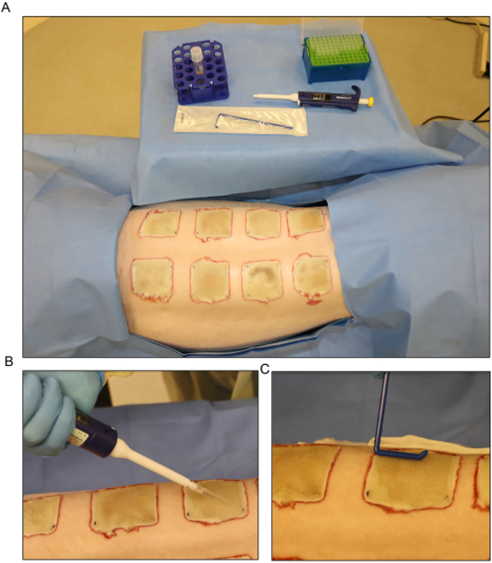
Biofilm infection is a major contributor to wound chronicity. The establishment of clinically relevant experimental wound biofilm infection requires the involvement of the host immune system. Iterative changes in the host and pathogen during the formation of such clinically relevant biofilm can only occur in vivo. The swine wound model is recognized for its advantages as a powerful pre-clinical model. There are several reported approaches for studying wound biofilms. In vitro and ex vivo systems are deficient in terms of the host immune response. Short-term in vivo studies involve acute responses and, thus, do not allow for biofilm maturation, as is known to occur clinically. The first long-term swine wound biofilm study was reported in 2014. The study recognized that biofilm-infected wounds may close as determined by planimetry, but the skin barrier function of the affected site may fail to be restored. Later, this observation was validated clinically. The concept of functional wound closure was thus born. Wounds closed but deficient in skin barrier function may be viewed as invisible wounds. In this work, we seek to report the methodological details necessary to reproduce the long-term swine model of biofilm-infected severe burn injury, which is clinically relevant and has translational value. This protocol provides detailed guidance on establishing an 8 week wound biofilm infection using P. aeruginosa (PA01). Eight full-thickness burn wounds were created symmetrically on the dorsum of domestic white pigs, which were inoculated with (PA01) at day 3 post-burn; subsequently, noninvasive assessments of the wound healing were conducted at different time points using laser speckle imaging (LSI), high-resolution ultrasound (HUSD), and transepidermal water loss (TEWL). The inoculated burn wounds were covered with a four-layer dressing. Biofilms, as established and confirmed structurally by SEM at day 7 post-inoculation, compromised the functional wound closure. Such an adverse outcome is subject to reversal in response to appropriate interventions.
生物膜感染是导致伤口慢性化的主要原因。建立具有临床相关性的实验性伤口生物膜感染需要宿主免疫系统的参与。在形成这种具有临床相关性的生物膜的过程中,宿主和病原体的反复变化只能在体内发生。猪伤口模型因其作为强大的临床前模型的优势而得到认可。目前有几种报道的方法可用于研究伤口生物膜。在体外和离体系统中,宿主的免疫反应存在缺陷。短期的体内研究涉及急性反应,因此,不允许生物膜成熟,正如临床上所知道的那样。第一个长期的猪伤口生物膜研究于 2014 年报道。该研究认识到,生物膜感染的伤口可能会按照平面测量法闭合,但受影响部位的皮肤屏障功能可能无法恢复。后来,这一观察结果在临床上得到了验证。因此,功能伤口闭合的概念诞生了。闭合但缺乏皮肤屏障功能的伤口可能被视为看不见的伤口。在这项工作中,我们旨在报告复制具有临床相关性和转化价值的长期猪生物膜感染严重烧伤模型所需的方法学细节。该方案提供了使用 P. aeruginosa (PA01) 建立 8 周伤口生物膜感染的详细指导。在家养白猪的背部对称地创建了 8 个全层烧伤伤口,在烧伤后第 3 天用(PA01)接种;随后,使用激光散斑成像(LSI)、高分辨率超声(HUSD)和经皮水分丢失(TEWL)在不同时间点对伤口愈合进行非侵入性评估。接种的烧伤伤口用四层敷料覆盖。生物膜在接种后第 7 天通过 SEM 建立并在结构上确认,破坏了功能伤口闭合。这种不良后果可以通过适当的干预来逆转。