Oniris, INRAE, PAnTher, 44300, Nantes, France.
IMT Atlantique, Lab-STICC, UMR CNRS 6285, 29238, Brest, France.
Sci Rep. 2023 Jul 4;13(1):10808. doi: 10.1038/s41598-023-37762-1.
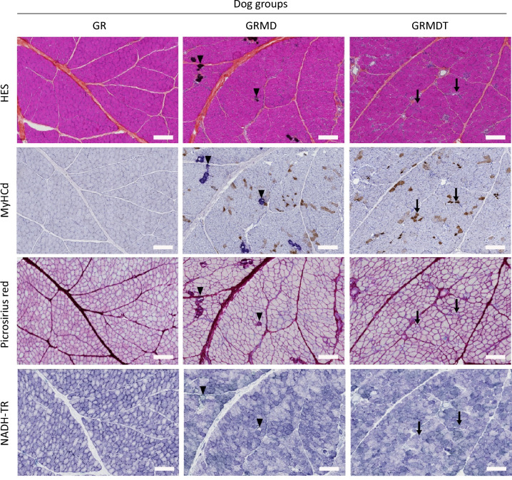
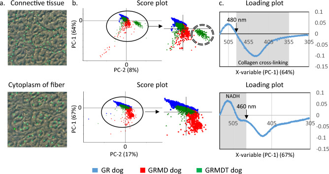
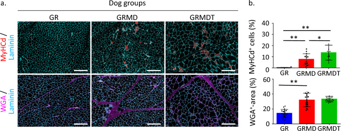
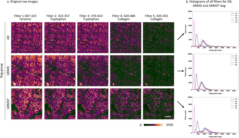
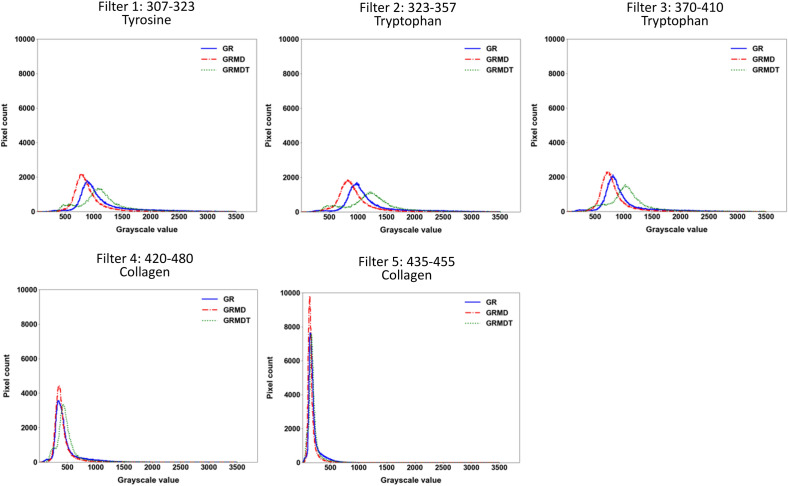
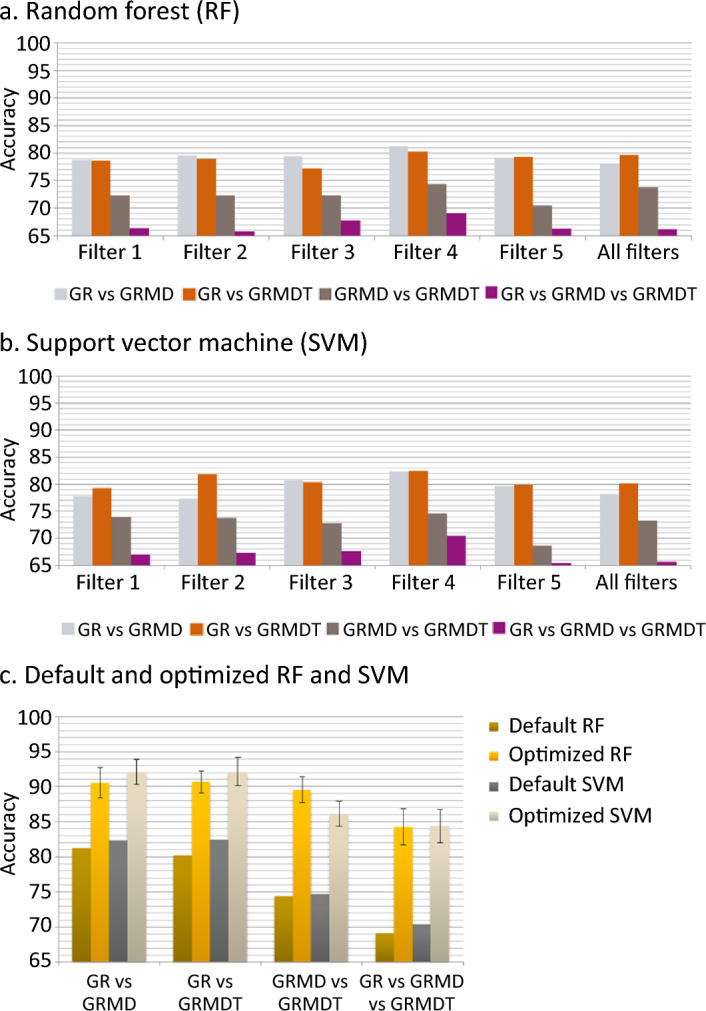
Dystrophic muscle is characterized by necrosis/regeneration cycles, inflammation, and fibro-adipogenic development. Conventional histological stainings provide essential topographical data of this remodeling but may be limited to discriminate closely related pathophysiological contexts. They fail to mention microarchitecture changes linked to the nature and spatial distribution of tissue compartment components. We investigated whether label-free tissue autofluorescence revealed by Synchrotron deep ultraviolet (DUV) radiation could serve as an additional tool for monitoring dystrophic muscle remodeling. Using widefield microscopy with specific emission fluorescence filters and microspectroscopy defined by high spectral resolution, we analyzed samples from healthy dogs and two groups of dystrophic dogs: naïve (severely affected) and MuStem cell-transplanted (clinically stabilized) animals. Multivariate statistical analysis and machine learning approaches demonstrated that autofluorescence emitted at 420-480 nm by the Biceps femoris muscle effectively discriminates between healthy, dystrophic, and transplanted dog samples. Microspectroscopy showed that dystrophic dog muscle displays higher and lower autofluorescence due to collagen cross-linking and NADH respectively than that of healthy and transplanted dogs, defining biomarkers to evaluate the impact of cell transplantation. Our findings demonstrate that DUV radiation is a sensitive, label-free method to assess the histopathological status of dystrophic muscle using small amounts of tissue, with potential applications in regenerative medicine.
营养不良的肌肉以坏死/再生循环、炎症和纤维脂肪生成发展为特征。传统的组织学染色提供了这种重构的基本拓扑数据,但可能仅限于区分密切相关的病理生理情况。它们未能提及与组织隔室成分的性质和空间分布相关的微结构变化。我们研究了无标记组织自发荧光是否可以通过同步加速器深紫外线(DUV)辐射作为监测营养不良肌肉重构的附加工具。我们使用具有特定发射荧光滤波器的宽场显微镜和通过高光谱分辨率定义的微光谱学,分析了来自健康犬和两组营养不良犬的样本:未处理(严重受影响)和 MuStem 细胞移植(临床稳定)动物。多元统计分析和机器学习方法表明,由比目鱼肌发出的 420-480nm 的自发荧光可有效区分健康、营养不良和移植犬样本。微光谱学显示,与健康和移植犬相比,营养不良犬的肌肉由于胶原交联和 NADH 而分别显示出更高和更低的自发荧光,定义了评估细胞移植影响的生物标志物。我们的研究结果表明,DUV 辐射是一种敏感的、无标记的方法,可使用少量组织评估营养不良肌肉的组织病理学状态,在再生医学中有潜在的应用。