Department of Clinical Medicine, UiT-The Arctic University of Norway, Tromsø, Norway.
Department of Medical Biology, UiT-The Arctic University of Norway, Tromsø, Norway.
Autophagy. 2023 Oct;19(10):2769-2788. doi: 10.1080/15548627.2023.2230837. Epub 2023 Jul 5.
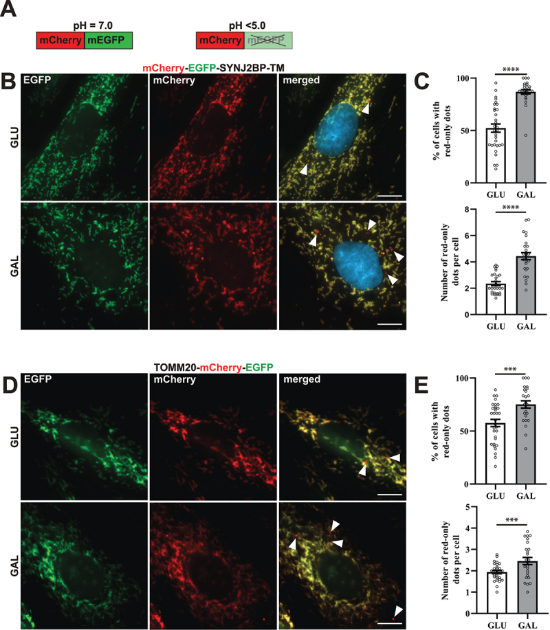
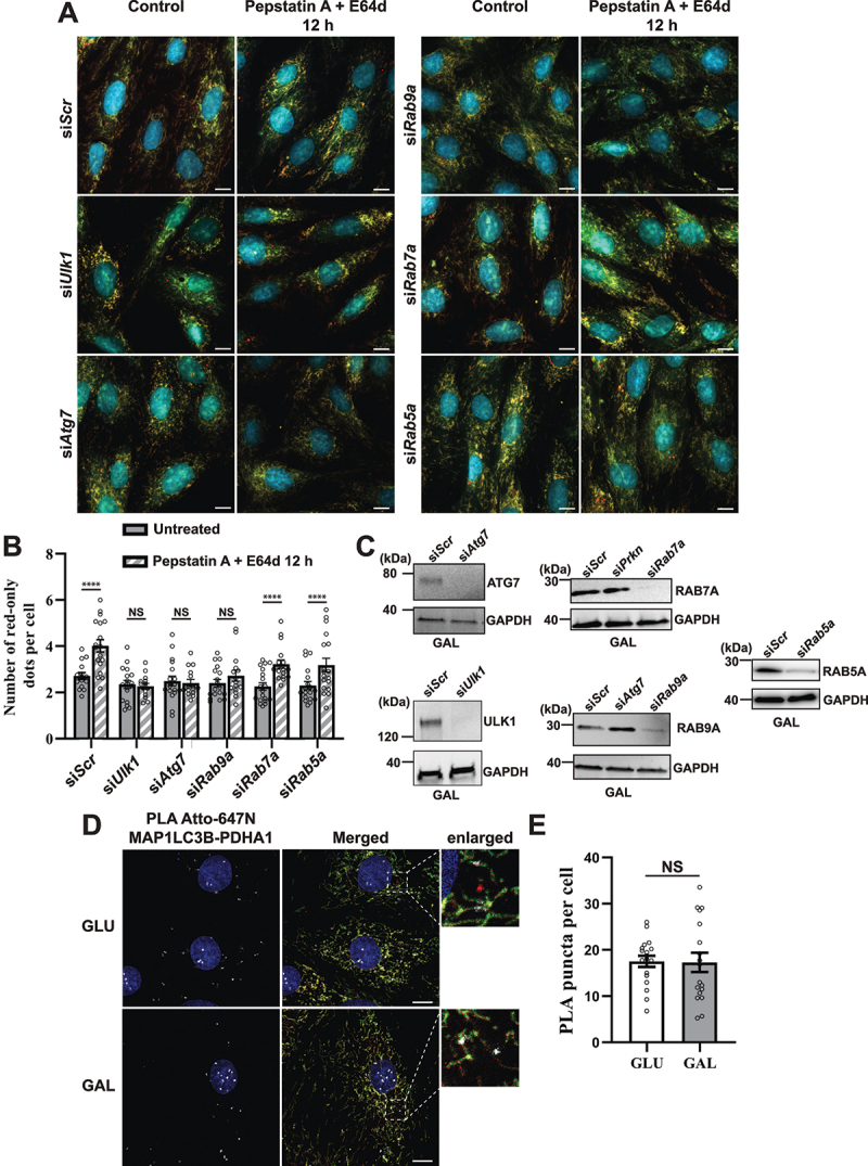
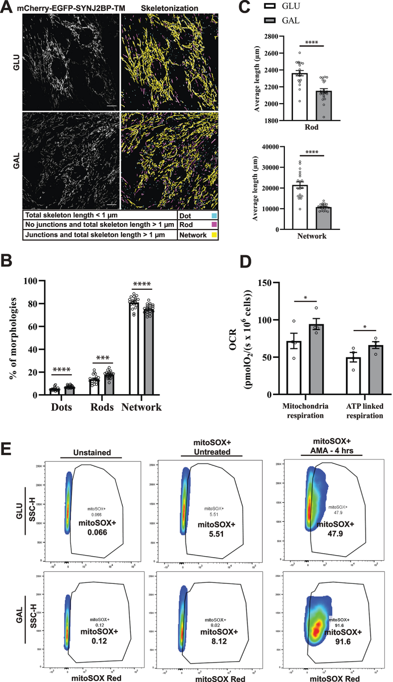
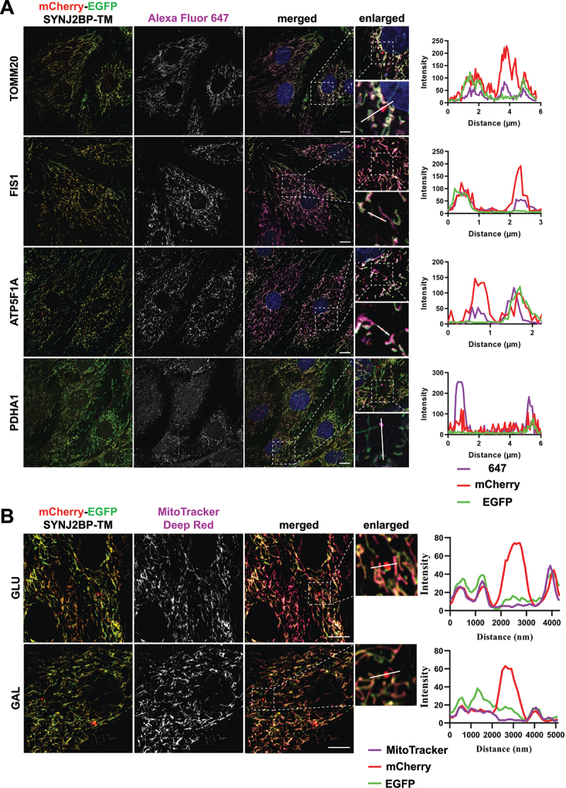
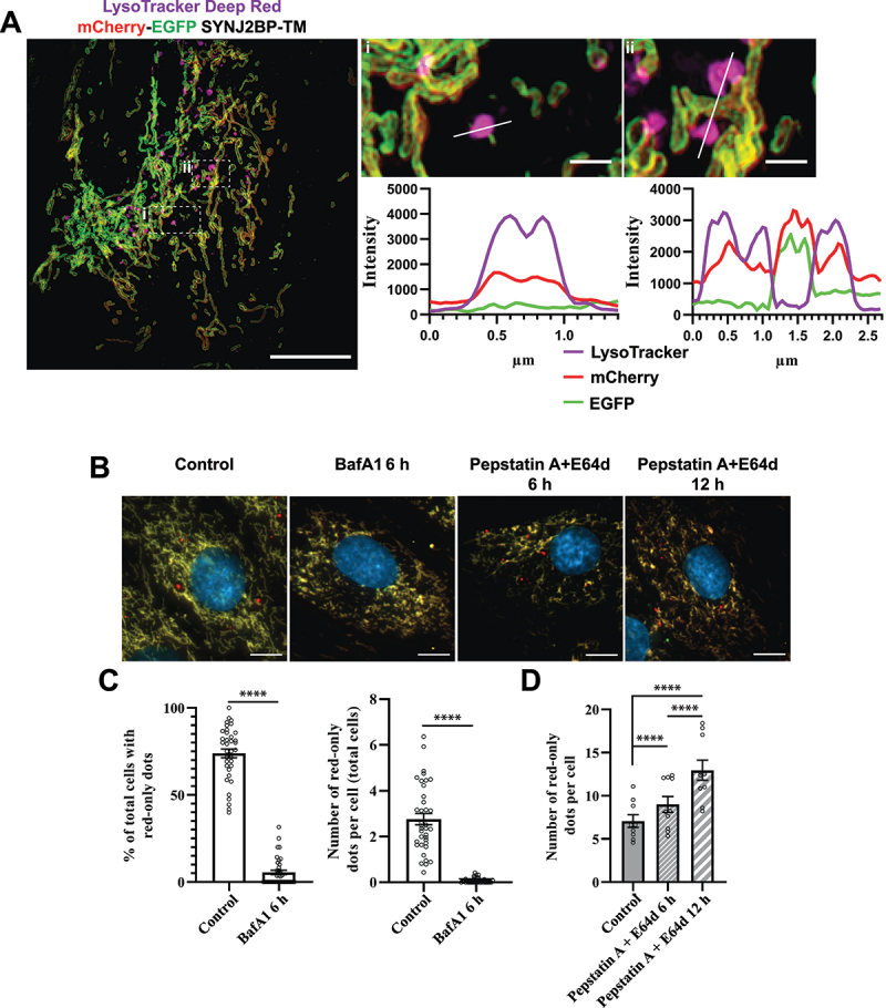
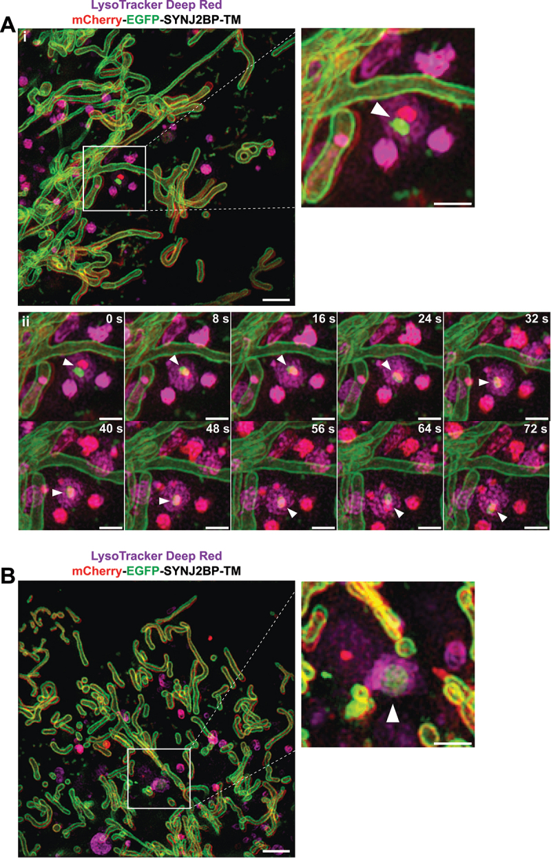
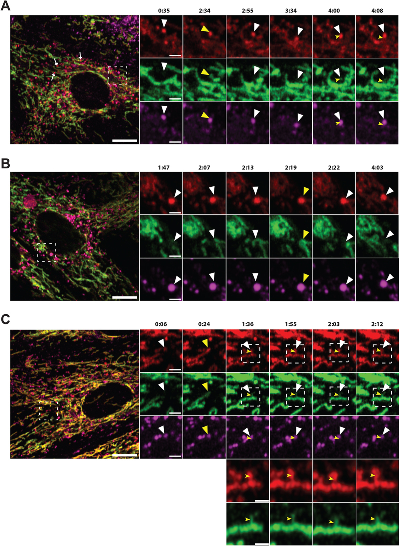
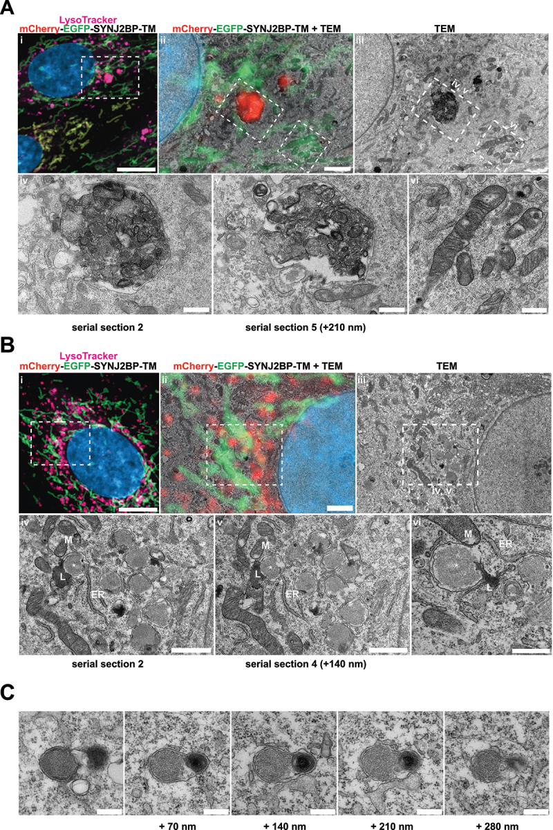
Mitochondria are susceptible to damage resulting from their activity as energy providers. Damaged mitochondria can cause harm to the cell and thus mitochondria are subjected to elaborate quality-control mechanisms including elimination via lysosomal degradation in a process termed mitophagy. Basal mitophagy is a house-keeping mechanism fine-tuning the number of mitochondria according to the metabolic state of the cell. However, the molecular mechanisms underlying basal mitophagy remain largely elusive. In this study, we visualized and assessed the level of mitophagy in H9c2 cardiomyoblasts at basal conditions and after OXPHOS induction by galactose adaptation. We used cells with a stable expression of a pH-sensitive fluorescent mitochondrial reporter and applied state-of-the-art imaging techniques and image analysis. Our data showed a significant increase in acidic mitochondria after galactose adaptation. Using a machine-learning approach we also demonstrated increased mitochondrial fragmentation by OXPHOS induction. Furthermore, super-resolution microscopy of live cells enabled capturing of mitochondrial fragments within lysosomes as well as dynamic transfer of mitochondrial contents to lysosomes. Applying correlative light and electron microscopy we revealed the ultrastructure of the acidic mitochondria confirming their proximity to the mitochondrial network, ER and lysosomes. Finally, exploiting siRNA knockdown strategy combined with flux perturbation with lysosomal inhibitors, we demonstrated the importance of both canonical as well as non-canonical autophagy mediators in lysosomal degradation of mitochondria after OXPHOS induction. Taken together, our high-resolution imaging approaches applied on H9c2 cells provide novel insights on mitophagy during physiologically relevant conditions. The implication of redundant underlying mechanisms highlights the fundamental importance of mitophagy. ATG: autophagy related; ATG7: autophagy related 7; ATP: adenosine triphosphate; BafA1: bafilomycin A; CLEM: correlative light and electron microscopy; EGFP: enhanced green fluorescent protein; MAP1LC3B: microtubule associated protein 1 light chain 3 beta; OXPHOS: oxidative phosphorylation; PepA: pepstatin A; PLA: proximity ligation assay; PRKN: parkin RBR E3 ubiquitin protein ligase; RAB5A: RAB5A, member RAS oncogene family; RAB7A: RAB7A, member RAS oncogene family; RAB9A: RAB9A, member RAS oncogene family; ROS: reactive oxygen species; SIM: structured illumination microscopy; siRNA: short interfering RNA; SYNJ2BP: synaptojanin 2 binding protein; TEM: transmission electron microscopy; TOMM20: translocase of outer mitochondrial membrane 20; ULK1: unc-51 like kinase 1.
线粒体容易受到其作为能量提供者的活性所导致的损伤。受损的线粒体可能会对细胞造成伤害,因此线粒体受到了精细的质量控制机制的调节,包括通过溶酶体降解进行消除,这个过程被称为线粒体自噬。基础线粒体自噬是一种根据细胞代谢状态微调线粒体数量的管家机制。然而,基础线粒体自噬的分子机制在很大程度上仍然难以捉摸。在这项研究中,我们在基础条件下和通过半乳糖适应诱导氧化磷酸化(OXPHOS)后,可视化和评估了 H9c2 心肌细胞中的线粒体自噬水平。我们使用稳定表达 pH 敏感荧光线粒体报告基因的细胞,并应用最先进的成像技术和图像分析。我们的数据显示,半乳糖适应后酸性线粒体的数量显著增加。使用机器学习方法,我们还证明了 OXPHOS 诱导会增加线粒体碎片化。此外,活细胞的超分辨率显微镜能够捕获溶酶体内的线粒体片段,并观察到线粒体内容物向溶酶体的动态转移。应用相关的光和电子显微镜,我们揭示了酸性线粒体的超微结构,证实了它们与线粒体网络、内质网和溶酶体的接近。最后,利用 siRNA 敲低策略结合溶酶体抑制剂的通量扰动,我们证明了在 OXPHOS 诱导后,经典和非经典自噬介体在溶酶体降解线粒体中的重要性。总之,我们在 H9c2 细胞上应用的高分辨率成像方法为生理相关条件下的线粒体自噬提供了新的见解。冗余基础机制的重要性突出了线粒体自噬的基本重要性。ATG:自噬相关;ATG7:自噬相关 7;ATP:三磷酸腺苷;BafA1:巴弗霉素 A;CEM:相关光和电子显微镜;EGFP:增强型绿色荧光蛋白;MAP1LC3B:微管相关蛋白 1 轻链 3β;OXPHOS:氧化磷酸化;PepA:胃蛋白酶抑制剂 A;PLA:接近连接测定;PRKN:parkin RBR E3 泛素蛋白连接酶;RAB5A:RAB5A,RAS 癌基因家族成员;RAB7A:RAB7A,RAS 癌基因家族成员;RAB9A:RAB9A,RAS 癌基因家族成员;ROS:活性氧;SIM:结构照明显微镜;siRNA:短发夹 RNA;SYNJ2BP:突触结合蛋白 2 结合蛋白;TEM:透射电子显微镜;TOMM20:外线粒体膜转位酶 20;ULK1:UNC-51 样激酶 1。