Barnden Leighton, Thapaliya Kiran, Eaton-Fitch Natalie, Barth Markus, Marshall-Gradisnik Sonya
National Centre for Neuroimmunology and Emerging Diseases, Menzies Health Institute Queensland, Griffith University, Southport, QLD, Australia.
Centre for Advanced Imaging, The University of Queensland, Brisbane, QLD, Australia.
Front Neurosci. 2023 Jun 22;17:1182607. doi: 10.3389/fnins.2023.1182607. eCollection 2023.
Debilitating Long-Covid symptoms occur frequently after SARS-COVID-19 infection.
Functional MRI was acquired in 10 Long Covid (LCov) and 13 healthy controls (HC) with a 7 Tesla scanner during a cognitive (Stroop color-word) task. BOLD time series were computed for 7 salience and 4 default-mode network hubs, 2 hippocampus and 7 brainstem regions (ROIs). Connectivity was characterized by the correlation coefficient between each pair of ROI BOLD time series. We tested for HC versus LCov differences in connectivity between each pair of the 20 regions (ROI-to-ROI) and between each ROI and the rest of the brain (ROI-to-voxel). For LCov, we also performed regressions of ROI-to-ROI connectivity with clinical scores.
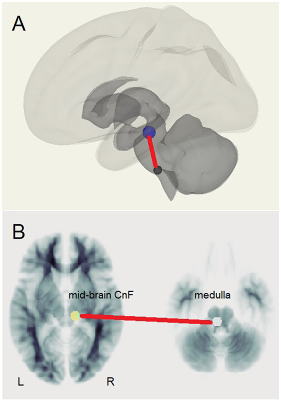
Two ROI-to-ROI connectivities differed between HC and LCov. Both involved the brainstem rostral medulla, one connection to the midbrain, another to a DM network hub. Both were stronger in LCov than HC. ROI-to-voxel analysis detected multiple other regions where LCov connectivity differed from HC located in all major lobes. Most, but not all connections, were weaker in LCov than HC. LCov, but not HC connectivity, was correlated with clinical scores for disability and autonomic function and involved brainstem ROI.
Multiple connectivity differences and clinical correlations involved brainstem ROIs. Stronger connectivity in LCov between the medulla and midbrain may reflect a compensatory response. This brainstem circuit regulates cortical arousal, autonomic function and the sleep-wake cycle. In contrast, this circuit exhibited weaker connectivity in ME/CFS. LCov connectivity regressions with disability and autonomic scores were consistent with altered brainstem connectivity in LCov.
严重的长新冠症状在感染SARS-CoV-19后经常出现。
在认知(斯特鲁普颜色-文字)任务期间,使用7特斯拉扫描仪对10名长新冠(LCov)患者和13名健康对照者(HC)进行了功能磁共振成像。计算了7个显著性和4个默认模式网络枢纽、2个海马体和7个脑干区域(感兴趣区)的血氧水平依赖(BOLD)时间序列。连通性通过每对感兴趣区BOLD时间序列之间的相关系数来表征。我们测试了20个区域中每对区域之间(感兴趣区到感兴趣区)以及每个感兴趣区与大脑其余部分之间(感兴趣区到体素)HC与LCov之间连通性的差异。对于LCov,我们还进行了感兴趣区到感兴趣区连通性与临床评分的回归分析。
HC和LCov之间有两种感兴趣区到感兴趣区的连通性不同。两者都涉及脑干延髓头端,一种连接到中脑,另一种连接到默认模式网络枢纽。两者在LCov中都比HC中更强。感兴趣区到体素分析在所有主要脑叶中检测到多个其他区域,其中LCov的连通性与HC不同。大多数但不是所有连接在LCov中比HC中更弱。LCov的连通性而非HC的连通性与残疾和自主神经功能的临床评分相关,且涉及脑干感兴趣区。
多种连通性差异和临床相关性涉及脑干感兴趣区。LCov中延髓和中脑之间更强的连通性可能反映了一种代偿反应。该脑干回路调节皮质觉醒、自主神经功能和睡眠-觉醒周期。相比之下,该回路在肌痛性脑脊髓炎/慢性疲劳综合征(ME/CFS)中表现出较弱的连通性。LCov连通性与残疾和自主神经评分的回归分析与LCov中脑干连通性改变一致。