Department of Endoscopy, The First Hospital Affiliated to China Medical University, Shenyang 110001, Liaoning Province, China.
World J Gastroenterol. 2023 Jun 28;29(24):3770-3792. doi: 10.3748/wjg.v29.i24.3770.
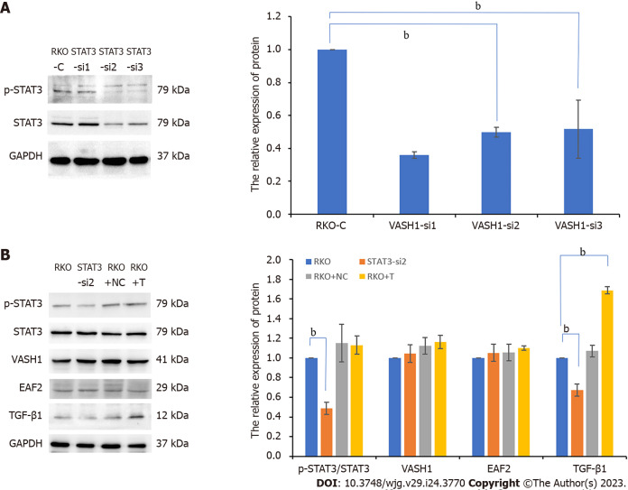
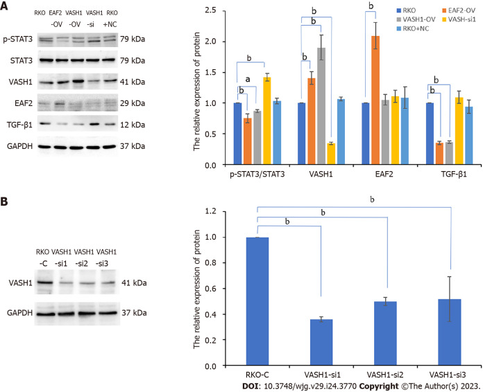
As a novel endogenous anti-angiogenic molecule, vasohibin 1 (VASH1) is not only expressed in tumor stroma, but also in tumor tissue. Moreover, studies have shown that VASH1 may be a prognostic marker in colorectal cancer (CRC). Knockdown of VASH1 enhanced transforming growth factor-β1 (TGF-β1)/Smad3 pathway activity and type I/III collagen production. Our previous findings suggest that ELL-associated factor 2 (EAF2) may play a tumor suppressor and protective role in the development and progression of CRC by regulating signal transducer and activator of transcription 3 (STAT3)/TGF-β1 signaling pathway. However, the functional role and mechanism of VASH1-mediated TGF-β1 related pathway in CRC has not been elucidated.
To investigate the expression of VASH1 in CRC and its correlation with the expression of EAF2. Furthermore, we studied the functional role and mechanism of VASH1 involved in the regulation and protection of EAF2 in CRC cells .
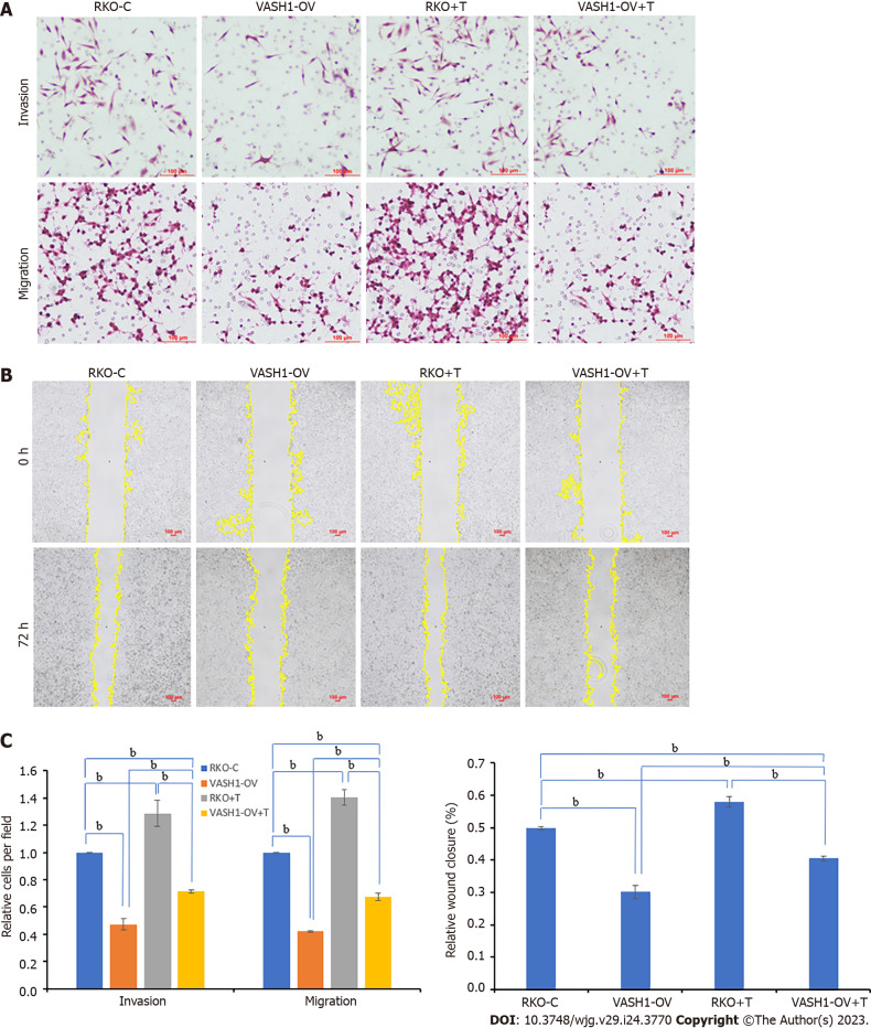
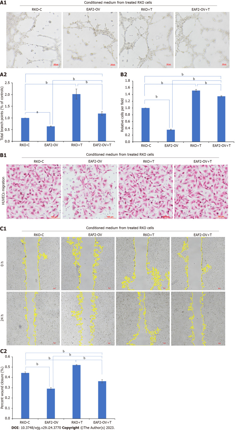
We collected colorectal adenocarcinoma and corresponding adjacent tissues to investigate the clinical expression of EAF2 protein and VASH1 protein in patients with advanced CRC. Following, we investigated the effect and mechanism of EAF2 and VASH1 on the invasion, migration and angiogenesis of CRC cells using plasmid transfection.
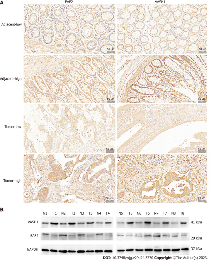
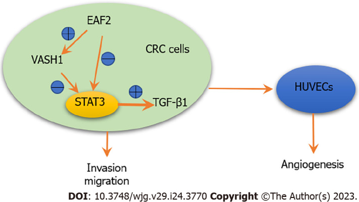
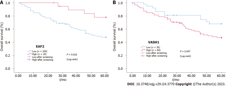
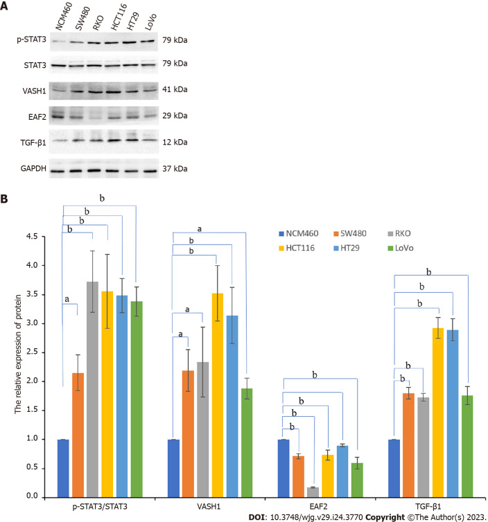
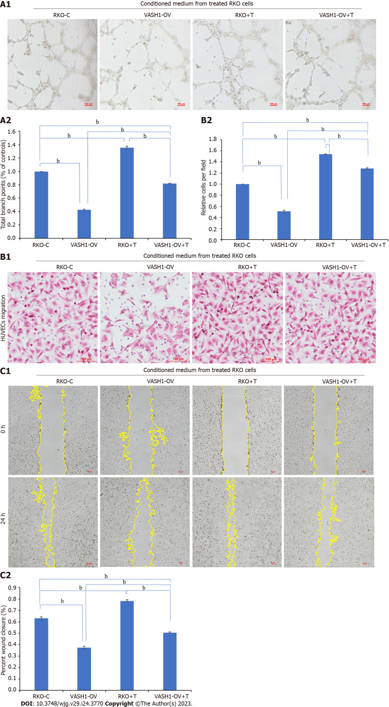
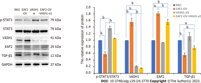
Our findings indicated that EAF2 was down-regulated and VASH1 was up-regulated in advanced CRC tissue compared to normal colorectal tissue. Kaplan-Meier survival analysis showed that the higher EAF2 Level group and the lower VASH1 Level group had a higher survival rate. Overexpression of EAF2 might inhibit the activity of STAT3/TGF-β1 pathway by up-regulating the expression of VASH1, and then weaken the invasion, migration and angiogenesis of CRC cells.
This study suggests that EAF2 and VASH1 may serve as new diagnostic and prognostic markers for CRC, and provide a clinical basis for exploring new biomarkers for CRC. This study complements the mechanism of EAF2 in CRC cells, enriches the role and mechanism of CRC cell-derived VASH1, and provides a new possible subtype of CRC as a therapeutic target of STAT3/TGF-β1 pathway.
作为一种新型的内源性抗血管生成分子,血管生成抑制素 1(VASH1)不仅在肿瘤基质中表达,也在肿瘤组织中表达。此外,研究表明 VASH1 可能是结直肠癌(CRC)的预后标志物。VASH1 的敲低增强了转化生长因子-β1(TGF-β1)/Smad3 通路的活性和 I/III 型胶原的产生。我们之前的研究结果表明,ELL 相关因子 2(EAF2)可能通过调节信号转导子和转录激活子 3(STAT3)/TGF-β1 信号通路,在 CRC 的发生和发展中发挥肿瘤抑制和保护作用。然而,VASH1 介导的 TGF-β1 相关通路在 CRC 中的功能作用和机制尚未阐明。
检测 VASH1 在 CRC 中的表达及其与 EAF2 表达的相关性。此外,我们研究了 VASH1 在 CRC 细胞中对 EAF2 调节和保护的功能作用和机制。
我们收集了结直肠腺癌和相应的癌旁组织,以检测晚期 CRC 患者中 EAF2 蛋白和 VASH1 蛋白的临床表达。然后,我们通过质粒转染研究了 EAF2 和 VASH1 对 CRC 细胞侵袭、迁移和血管生成的影响及其机制。
我们的研究结果表明,与正常结直肠组织相比,晚期 CRC 组织中 EAF2 下调,VASH1 上调。Kaplan-Meier 生存分析表明,EAF2 水平较高和 VASH1 水平较低的组具有更高的生存率。EAF2 的过表达可能通过上调 VASH1 的表达来抑制 STAT3/TGF-β1 通路的活性,从而削弱 CRC 细胞的侵袭、迁移和血管生成。
本研究提示 EAF2 和 VASH1 可能作为 CRC 的新的诊断和预后标志物,并为探索 CRC 的新生物标志物提供临床依据。本研究补充了 EAF2 在 CRC 细胞中的作用机制,丰富了 CRC 细胞来源的 VASH1 的作用和机制,并为 STAT3/TGF-β1 通路的治疗靶点提供了一种新的 CRC 可能的亚型。