Cardiovascular Imaging Research Center (CIRC), Department of Radiology and Division of Cardiology, Massachusetts General Hospital Department of Radiology, Boston, Massachusetts, USA
Heart and Vascular Center, Semmelweis University, Budapest, Hungary.
J Immunother Cancer. 2023 Jul;11(7). doi: 10.1136/jitc-2023-007307.
Patients with lung cancer face a heightened risk of atherosclerosis-related cardiovascular events. Despite the strong scientific rationale, there is currently a lack of clinical evidence examining the impact of immune checkpoint inhibitors (ICIs) on the advancement of atherosclerosis in patients with lung cancer. The objective of our study was to investigate whether there is a correlation between ICIs and the accelerated progression of atherosclerosis among individuals with lung cancer.
In this case-control (2:1 matched by age and gender) study, total, non-calcified, and calcified plaque volumes were measured in the thoracic aorta using sequential contrast-enhanced chest CT scans. Univariate and multivariate rank-based estimation regression models were developed to estimate the effect of ICI therapy on plaque progression in 40 cases (ICI) and 20 controls (non-ICI).
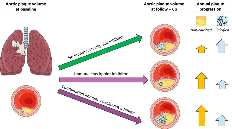
The patients had a median age of 66 years (IQR: 58-69), with 50% of them being women. At baseline, there were no significant differences in plaque volumes between the groups, and their cardiovascular risk profiles were similar. However, the annual progression rate for non-calcified plaque volume was 7 times higher in the ICI group compared with the controls (11.2% vs 1.6% per year, p=0.001). Conversely, the controls showed a greater progression in calcified plaque volume compared with the ICI group (25% vs 2% per year, p=0.017). In a multivariate model that considered cardiovascular risk factors, the use of an ICI was associated with a more substantial progression of non-calcified plaque volume. Additionally, individuals treated with combination ICI therapy exhibited greater plaque progression.
ICI therapy was associated with more non-calcified plaque progression. These findings underscore the importance of conducting studies aimed at identifying the underlying mechanisms responsible for plaque advancement in patients undergoing ICI treatment.
NCT04430712.
肺癌患者面临着与动脉粥样硬化相关的心血管事件风险增加。尽管有强有力的科学依据,但目前缺乏临床证据来研究免疫检查点抑制剂 (ICI) 对肺癌患者动脉粥样硬化进展的影响。我们的研究目的是探讨 ICI 是否与肺癌患者的动脉粥样硬化加速进展有关。
在这项病例对照研究(按年龄和性别 2:1 匹配)中,使用连续对比增强胸部 CT 扫描测量胸主动脉的总、非钙化和钙化斑块体积。采用单变量和多变量基于秩的估计回归模型,对 40 例接受 ICI 治疗的患者(ICI 组)和 20 例接受非 ICI 治疗的患者(非 ICI 组)的斑块进展情况进行估计。
患者的中位年龄为 66 岁(IQR:58-69),其中 50%为女性。在基线时,两组的斑块体积无显著差异,且心血管风险状况相似。然而,ICI 组的非钙化斑块体积年增长率是非 ICI 组的 7 倍(11.2% vs. 1.6%/年,p=0.001)。相反,非 ICI 组的钙化斑块体积增长速度快于 ICI 组(25% vs. 2%/年,p=0.017)。在考虑心血管危险因素的多变量模型中,使用 ICI 与非钙化斑块体积的更大进展相关。此外,接受联合 ICI 治疗的个体表现出更大的斑块进展。
ICI 治疗与更多的非钙化斑块进展有关。这些发现强调了进行研究的重要性,旨在确定接受 ICI 治疗的患者斑块进展的潜在机制。
NCT04430712。