Hubei Key Laboratory of Edible Wild Plants Conservation and Utilization, College of Life Sciences, Hubei Normal University, Huangshi, China.
Key Laboratory of Cognitive Science, Hubei Key Laboratory of Medical Information Analysis and Tumor Diagnosis & Treatment, Laboratory of Membrane Ion Channels and Medicine, College of Biomedical Engineering, South-Central Minzu University, Wuhan, China.
BMC Biol. 2023 Jul 13;21(1):158. doi: 10.1186/s12915-023-01655-6.
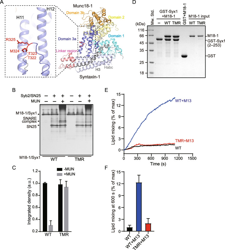
Neurotransmitter release depends on the fusion of synaptic vesicles with the presynaptic membrane and is mainly mediated by SNARE complex assembly. During the transition of Munc18-1/Syntaxin-1 to the SNARE complex, the opening of the Syntaxin-1 linker region catalyzed by Munc13-1 leads to the extension of the domain 3a hinge loop, which enables domain 3a to bind SNARE motifs in Synaptobrevin-2 and Syntaxin-1 and template the SNARE complex assembly. However, the exact mechanism of domain 3a extension remains elusive.
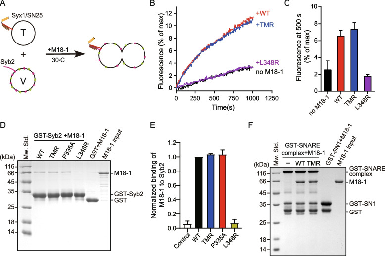
Here, we characterized residues on the domain 3a hinge loop that are crucial for the extension of domain 3a by using biophysical and biochemical approaches and electrophysiological recordings. We showed that the mutation of residues T323/M324/R325 disrupted Munc13-1-mediated SNARE complex assembly and membrane fusion starting from Munc18-1/Syntaxin-1 in vitro and caused severe defects in the synaptic exocytosis of mouse cortex neurons in vivo. Moreover, the mutation had no effect on the binding of Synaptobrevin-2 to isolated Munc18-1 or the conformational change of the Syntaxin-1 linker region catalyzed by the Munc13-1 MUN domain. However, the extension of the domain 3a hinge loop in Munc18-1/Syntaxin-1 was completely disrupted by the mutation, leading to the failure of Synaptobrevin-2 binding to Munc18-1/Syntaxin-1.
Together with previous results, our data further support the model that the template function of Munc18-1 in SNARE complex assembly requires the extension of domain 3a, and particular residues in the domain 3a hinge loop are crucial for the autoinhibitory release of domain 3a after the MUN domain opens the Syntaxin-1 linker region.
神经递质的释放依赖于突触小泡与突触前膜的融合,主要由 SNARE 复合物的组装介导。在 Munc18-1/Syntaxin-1 向 SNARE 复合物转变过程中,Munc13-1 催化的 Syntaxin-1 连接区的打开导致结构域 3a 铰链环的延伸,从而使结构域 3a 能够结合突触融合蛋白-2 和 Syntaxin-1 的 SNARE 基序,并模板 SNARE 复合物的组装。然而,结构域 3a 延伸的确切机制仍不清楚。
在这里,我们使用生物物理和生化方法以及电生理记录来表征结构域 3a 铰链环上对结构域 3a 延伸至关重要的残基。我们表明,残基 T323/M324/R325 的突变破坏了 Munc13-1 介导的体外从 Munc18-1/Syntaxin-1 开始的 SNARE 复合物组装和膜融合,并且在体内导致小鼠皮质神经元的突触胞吐严重缺陷。此外,该突变对突触融合蛋白-2 与分离的 Munc18-1 的结合或 Munc13-1 MUN 结构域催化的 Syntaxin-1 连接区构象变化没有影响。然而,该突变完全破坏了结构域 3a 铰链环的延伸,导致突触融合蛋白-2 无法与 Munc18-1/Syntaxin-1 结合。
与以前的结果一起,我们的数据进一步支持了这样的模型,即 Munc18-1 在 SNARE 复合物组装中的模板功能需要结构域 3a 的延伸,并且结构域 3a 铰链环中的特定残基对于 MUN 结构域打开 Syntaxin-1 连接区后结构域 3a 的自动抑制释放至关重要。