Scientific Research Department, Armed Forces Radiobiology Research Institute, Uniformed Services University of the Health Sciences, Bethesda, MD 20889, USA.
Henry M. Jackson Foundation for the Advancement of Military Medicine, Bethesda, MD 20817, USA.
Int J Mol Sci. 2023 Jun 27;24(13):10701. doi: 10.3390/ijms241310701.
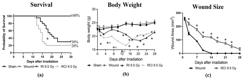
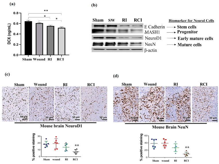
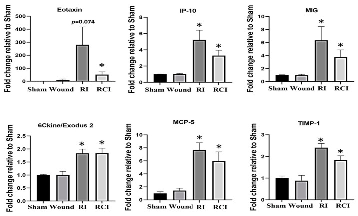
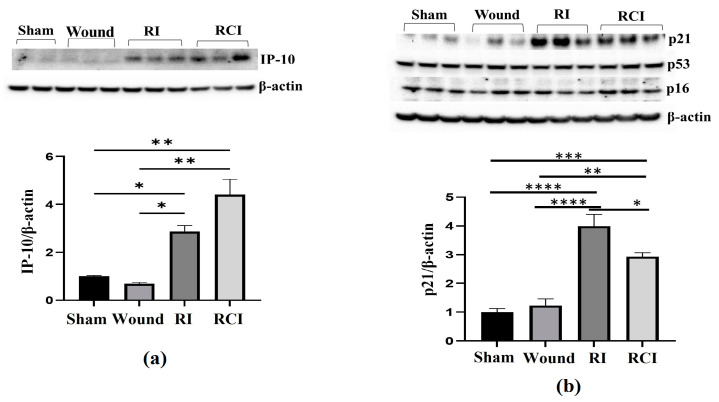
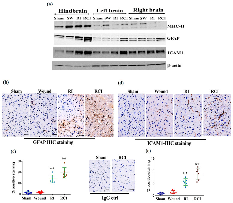
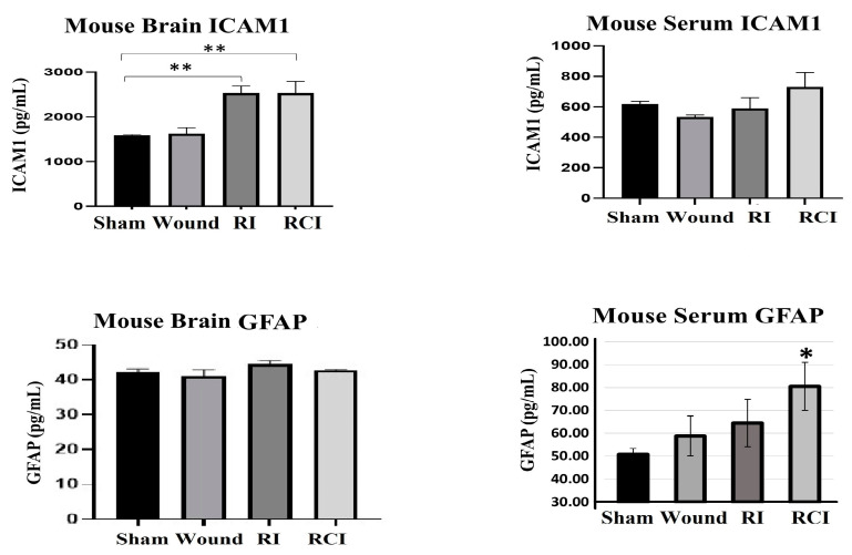
Radiation injury- and radiation combined with skin injury-induced inflammatory responses in the mouse brain were evaluated in this study. Female B6D2F1/J mice were subjected to a sham, a skin wound (SW), 9.5 Gy Co total-body gamma irradiation (RI), or 9.5 Gy RI combined with a skin puncture wound (RCI). Survival, body weight, and wound healing were tracked for 30 days, and mouse brain samples were collected on day 30 after SW, RI, RCI, and the sham control. Our results showed that RCI caused more severe animal death and body weight loss compared with RI, and skin wound healing was significantly delayed by RCI compared to SW. RCI and RI increased the chemokines Eotaxin, IP-10, MIG, 6Ckine/Exodus2, MCP-5, and TIMP-1 in the brain compared to SW and the sham control mice, and the Western blot results showed that IP-10 and p21 were significantly upregulated in brain cells post-RI or -RCI. RI and RCI activated both astrocytes and endothelial cells in the mouse brain, subsequently inducing blood-brain barrier (BBB) leakage, as shown by the increased ICAM1 and GFAP proteins in the brain and GFAP in the serum. The Doublecortin (DCX) protein, the "gold standard" for measuring neurogenesis, was significantly downregulated by RI and RCI compared with the sham group. Furthermore, RI and RCI decreased the expression of the neural stem cell marker E-cadherin, the intermediate progenitor marker MASH1, the immature neuron cell marker NeuroD1, and the mature neuron cell marker NeuN, indicating neural cell damage in all development stages after RI and RCI. Immunohistochemistry (IHC) staining further confirmed the significant loss of neural cells in RCI. Our data demonstrated that RI and RCI induced brain injury through inflammatory pathways, and RCI exacerbated neural cell damage more than RI.
本研究评估了小鼠脑内辐射损伤和辐射合并皮肤损伤诱导的炎症反应。将雌性 B6D2F1/J 小鼠分为假手术组、皮肤伤口组(SW)、9.5Gy 全身γ射线照射组(RI)和 9.5GyRI 联合皮肤穿刺伤组(RCI)。观察 30 天内的生存情况、体重变化和伤口愈合情况,于 SW、RI、RCI 和假手术对照组后第 30 天采集小鼠脑样本。结果显示,与 RI 相比,RCI 导致更严重的动物死亡和体重减轻,与 SW 相比,RCI 显著延迟皮肤伤口愈合。与 SW 和假手术对照组相比,RCI 和 RI 增加了脑中趋化因子 Eotaxin、IP-10、MIG、6Ckine/Exodus2、MCP-5 和 TIMP-1 的表达,Western blot 结果显示,IP-10 和 p21 在 RI 或 RCI 后大脑细胞中显著上调。RI 和 RCI 激活了小鼠脑内的星形胶质细胞和内皮细胞,随后导致血脑屏障(BBB)渗漏,脑内 ICAM1 和 GFAP 蛋白以及血清中 GFAP 增加。神经发生的“金标准”蛋白 Doublecortin(DCX)与假手术组相比,RI 和 RCI 显著下调。此外,RI 和 RCI 降低了神经干细胞标志物 E-cadherin、中间祖细胞标志物 MASH1、未成熟神经元细胞标志物 NeuroD1 和成熟神经元细胞标志物 NeuN 的表达,表明 RI 和 RCI 后所有发育阶段的神经细胞损伤。免疫组织化学(IHC)染色进一步证实了 RCI 中神经细胞的显著丢失。本研究数据表明,RI 和 RCI 通过炎症途径诱导脑损伤,RCI 比 RI 更严重地加剧神经细胞损伤。