Sharifian Rasoul, Nazari Behzad, Sadri Saeed, Adibi Peyman
Department of Electrical and Computer Engineering, Digital Signal Processing Lab., Isfahan University of Technology, Isfahan, Iran.
Department of Internal Medicine, Faculty of Medicine, Isfahan University of Medical Science, Isfahan, Iran.
J Med Signals Sens. 2023 May 29;13(2):73-83. doi: 10.4103/jmss.jmss_182_21. eCollection 2023 Apr-Jun.
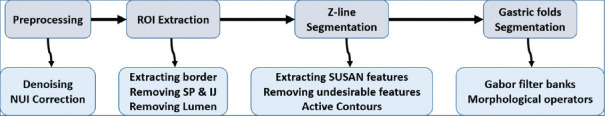
The endoscopic diagnosis of pathological changes in the gastroesophageal junction including esophagitis and Barrett's mucosa is based on the visual detection of two boundaries: mucosal color change between esophagus and stomach, and top endpoint of gastric folds. The presence and pattern of mucosal breaks in the gastroesophageal mucosal junction (Z line) classify esophagitis in patients and the distance between the two boundaries points to the possible columnar lined epithelium. Since visual detection may suffer from intra- and interobserver variability, our objective was to define the boundaries automatically based on image processing algorithms, which may enable us to measure the detentions of changes in future studies.
To demarcate the Z-line, first the artifacts of endoscopy images are eliminated. In the second step, using SUSAN edge detector, Mahalanobis distance criteria, and Gabor filter bank, an initial contour is estimated for the Z-line. Using region-based active contours, this initial contour converges to the Z-line. Finally, by applying morphological operators and Gabor Filter Bank to the region inside of the Z-line, gastric folds are segmented.
To evaluate the results, a database consisting of 50 images and their ground truths were collected. The average dice coefficient and mean square error of Z-line segmentation were 0.93 and 3.3, respectively. Furthermore, the average boundary distance criteria are 12.3 pixels. In addition, two other criteria that compare the segmentation of folds with several ground truths, i.e., Sweet-Spot Coverage and Jaccard Index for Golden Standard, are 0.90 and 0.84, respectively.
Considering the results, automatic segmentation of Z-line and gastric folds are matched to the ground truths with appropriate accuracy.
胃食管交界处病理变化(包括食管炎和巴雷特黏膜)的内镜诊断基于对两个边界的视觉检测:食管与胃之间的黏膜颜色变化,以及胃皱襞的顶端终点。胃食管黏膜交界处(Z线)黏膜破损的存在及模式对患者的食管炎进行分类,而两个边界之间的距离则指向可能的柱状上皮化生。由于视觉检测可能存在观察者内和观察者间的差异,我们的目标是基于图像处理算法自动定义边界,这可能使我们能够在未来的研究中测量变化的程度。
为了划定Z线,首先消除内镜图像的伪影。第二步,使用SUSAN边缘检测器、马氏距离准则和Gabor滤波器组,估计Z线的初始轮廓。使用基于区域的活动轮廓,该初始轮廓收敛到Z线。最后,通过对Z线内部区域应用形态学算子和Gabor滤波器组,分割胃皱襞。
为了评估结果,收集了一个由50幅图像及其真实边界组成的数据库。Z线分割的平均骰子系数和均方误差分别为0.93和3.3。此外,平均边界距离准则为12.3像素。此外,另外两个将皱襞分割与多个真实边界进行比较的准则,即黄金标准的最佳点覆盖率和杰卡德指数,分别为0.90和0.84。
考虑到结果,Z线和胃皱襞的自动分割与真实边界具有适当的匹配精度。