Laboratory for Cell Biology and Signalling, Division of Molecular Biology, Ruđer Bošković Institute, Zagreb, Croatia.
Department of Life Science, Manchester Metropolitan University, Manchester, United Kingdom.
Cell Mol Biol Lett. 2023 Jul 17;28(1):56. doi: 10.1186/s11658-023-00473-6.
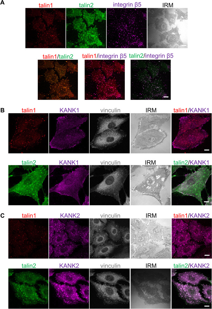
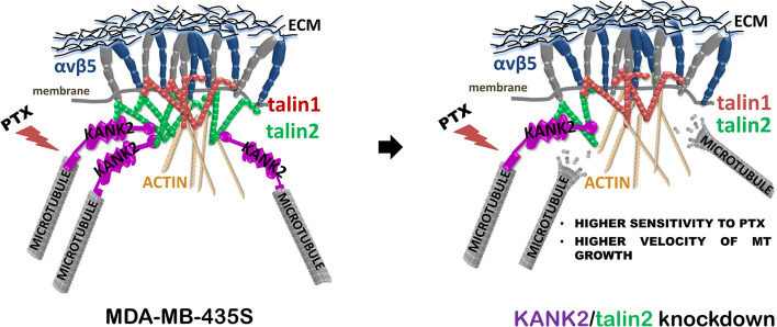
Focal adhesions (FAs) are integrin-containing, multi-protein structures that link intracellular actin to the extracellular matrix and trigger multiple signaling pathways that control cell proliferation, differentiation, survival and motility. Microtubules (MTs) are stabilized in the vicinity of FAs through interaction with the components of the cortical microtubule stabilizing complex (CMSC). KANK (KN motif and ankyrin repeat domains) family proteins within the CMSC, KANK1 or KANK2, bind talin within FAs and thus mediate actin-MT crosstalk. We previously identified in MDA-MB-435S cells, which preferentially use integrin αVβ5 for adhesion, KANK2 as a key molecule enabling the actin-MT crosstalk. KANK2 knockdown also resulted in increased sensitivity to MT poisons, paclitaxel (PTX) and vincristine and reduced migration. Here, we aimed to analyze whether KANK1 has a similar role and to distinguish which talin isoform binds KANK2.
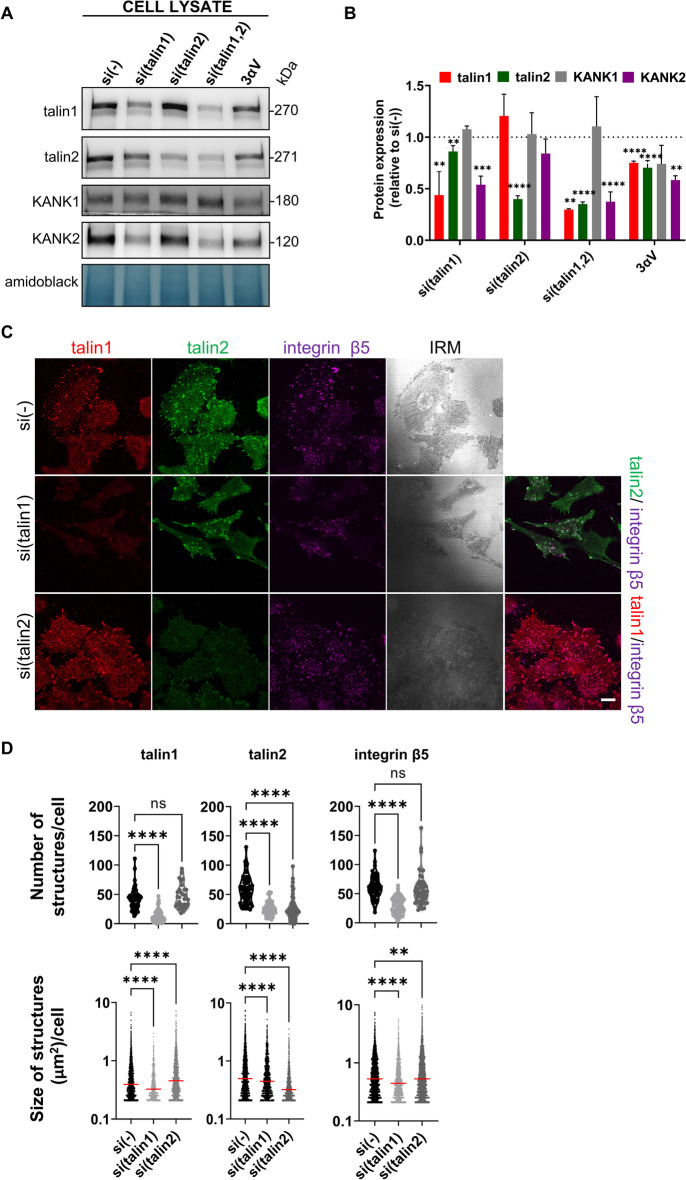
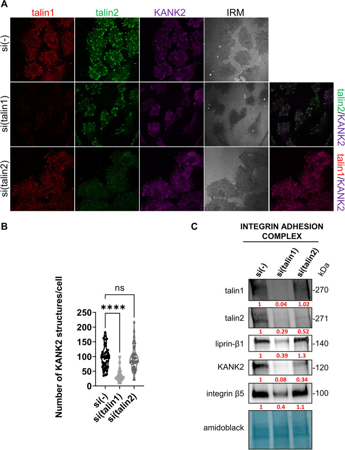
The cell model consisted of human melanoma cell line MDA-MB-435S and stably transfected clone with decreased expression of integrin αV (3αV). For transient knockdown of talin1, talin2, KANK1 or KANK2 we used gene-specific siRNAs transfection. Using previously standardized protocol we isolated integrin adhesion complexes. SDS-PAGE and Western blot was used for protein expression analysis. The immunofluorescence analysis and live cell imaging was done using confocal microscopy. Cell migration was analyzed with Transwell Cell Culture Inserts. Statistical analysis using GraphPad Software consisted of either one-way analysis of variance (ANOVA), unpaired Student's t-test or two-way ANOVA analysis.
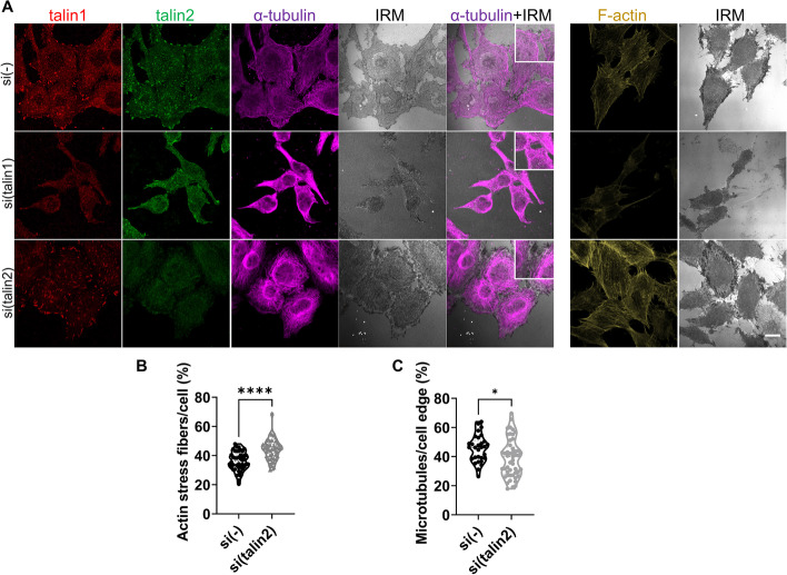
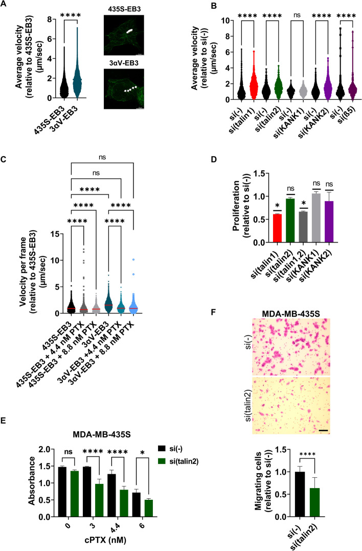
We show that KANK1 is not a part of the CMSC associated with integrin αVβ5 FAs and its knockdown did not affect the velocity of MT growth or cell sensitivity to PTX. The talin2 knockdown mimicked KANK2 knockdown i.e. led to the perturbation of actin-MT crosstalk, which is indicated by the increased velocity of MT growth and increased sensitivity to PTX and also reduced migration.
We conclude that KANK2 functionally interacts with talin2 and that the mechanism of increased sensitivity to PTX involves changes in microtubule dynamics. These data elucidate a cell-type-specific role of talin2 and KANK2 isoforms and we propose that talin2 and KANK2 are therefore potential therapeutic targets for improved cancer therapy.
粘着斑(FA)是整合素含有,多蛋白结构,它将细胞内肌动蛋白与细胞外基质连接起来,并触发多个信号通路,控制细胞增殖、分化、存活和迁移。微管(MTs)通过与皮质微管稳定复合物(CMSC)的成分相互作用而在 FA 附近稳定。CMSC 内的 KANK(KN 基序和锚蛋白重复结构域)家族蛋白,KANK1 或 KANK2,与 FA 内的 talin 结合,从而介导肌动蛋白-MT 串扰。我们之前在 MDA-MB-435S 细胞中鉴定到,该细胞优先使用整合素 αVβ5 进行黏附,KANK2 是使肌动蛋白-MT 串扰成为可能的关键分子。KANK2 敲低也导致对 MT 毒物紫杉醇(PTX)和长春新碱的敏感性增加,迁移减少。在这里,我们旨在分析 KANK1 是否具有类似的作用,并区分结合 KANK2 的 talin 同工型。
细胞模型由人黑色素瘤细胞系 MDA-MB-435S 和稳定转染的整合素 αV 表达降低的克隆(3αV)组成。为了瞬时敲低 talin1、talin2、KANK1 或 KANK2,我们使用了基因特异性 siRNAs 转染。使用以前标准化的方案,我们分离了整合素黏附复合物。SDS-PAGE 和 Western blot 用于蛋白质表达分析。免疫荧光分析和活细胞成像使用共聚焦显微镜进行。使用 Transwell 细胞培养插入物分析细胞迁移。GraphPad 软件中的统计分析包括单向方差分析(ANOVA)、未配对学生 t 检验或双向 ANOVA 分析。
我们表明 KANK1 不是与整合素 αVβ5 FA 相关的 CMSC 的一部分,其敲低并不影响 MT 生长的速度或细胞对 PTX 的敏感性。talin2 敲低模拟了 KANK2 敲低,即导致肌动蛋白-MT 串扰的扰动,这表现为 MT 生长速度的增加以及对 PTX 的敏感性增加,同时迁移减少。
我们得出结论,KANK2 与 talin2 具有功能相互作用,对 PTX 敏感性增加的机制涉及微管动力学的变化。这些数据阐明了 talin2 和 KANK2 同工型的细胞类型特异性作用,我们提出 talin2 和 KANK2 是改善癌症治疗的潜在治疗靶点。