• 文献检索
  • 文档翻译
  • 深度研究
  • 学术资讯
  • Suppr Zotero 插件Zotero 插件
  • 邀请有礼
  • 套餐&价格
  • 历史记录
应用&插件
Suppr Zotero 插件Zotero 插件浏览器插件Mac 客户端Windows 客户端微信小程序
定价
高级版会员购买积分包购买API积分包
服务
文献检索文档翻译深度研究API 文档MCP 服务
关于我们
关于 Suppr公司介绍联系我们用户协议隐私条款
关注我们

Suppr 超能文献

核心技术专利:CN118964589B侵权必究
粤ICP备2023148730 号-1Suppr @ 2026

文献检索

告别复杂PubMed语法,用中文像聊天一样搜索,搜遍4000万医学文献。AI智能推荐,让科研检索更轻松。

立即免费搜索

文件翻译

保留排版,准确专业,支持PDF/Word/PPT等文件格式,支持 12+语言互译。

免费翻译文档

深度研究

AI帮你快速写综述,25分钟生成高质量综述,智能提取关键信息,辅助科研写作。

立即免费体验

结节性硬化症患者的眼眶血管周围上皮样细胞瘤

Orbital Perivascular Epithelioid Cell Tumor in a Case of Tuberous Sclerosis.

作者信息

Aljneibi Shaikha H, Aldhanhani Aisha A, Abuhaleeqa Khaled, Pichi Francesco

机构信息

Cleveland Clinic AbuFcoi Dhabi, Eye Institute, Abu Dhabi, United Arab Emirates.

Cleveland Clinic Lerner College of Medicine, Case Western Reserve University, Cleveland, OH, USA.

出版信息

Case Rep Ophthalmol. 2023 Jul 7;14(1):282-287. doi: 10.1159/000530036. eCollection 2023 Jan-Dec.

DOI:10.1159/000530036
PMID:37485243
原文链接:https://pmc.ncbi.nlm.nih.gov/articles/PMC10359681/
Abstract

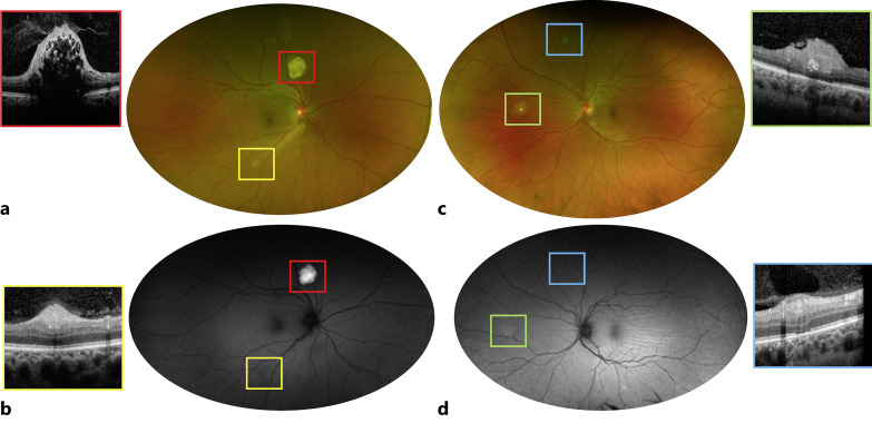
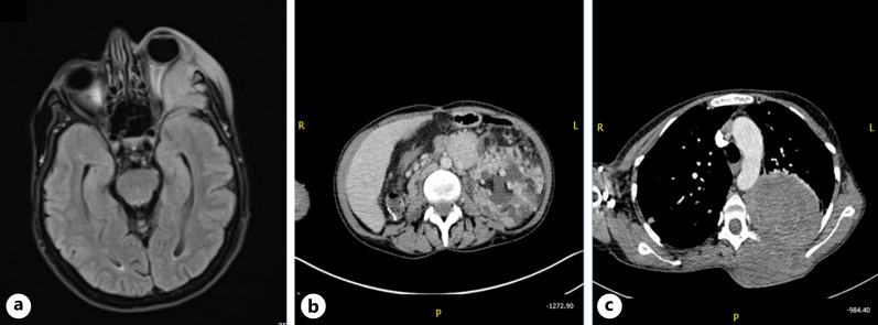
The aim of the study was to report a case of orbital perivascular epithelioid cell tumor (PEComa) in a known diagnosed patient of tuberous sclerosis and retinal astrocytic hamartoma. 43-year-old female presented with rapid progressive painful proptosis in the left eye, also reported new mass growing in her upper back. The patient past medical history is significant for left renal angiomyolipoma and multiple bilateral lung cysts of which she underwent right nephrectomy and lung biopsy, respectively. The lung biopsy turned diagnostic for lymphangiomyomatosis. On external examination, the left eye was grossly proptotic with hypoglobus. A typical butterfly distribution of sebaceous adenoma was noted across the patient cheeks and nose. Visual acuity in the right eye was 20/20 and the left eye, 20/25. Funduscopic examination identified type 1, 2, and 3 retinal astrocytic hamartomas. MRI brain and orbit was significant for a lesion arising from the lateral orbital wall with extensive bone destruction, displacing the left optic nerve medially. CT chest showed left extrathoracic mass had same radiological features as the orbital lesion; thus, an incisional biopsy performed on the former was diagnostic for PEComa with atypical features. This is the first observed case of PEComa in a known diagnosed patent with TS and retinal astrocytic hamartoma. The association of tuberous sclerosis complex and orbital PEComa is rarely and poorly reported in the literature compared to extraocular PEComa.

摘要

本研究的目的是报告1例患有结节性硬化症和视网膜星形细胞错构瘤的确诊患者发生眼眶血管周上皮样细胞瘤(PEComa)的病例。一名43岁女性因左眼迅速进展的疼痛性眼球突出就诊,还报告其背部上方有新肿物生长。该患者既往病史包括左肾血管平滑肌脂肪瘤和双侧多发肺囊肿,她分别接受了右肾切除术和肺活检。肺活检诊断为淋巴管平滑肌瘤病。体格检查时,左眼明显眼球突出且眼球下转。在患者的脸颊和鼻子上可见典型的皮脂腺腺瘤蝶形分布。右眼视力为20/20,左眼视力为20/25。眼底检查发现1型、2型和3型视网膜星形细胞错构瘤。头颅和眼眶MRI显示有一个起源于眶外侧壁的病变,伴有广泛骨质破坏,将左侧视神经向内移位。胸部CT显示左侧胸壁肿物与眼眶病变具有相同的影像学特征;因此,对前者进行的切开活检诊断为具有非典型特征的PEComa。这是首例在已知患有结节性硬化症和视网膜星形细胞错构瘤的确诊患者中观察到的PEComa病例。与眼外PEComa相比,结节性硬化症复合体与眼眶PEComa的关联在文献中报道较少且不充分。

https://cdn.ncbi.nlm.nih.gov/pmc/blobs/4aec/10359681/2aef93204d4a/cop-2023-0014-0001-530036_F02.jpg
https://cdn.ncbi.nlm.nih.gov/pmc/blobs/4aec/10359681/a1475b89d5d3/cop-2023-0014-0001-530036_F01.jpg
https://cdn.ncbi.nlm.nih.gov/pmc/blobs/4aec/10359681/2aef93204d4a/cop-2023-0014-0001-530036_F02.jpg
https://cdn.ncbi.nlm.nih.gov/pmc/blobs/4aec/10359681/a1475b89d5d3/cop-2023-0014-0001-530036_F01.jpg
https://cdn.ncbi.nlm.nih.gov/pmc/blobs/4aec/10359681/2aef93204d4a/cop-2023-0014-0001-530036_F02.jpg

相似文献

1
Orbital Perivascular Epithelioid Cell Tumor in a Case of Tuberous Sclerosis.结节性硬化症患者的眼眶血管周围上皮样细胞瘤
Case Rep Ophthalmol. 2023 Jul 7;14(1):282-287. doi: 10.1159/000530036. eCollection 2023 Jan-Dec.
2
EXUDATIVE RETINAL DETACHMENT AS THE PRESENTING FEATURE OF TUBEROUS SCLEROSIS COMPLEX.渗出性视网膜脱离作为结节性硬化症复合体的首发特征
Retin Cases Brief Rep. 2016 Spring;10(2):121-6. doi: 10.1097/ICB.0000000000000155.
3
[A Case of Perivascular Epithelioid Cell Tumor (PEComa) of the Uterus Associated with Tuberous Sclerosis Complex Diagnosed by Intra-Abdominal Bleeding from the Uterus].[一例因子宫腹腔内出血诊断为与结节性硬化症相关的子宫血管周上皮样细胞肿瘤(PEComa)]
Hinyokika Kiyo. 2019 Nov;65(11):479-484. doi: 10.14989/ActaUrolJap_65_11_479.
4
Fibroma-like PEComa: a newly recognized soft tissue neoplasm in tuberous sclerosis patients-imaging features and review of literature.纤维瘤样错构瘤:结节性硬化症患者中一种新认识的软组织肿瘤-影像学特征及文献复习。
Skeletal Radiol. 2022 Apr;51(4):881-887. doi: 10.1007/s00256-021-03923-4. Epub 2021 Sep 29.
5
Leiomyomatosis-like lymphangioleiomyomatosis: A case report of the colonic manifestation of tuberous sclerosis.平滑肌脂肪瘤样淋巴管平滑肌瘤病:结节性硬化症结肠表现一例报告。
Medicine (Baltimore). 2021 Dec 17;100(50):e27723. doi: 10.1097/MD.0000000000027723.
6
Optic coherence tomography appearances of retinal astrocytic hamartoma and systemic features in tuberous sclerosis of Japanese patients.日本结节性硬化症患者视网膜星形细胞错构瘤的光学相干断层扫描表现及全身特征
Eur J Ophthalmol. 2019 May;29(3):330-337. doi: 10.1177/1120672118787441. Epub 2018 Jul 11.
7
Retinal astrocytic hamartoma in tuberous sclerosis complex in an elderly person: a case report.老年结节性硬化症患者的视网膜星形细胞错构瘤:一例报告
BMC Ophthalmol. 2018 Dec 12;18(1):319. doi: 10.1186/s12886-018-0991-z.
8
Case report: Liver PEComa after kidney transplantation in recipient with tuberous sclerosis complex.病例报告:结节性硬化症复合体患者肾移植后发生肝脏血管周上皮样细胞瘤。
Front Oncol. 2024 Jul 4;14:1386569. doi: 10.3389/fonc.2024.1386569. eCollection 2024.
9
Improving recognition of hepatic perivascular epithelioid cell tumor: Case report and literature review.提高肝脏血管周上皮样细胞肿瘤的认识:病例报告及文献复习
World J Gastroenterol. 2015 May 7;21(17):5432-41. doi: 10.3748/wjg.v21.i17.5432.
10
Fibroma-like perivascular epithelioid cell tumor: a rare case in a long bone.纤维瘤样血管周上皮样细胞肿瘤:长骨中的罕见病例。
Skeletal Radiol. 2021 Apr;50(4):821-825. doi: 10.1007/s00256-020-03610-w. Epub 2020 Sep 18.

本文引用的文献

1
Orbital TFE3-Rearranged Perivascular Epithelioid Cell Tumor: A Case Report and Review of the Literature.眼眶 TFE3 重排血管周上皮样细胞肿瘤:病例报告及文献复习。
Am J Dermatopathol. 2021 Dec 1;43(12):e263-e266. doi: 10.1097/DAD.0000000000002023.
2
Hepatic perivascular epithelioid cell tumours in children with tuberous sclerosis.儿童结节性硬化症相关肝血管周上皮样细胞瘤。
BMJ Case Rep. 2020 Dec 9;13(12):e236288. doi: 10.1136/bcr-2020-236288.
3
Giant Perivascular Epithelioid Cell Tumor of the Orbit: A Clinicopathological Analysis and Review of the Literature.
眼眶巨大血管周上皮样细胞瘤:临床病理分析及文献复习
Ocul Oncol Pathol. 2018 Sep;4(5):272-279. doi: 10.1159/000484425. Epub 2018 Feb 6.
4
Optic coherence tomography appearances of retinal astrocytic hamartoma and systemic features in tuberous sclerosis of Japanese patients.日本结节性硬化症患者视网膜星形细胞错构瘤的光学相干断层扫描表现及全身特征
Eur J Ophthalmol. 2019 May;29(3):330-337. doi: 10.1177/1120672118787441. Epub 2018 Jul 11.
5
Tuberous sclerosis complex: Recent advances in manifestations and therapy.结节性硬化症:临床表现与治疗的最新进展
Int J Urol. 2017 Sep;24(9):681-691. doi: 10.1111/iju.13390. Epub 2017 Jul 1.
6
OPTICAL COHERENCE TOMOGRAPHY AND INFRARED IMAGES OF ASTROCYTIC HAMARTOMAS NOT REVEALED BY FUNDUSCOPY IN TUBEROUS SCLEROSIS COMPLEX.结节性硬化症中眼底检查未发现的星形细胞瘤的光学相干断层扫描和红外图像
Retina. 2017 Jul;37(7):1383-1392. doi: 10.1097/IAE.0000000000001373.
7
RETINAL ASTROCYTIC HAMARTOMA: Optical Coherence Tomography Classification and Correlation With Tuberous Sclerosis Complex.视网膜星形细胞错构瘤:光学相干断层扫描分类及其与结节性硬化症的相关性
Retina. 2016 Jun;36(6):1199-208. doi: 10.1097/IAE.0000000000000829.
8
PEComa: morphology and genetics of a complex tumor family.血管周上皮样细胞瘤:一个复杂肿瘤家族的形态学与遗传学
Ann Diagn Pathol. 2015 Oct;19(5):359-68. doi: 10.1016/j.anndiagpath.2015.06.003. Epub 2015 Jun 9.
9
Improving recognition of hepatic perivascular epithelioid cell tumor: Case report and literature review.提高肝脏血管周上皮样细胞肿瘤的认识:病例报告及文献复习
World J Gastroenterol. 2015 May 7;21(17):5432-41. doi: 10.3748/wjg.v21.i17.5432.
10
Ocular perivascular epithelioid cell tumor: report of 2 cases with distinct clinical presentations.眼周血管外皮细胞瘤:2 例临床表现不同的病例报告。
Hum Pathol. 2010 May;41(5):768-72. doi: 10.1016/j.humpath.2009.12.006. Epub 2010 Mar 17.