Department of Radiology, Ospedale del Mare-ASL NA1 Centro, Via Enrico Russo 11, 80147 Naples, Italy.
Division of Radiology, Università degli Studi della Campania "Luigi Vanvitelli", Piazza Luigi Miraglia 2, 80138 Naples, Italy.
Tomography. 2023 Jul 12;9(4):1356-1368. doi: 10.3390/tomography9040108.
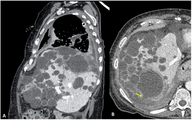
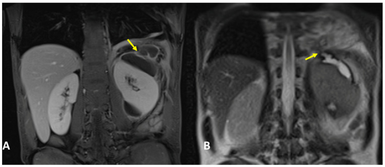
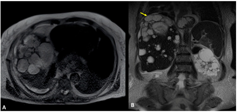
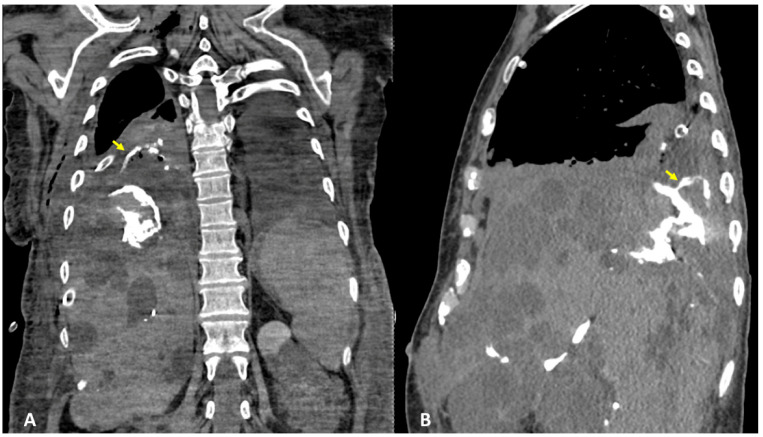
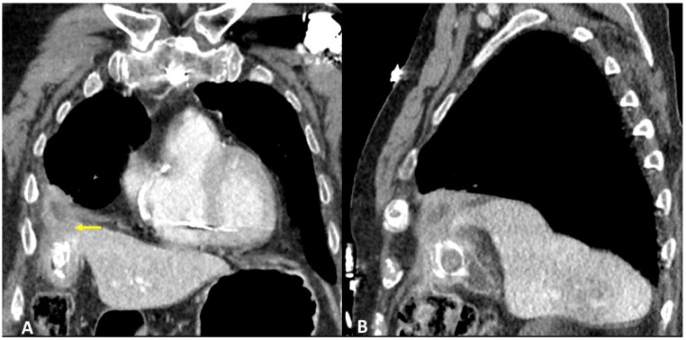
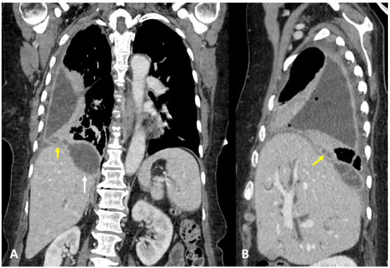
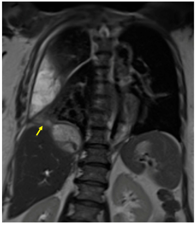
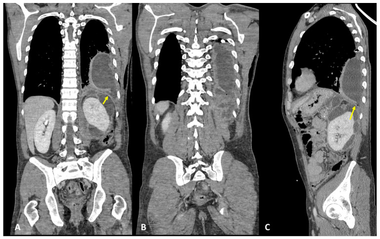
Transdiaphragmatic fistulae are rare conditions characterized by pathological communication between two epithelium-lined surfaces. Hepato-thoracic fistula consists of abnormal communication between the liver and/or the biliary system and the thorax; while the pancreaticopleural fistula consists of abnormal communication between the pancreas and the thorax, the pleuro-biliary fistula represents the more common type. Clinical symptoms and laboratory findings are generally non-specific (e.g., thoracic and abdominal pain, dyspnea, cough, neutrophilia, elevated CPR, and bilirubin values) and initially, first-level investigations, such as chest RX and abdominal ultrasound, are generally inconclusive for the diagnosis. Contrast-enhanced CT represents the first two-level radiological imaging technique, usually performed to identify and evaluate the underlying pathology sustained by transdiaphragmatic fistulae, their complications, and the evaluation of the fistulous tract. When the CT remains inconclusive, other techniques such as MRI and MRCP can be performed. A prompt and accurate diagnosis is crucial because the recognition of fistulae and the precise definition of the fistulous tract have a major impact on the management acquisition process.
横膈膜瘘是一种罕见的疾病,其特征是两个上皮衬里表面之间存在病理性的沟通。肝胸瘘是指肝脏和/或胆道系统与胸腔之间的异常沟通;而胰胸膜瘘是指胰腺和胸腔之间的异常沟通,其中更常见的是胆胸瘘。临床症状和实验室发现通常是非特异性的(例如,胸痛和腹痛、呼吸困难、咳嗽、中性粒细胞增多、CPR 和胆红素值升高),最初,如胸部 X 光和腹部超声等一级检查通常无法明确诊断。增强 CT 是前两级影像学检查技术,通常用于识别和评估横膈膜瘘及其并发症的潜在病理学,并评估瘘管。当 CT 结果不确定时,可以进行其他技术,如 MRI 和 MRCP。及时准确的诊断至关重要,因为识别瘘管和准确定义瘘管对管理过程有重大影响。