• 文献检索
  • 文档翻译
  • 深度研究
  • 学术资讯
  • Suppr Zotero 插件Zotero 插件
  • 邀请有礼
  • 套餐&价格
  • 历史记录
应用&插件
Suppr Zotero 插件Zotero 插件浏览器插件Mac 客户端Windows 客户端微信小程序
定价
高级版会员购买积分包购买API积分包
服务
文献检索文档翻译深度研究API 文档MCP 服务
关于我们
关于 Suppr公司介绍联系我们用户协议隐私条款
关注我们

Suppr 超能文献

核心技术专利:CN118964589B侵权必究
粤ICP备2023148730 号-1Suppr @ 2026

文献检索

告别复杂PubMed语法,用中文像聊天一样搜索,搜遍4000万医学文献。AI智能推荐,让科研检索更轻松。

立即免费搜索

文件翻译

保留排版,准确专业,支持PDF/Word/PPT等文件格式,支持 12+语言互译。

免费翻译文档

深度研究

AI帮你快速写综述,25分钟生成高质量综述,智能提取关键信息,辅助科研写作。

立即免费体验

早期乳腺癌患者放疗后弥漫性心肌纤维化的多模态影像学研究-六年随访。

Radiotherapy-induced diffuse myocardial fibrosis in early-stage breast cancer patients - multimodality imaging study with six-year follow-up.

机构信息

Faculty of Medicine and Health Technology, Tampere University, PO Box 100, Tampere, 33014, Finland.

Department of Oncology, Tampere University Hospital, Sädetie 6, PO Box 2000, Tampere, 33521, Finland.

出版信息

Radiat Oncol. 2023 Jul 26;18(1):124. doi: 10.1186/s13014-023-02319-z.

DOI:10.1186/s13014-023-02319-z
PMID:37496091
原文链接:https://pmc.ncbi.nlm.nih.gov/articles/PMC10373367/
Abstract

BACKGROUND

Breast radiotherapy (RT) induces diffuse myocardial changes, which may increase the incidence of heart failure with preserved ejection fraction. This study aimed to evaluate the early signs of diffuse fibrosis after RT and their evolution during a six-year follow-up.

METHODS

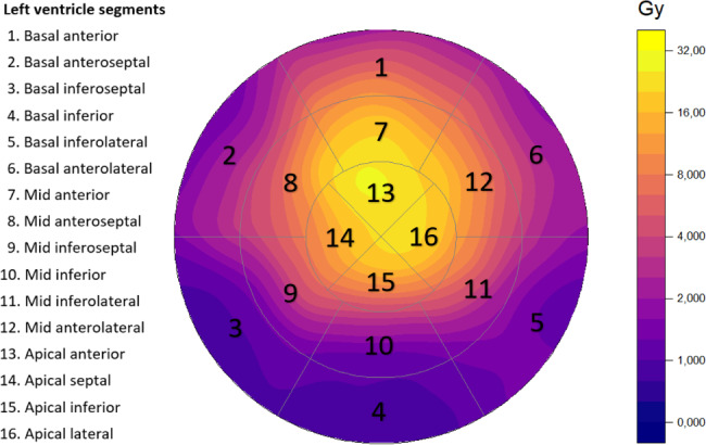
Thirty patients with early-stage left-sided breast cancer were studied with echocardiography and electrocardiography (ECG) at baseline, after RT, and at three-year and six-year follow-up visits. Echocardiography analysis included an off-line analysis of integrated backscatter (IBS). ECG was analysed for fragmented QRS (fQRS). In addition, cardiac magnetic resonance (CMR) imaging was performed at the six-year control. The left ventricle 16-segment model was used in cardiac imaging, and respective local radiation doses were analysed.

RESULTS

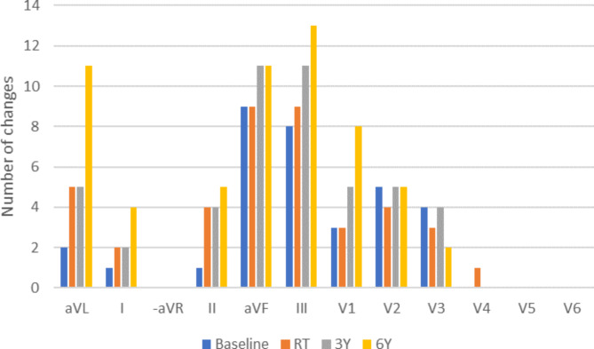
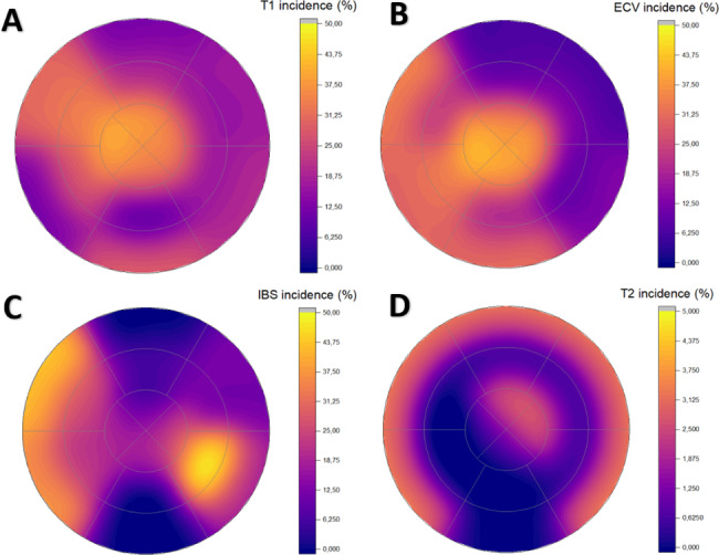
Regional myocardial reflectivity in inferoseptal segments increased by 2.02 (4.53) dB (p = 0.026) and the percentage of leads with fQRS increased from 9.2 to 16.4% (p = 0.002) during the follow-up. In CMR imaging, abnormal extracellular volume (ECV) and T1 mapping values were found with anteroseptal and apical localization in a median of 3.5 (1.00-5.75) and 3 (1.25-4.00) segments, respectively. A higher left ventricle radiation dose was associated with an increased likelihood of having changes simultaneously in CMR and echocardiography (OR 1.26, 95% Cl. 1.00-1.59, p = 0.047).

CONCLUSIONS

After radiotherapy, progressive changes in markers of diffuse myocardial fibrosis were observed in a multimodal manner in ECG and echocardiography. Changes in echocardiography and abnormal values in CMR were localized in the septal and apical regions, and multiple changes were associated with higher radiation doses.

摘要

背景

乳腺癌放射治疗(RT)会引起弥漫性心肌变化,从而增加射血分数保留型心力衰竭的发病率。本研究旨在评估 RT 后弥漫性纤维化的早期迹象及其在六年随访期间的演变。

方法

对 30 例早期左侧乳腺癌患者进行超声心动图和心电图(ECG)检查,分别在基线、RT 后以及三年和六年随访时进行检查。超声心动图分析包括背向散射积分(IBS)的离线分析。ECG 分析为碎裂 QRS(fQRS)。此外,在六年对照时还进行心脏磁共振(CMR)成像。心脏成像采用左心室 16 节段模型,分析各自的局部辐射剂量。

结果

在随访期间,下间隔节段的区域性心肌反射性增加了 2.02(4.53)dB(p=0.026),fQRS 的导联百分比从 9.2%增加到 16.4%(p=0.002)。在 CMR 成像中,在前间隔和心尖定位发现异常细胞外容积(ECV)和 T1 映射值,中位数分别为 3.5(1.00-5.75)和 3(1.25-4.00)个节段。左心室辐射剂量较高与 CMR 和超声心动图同时发生变化的可能性增加相关(OR 1.26,95%CI. 1.00-1.59,p=0.047)。

结论

放射治疗后,心电图和超声心动图以多模态方式观察到弥漫性心肌纤维化标志物的进行性变化。超声心动图的变化和 CMR 的异常值定位于间隔和心尖区域,多个变化与较高的辐射剂量相关。

https://cdn.ncbi.nlm.nih.gov/pmc/blobs/883d/10373367/42b5d52c60a7/13014_2023_2319_Fig4_HTML.jpg
https://cdn.ncbi.nlm.nih.gov/pmc/blobs/883d/10373367/feaf3cc230a5/13014_2023_2319_Fig1_HTML.jpg
https://cdn.ncbi.nlm.nih.gov/pmc/blobs/883d/10373367/33bdb9ecbf4c/13014_2023_2319_Fig2_HTML.jpg
https://cdn.ncbi.nlm.nih.gov/pmc/blobs/883d/10373367/556ffa65ee8d/13014_2023_2319_Fig3_HTML.jpg
https://cdn.ncbi.nlm.nih.gov/pmc/blobs/883d/10373367/42b5d52c60a7/13014_2023_2319_Fig4_HTML.jpg

相似文献

1
Radiotherapy-induced diffuse myocardial fibrosis in early-stage breast cancer patients - multimodality imaging study with six-year follow-up.早期乳腺癌患者放疗后弥漫性心肌纤维化的多模态影像学研究-六年随访。
Radiat Oncol. 2023 Jul 26;18(1):124. doi: 10.1186/s13014-023-02319-z.
2
Quantification of cardiac subvolume dosimetry using a 17 segment model of the left ventricle in breast cancer patients receiving tangential beam radiotherapy.使用乳腺癌患者接受切线束放射治疗的左心室 17 节段模型对心脏亚体积剂量进行定量。
Radiother Oncol. 2019 Mar;132:257-265. doi: 10.1016/j.radonc.2018.09.021. Epub 2018 Nov 13.
3
Fragmented QRS on electrocardiography as a predictor of myocardial scar in patients with hypertrophic cardiomyopathy.心电图上的碎裂QRS波作为肥厚型心肌病患者心肌瘢痕的预测指标
Acta Cardiol. 2020 Feb;75(1):42-46. doi: 10.1080/00015385.2018.1547355. Epub 2019 Jan 3.
4
Myocardial injury detected by T1 and T2 mapping on CMR predicts subsequent cancer therapy-related cardiac dysfunction in patients with breast cancer treated by epirubicin-based chemotherapy or left-sided RT.CMR 上 T1 和 T2 映射检测到的心肌损伤可预测接受表阿霉素为基础的化疗或左侧 RT 治疗的乳腺癌患者随后发生与癌症治疗相关的心脏功能障碍。
Eur Radiol. 2022 Mar;32(3):1853-1865. doi: 10.1007/s00330-021-08260-7. Epub 2021 Sep 18.
5
Radiotherapy-induced global and regional differences in early-stage left-sided versus right-sided breast cancer patients: speckle tracking echocardiography study.放疗引起的早期左侧与右侧乳腺癌患者的整体和局部差异:斑点追踪超声心动图研究
Int J Cardiovasc Imaging. 2017 Apr;33(4):463-472. doi: 10.1007/s10554-016-1021-y. Epub 2016 Nov 21.
6
CMR-verified diffuse myocardial fibrosis is associated with diastolic dysfunction in HFpEF.CMR 证实的弥漫性心肌纤维化与 HFpEF 的舒张功能障碍有关。
JACC Cardiovasc Imaging. 2014 Oct;7(10):991-7. doi: 10.1016/j.jcmg.2014.04.022. Epub 2014 Sep 17.
7
Troponin T-release associates with cardiac radiation doses during adjuvant left-sided breast cancer radiotherapy.在辅助性左侧乳腺癌放疗期间,肌钙蛋白T释放与心脏辐射剂量相关。
Radiat Oncol. 2015 Jul 10;10:141. doi: 10.1186/s13014-015-0436-2.
8
Radiotherapy-induced Early ECG Changes and Their Comparison with Echocardiography in Patients with Early-stage Breast Cancer.早期乳腺癌患者放疗引起的早期心电图变化及其与超声心动图的比较
Anticancer Res. 2018 Apr;38(4):2207-2215. doi: 10.21873/anticanres.12463.
9
Diffuse Myocardial Fibrosis and Inflammation in Rheumatoid Arthritis: Insights From CMR T1 Mapping.类风湿关节炎的弥漫性心肌纤维化和炎症:CMR T1 映射的见解。
JACC Cardiovasc Imaging. 2015 May;8(5):526-536. doi: 10.1016/j.jcmg.2014.12.025. Epub 2015 Apr 15.
10
Diffuse myocardial fibrosis and the prognosis of heart failure with reduced ejection fraction in Chinese patients: a cohort study.弥漫性心肌纤维化与射血分数降低的心力衰竭中国患者的预后:一项队列研究。
Int J Cardiovasc Imaging. 2020 Apr;36(4):671-689. doi: 10.1007/s10554-019-01752-0. Epub 2020 Jan 1.

引用本文的文献

1
Research progress of cardiotoxicity caused by radiotherapy in breast cancer.乳腺癌放疗所致心脏毒性的研究进展
Precis Radiat Oncol. 2024 Sep 21;8(3):153-158. doi: 10.1002/pro6.1241. eCollection 2024 Sep.
2
Heart is a heavy burden: cardiac toxicity in radiation oncology.心脏是一个沉重的负担:放射肿瘤学中心脏毒性。
Support Care Cancer. 2024 Nov 4;32(11):769. doi: 10.1007/s00520-024-08949-7.
3
Ultrasound Findings After Breast Cancer Radiation Therapy: Cutaneous, Pleural, Pulmonary, and Cardiac Changes.乳腺癌放疗后的超声表现:皮肤、胸膜、肺部和心脏改变。

本文引用的文献

1
Early benefit with empagliflozin in heart failure with preserved ejection fraction: insights from the EMPEROR-Preserved trial.恩格列净对射血分数保留的心力衰竭的早期获益:来自EMPEROR-Preserved试验的见解
Eur J Heart Fail. 2022 Feb;24(2):245-248. doi: 10.1002/ejhf.2420. Epub 2022 Jan 18.
2
Myocardial injury detected by T1 and T2 mapping on CMR predicts subsequent cancer therapy-related cardiac dysfunction in patients with breast cancer treated by epirubicin-based chemotherapy or left-sided RT.CMR 上 T1 和 T2 映射检测到的心肌损伤可预测接受表阿霉素为基础的化疗或左侧 RT 治疗的乳腺癌患者随后发生与癌症治疗相关的心脏功能障碍。
Eur Radiol. 2022 Mar;32(3):1853-1865. doi: 10.1007/s00330-021-08260-7. Epub 2021 Sep 18.
3
Korean J Radiol. 2024 Nov;25(11):982-991. doi: 10.3348/kjr.2024.0672.
4
Moving beyond mean heart dose: The importance of cardiac substructures in radiation therapy toxicity.超越平均心脏剂量:心脏亚结构在放射治疗毒性中的重要性。
J Med Imaging Radiat Oncol. 2024 Dec;68(8):974-986. doi: 10.1111/1754-9485.13737. Epub 2024 Sep 3.
5
Consequences of ionizing radiation exposure to the cardiovascular system.电离辐射对心血管系统的影响。
Nat Rev Cardiol. 2024 Dec;21(12):880-898. doi: 10.1038/s41569-024-01056-4. Epub 2024 Jul 10.
6
Early Effects of Modern Radiotherapy for Lung Cancer on Endothelial Damage and Myocardial Fibrosis: A Prospective Single-Center Study.肺癌现代放疗对血管内皮损伤和心肌纤维化的早期影响:一项前瞻性单中心研究。
Int J Mol Sci. 2024 Jun 18;25(12):6705. doi: 10.3390/ijms25126705.
Cardiac Magnetic Resonance Imaging and Blood Biomarkers for Evaluation of Radiation-Induced Cardiotoxicity in Patients With Breast Cancer: Results of a Phase 2 Clinical Trial.
心脏磁共振成像和血液生物标志物在乳腺癌患者放射性心脏毒性评估中的应用:一项 2 期临床试验结果。
Int J Radiat Oncol Biol Phys. 2022 Feb 1;112(2):417-425. doi: 10.1016/j.ijrobp.2021.08.039. Epub 2021 Sep 9.
4
Magnetic Resonance Imaging to Detect Cardiovascular Effects of Cancer Therapy: State-of-the-Art Review.磁共振成像检测癌症治疗的心血管效应:最新综述
JACC CardioOncol. 2020 Jun 16;2(2):270-292. doi: 10.1016/j.jaccao.2020.04.011. eCollection 2020 Jun.
5
Effect of Active Cancer on the Cardiac Phenotype: A Cardiac Magnetic Resonance Imaging-Based Study of Myocardial Tissue Health and Deformation in Patients With Chemotherapy-Naïve Cancer.活性癌症对心脏表型的影响:基于心脏磁共振成像的化疗初治癌症患者心肌组织健康和变形研究。
J Am Heart Assoc. 2021 May 4;10(9):e019811. doi: 10.1161/JAHA.120.019811. Epub 2021 Apr 21.
6
Radiation-Induced Myocardial Fibrosis in Long-Term Esophageal Cancer Survivors.长期食管癌幸存者的放射性心肌纤维化。
Int J Radiat Oncol Biol Phys. 2021 Jul 15;110(4):1013-1021. doi: 10.1016/j.ijrobp.2021.02.007. Epub 2021 Feb 10.
7
Diffuse myocardial fibrosis: mechanisms, diagnosis and therapeutic approaches.弥漫性心肌纤维化:机制、诊断与治疗方法。
Nat Rev Cardiol. 2021 Jul;18(7):479-498. doi: 10.1038/s41569-020-00504-1. Epub 2021 Feb 10.
8
Dose-Dependent Radiation-Induced Myocardial Damage in Esophageal Cancer Treated With Chemoradiotherapy: A Prospective Cardiac Magnetic Resonance Imaging Study.同步放化疗治疗食管癌中剂量依赖性辐射诱导的心肌损伤:一项前瞻性心脏磁共振成像研究
Adv Radiat Oncol. 2020 Aug 5;5(6):1170-1178. doi: 10.1016/j.adro.2020.07.012. eCollection 2020 Nov-Dec.
9
A Pilot Study of Cardiac MRI in Breast Cancer Survivors After Cardiotoxic Chemotherapy and Three-Dimensional Conformal Radiotherapy.心脏毒性化疗和三维适形放疗后乳腺癌幸存者的心脏磁共振成像初步研究。
Front Oncol. 2020 Oct 16;10:506739. doi: 10.3389/fonc.2020.506739. eCollection 2020.
10
Cardiac MRI utilizing late gadolinium enhancement (LGE) and T1 mapping in the detection of radiation induced heart disease.利用延迟钆增强(LGE)和T1映射的心脏磁共振成像在放射性心脏病检测中的应用
Cardiooncology. 2020 Jul 1;6:6. doi: 10.1186/s40959-020-00061-z. eCollection 2020.