• 文献检索
  • 文档翻译
  • 深度研究
  • 学术资讯
  • Suppr Zotero 插件Zotero 插件
  • 邀请有礼
  • 套餐&价格
  • 历史记录
应用&插件
Suppr Zotero 插件Zotero 插件浏览器插件Mac 客户端Windows 客户端微信小程序
定价
高级版会员购买积分包购买API积分包
服务
文献检索文档翻译深度研究API 文档MCP 服务
关于我们
关于 Suppr公司介绍联系我们用户协议隐私条款
关注我们

Suppr 超能文献

核心技术专利:CN118964589B侵权必究
粤ICP备2023148730 号-1Suppr @ 2026

文献检索

告别复杂PubMed语法,用中文像聊天一样搜索,搜遍4000万医学文献。AI智能推荐,让科研检索更轻松。

立即免费搜索

文件翻译

保留排版,准确专业,支持PDF/Word/PPT等文件格式,支持 12+语言互译。

免费翻译文档

深度研究

AI帮你快速写综述,25分钟生成高质量综述,智能提取关键信息,辅助科研写作。

立即免费体验

利用延迟钆增强(LGE)和T1映射的心脏磁共振成像在放射性心脏病检测中的应用

Cardiac MRI utilizing late gadolinium enhancement (LGE) and T1 mapping in the detection of radiation induced heart disease.

作者信息

Ricco Anthony, Slade Alexander, Canada Justin M, Grizzard John, Dana Franklin, Rezai Gharai Leila, Neiderer Keith, Vera Armando, Abbate Antonio, Weiss Elisabeth

机构信息

Department of Radiation Oncology, Virginia Commonwealth University Health System, 401 College Street, Richmond, VA 23298 USA.

Department of Cardiology, Virginia Commonwealth University Health System, Richmond, VA USA.

出版信息

Cardiooncology. 2020 Jul 1;6:6. doi: 10.1186/s40959-020-00061-z. eCollection 2020.

DOI:10.1186/s40959-020-00061-z
PMID:32626602
原文链接:https://pmc.ncbi.nlm.nih.gov/articles/PMC7329507/
Abstract

BACKGROUND AND PURPOSE

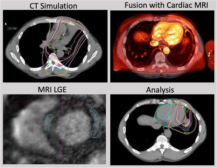
Radiotherapy has been associated with late dose-dependent cardiovascular toxicity. In this cross-sectional pilot study, radiation dose distributions were correlated with areas of localized and diffuse myocardial fibrosis as measured by novel cardiac MRI (CMR) sequences including late gadolinium enhancement (LGE) and T1 mapping with the goal to identify early markers of myocardial damage.

MATERIALS AND METHODS

Twenty-eight patients with chest tumors including lung, breast, esophagus, and lymphoma underwent CMR per study protocol on average 46.4 months (range 1.7-344.5) after radiotherapy. Patients without pretreatment cardiac history were included if the volume of heart receiving 5 Gy or more was at least 10% (V5Gy ≥ 10%). The association of LGE with cardiac dosimetric factors, clinical factors (e.g., tumor type, smoking history, BMI), and T1 values was analyzed.

RESULTS

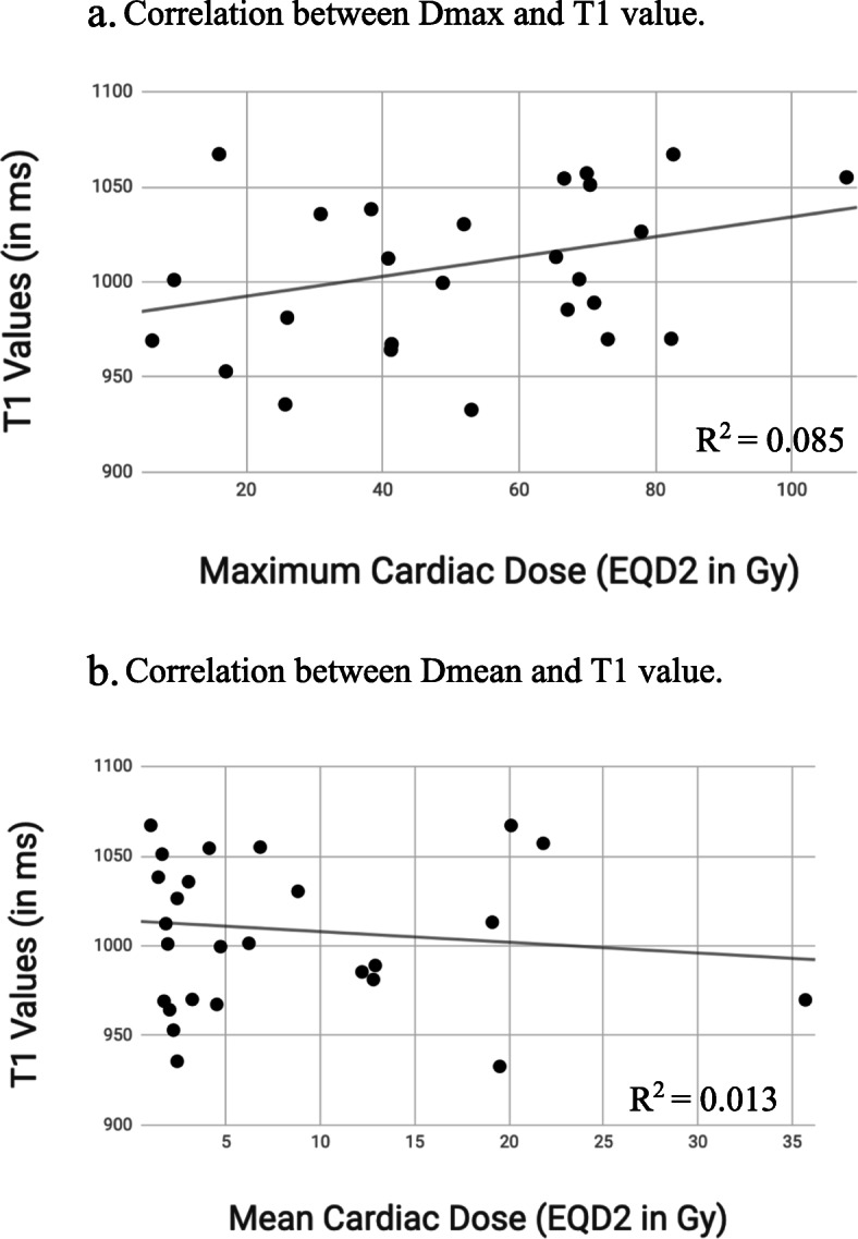
Cardiac maximum (Dmax) and mean dose (Dmean) equivalent to doses delivered in 2 Gy fractions (EQD2) were on average 50.9 Gy (range 6.2-108.0) and 8.2 Gy (range 1.0-35.7), respectively, compared to 60.8 Gy (40.8-108.0) and 6.8 Gy (1.8-21.8) among the 9 patients with LGE. Doses were not different between patients with and without LGE ( = 0.16 and 0.56, respectively). The average T1 value of the left ventricle myocardium was 1009 ms (range 933-1117). No significant correlation was seen for heart Dmax and Dmean and T1 values ( = 0.14 and 0.58, respectively). In addition, no significant association between clinical factors and the development of LGE was identified.

CONCLUSIONS

No relation between cardiac doses, the presence of LGE or T1 values was observed. Further study is needed to determine the benefit of CMR for detecting radiotherapy-related myocardial fibrosis.

摘要

背景与目的

放射治疗与晚期剂量依赖性心血管毒性相关。在这项横断面试点研究中,通过新型心脏磁共振成像(CMR)序列(包括延迟钆增强(LGE)和T1映射)测量的放射剂量分布与局部和弥漫性心肌纤维化区域相关,目的是识别心肌损伤的早期标志物。

材料与方法

28例患有胸部肿瘤(包括肺癌、乳腺癌、食管癌和淋巴瘤)的患者按照研究方案在放疗后平均46.4个月(范围1.7 - 344.5个月)接受CMR检查。如果接受5 Gy或更高剂量照射的心脏体积至少为10%(V5Gy≥10%),则纳入无预处理心脏病史的患者。分析LGE与心脏剂量学因素、临床因素(如肿瘤类型、吸烟史、体重指数)和T1值之间的关联。

结果

与9例出现LGE的患者相比,相当于2 Gy分次剂量的心脏最大剂量(Dmax)和平均剂量(Dmean)(EQD2)平均分别为50.9 Gy(范围6.2 - 108.0)和8.2 Gy(范围1.0 - 35.7),而这9例患者的相应剂量分别为60.8 Gy(40.8 - 108.0)和6.8 Gy(1.8 - 21.8)。有LGE和无LGE的患者之间剂量无差异(分别为 = 0.16和0.56)。左心室心肌的平均T1值为1009 ms(范围933 - 1117)。未观察到心脏Dmax和Dmean与T1值之间存在显著相关性(分别为 = 0.14和0.58)。此外,未发现临床因素与LGE的发生之间存在显著关联。

结论

未观察到心脏剂量、LGE的存在或T1值之间存在关联。需要进一步研究以确定CMR在检测放疗相关心肌纤维化方面的益处。

https://cdn.ncbi.nlm.nih.gov/pmc/blobs/1662/7329507/480ea4b1e74f/40959_2020_61_Fig2_HTML.jpg
https://cdn.ncbi.nlm.nih.gov/pmc/blobs/1662/7329507/81b62cc673e5/40959_2020_61_Fig1_HTML.jpg
https://cdn.ncbi.nlm.nih.gov/pmc/blobs/1662/7329507/480ea4b1e74f/40959_2020_61_Fig2_HTML.jpg
https://cdn.ncbi.nlm.nih.gov/pmc/blobs/1662/7329507/81b62cc673e5/40959_2020_61_Fig1_HTML.jpg
https://cdn.ncbi.nlm.nih.gov/pmc/blobs/1662/7329507/480ea4b1e74f/40959_2020_61_Fig2_HTML.jpg

相似文献

1
Cardiac MRI utilizing late gadolinium enhancement (LGE) and T1 mapping in the detection of radiation induced heart disease.利用延迟钆增强(LGE)和T1映射的心脏磁共振成像在放射性心脏病检测中的应用
Cardiooncology. 2020 Jul 1;6:6. doi: 10.1186/s40959-020-00061-z. eCollection 2020.
2
[Late gadolinium enhancement and T1 mapping for the diagnosis of cardiac amyloidosis].[延迟钆增强及T1成像在心脏淀粉样变性诊断中的应用]
Zhonghua Wei Zhong Bing Ji Jiu Yi Xue. 2019 Dec;31(12):1538-1541. doi: 10.3760/cma.j.issn.2095-4352.2019.12.021.
3
Quantitative T1 Mapping for Detecting Microvascular Obstruction in Reperfused Acute Myocardial Infarction: Comparison with Late Gadolinium Enhancement Imaging.定量 T1 映射检测再灌注急性心肌梗死中的微血管阻塞:与晚期钆增强成像的比较。
Korean J Radiol. 2020 Aug;21(8):978-986. doi: 10.3348/kjr.2019.0736.
4
[T1 mapping and late gadolinium enhancement for the diagnosis of dilated cardiomyopathy].用于扩张型心肌病诊断的T1映射和延迟钆增强
Zhonghua Wei Zhong Bing Ji Jiu Yi Xue. 2020 Dec;32(12):1506-1510. doi: 10.3760/cma.j.cn121430-20200413-00287.
5
Myocardial fibrosis imaging based on T1-mapping and extracellular volume fraction (ECV) measurement in muscular dystrophy patients: diagnostic value compared with conventional late gadolinium enhancement (LGE) imaging.基于T1映射和细胞外容积分数(ECV)测量的肌营养不良患者心肌纤维化成像:与传统钆延迟增强(LGE)成像相比的诊断价值
Eur Heart J Cardiovasc Imaging. 2014 Sep;15(9):1004-12. doi: 10.1093/ehjci/jeu050. Epub 2014 Mar 30.
6
T(1) mapping for the diagnosis of acute myocarditis using CMR: comparison to T2-weighted and late gadolinium enhanced imaging.CMR 用于诊断急性心肌炎的 T(1) 映射:与 T2 加权和晚期钆增强成像的比较。
JACC Cardiovasc Imaging. 2013 Oct;6(10):1048-1058. doi: 10.1016/j.jcmg.2013.03.008. Epub 2013 Sep 4.
7
Cardiac magnetic resonance in patients with mitral valve prolapse: Focus on late gadolinium enhancement and T1 mapping.心脏磁共振成像在二尖瓣脱垂患者中的应用:关注晚期钆增强和 T1 mapping。
Eur Radiol. 2019 Mar;29(3):1546-1554. doi: 10.1007/s00330-018-5634-5. Epub 2018 Aug 7.
8
Variations in native T1 values in patients with Duchenne muscular dystrophy with and without late gadolinium enhancement.杜氏肌营养不良症患者伴或不伴晚期钆增强的固有 T1 值变化。
Int J Cardiovasc Imaging. 2021 Feb;37(2):635-642. doi: 10.1007/s10554-020-02031-z. Epub 2020 Sep 20.
9
MRI findings of radiation-induced myocardial damage in patients with oesophageal cancer.食管癌患者放射性心肌损伤的磁共振成像表现
Clin Radiol. 2014 Dec;69(12):1273-9. doi: 10.1016/j.crad.2014.08.010. Epub 2014 Sep 20.
10
Characterization of Compacted Myocardial Abnormalities by Cardiac Magnetic Resonance With Native T1 Mapping in Left Ventricular Non-Compaction Patients - A Comparison With Late Gadolinium Enhancement.心脏磁共振成像的原生T1映射技术对左心室心肌致密化不全患者致密心肌异常的特征分析——与延迟钆增强的比较
Circ J. 2016 Apr 25;80(5):1210-6. doi: 10.1253/circj.CJ-15-1269. Epub 2016 Mar 23.

引用本文的文献

1
Respiratory navigator-guided multi-slice free-breathing cardiac T mapping on a magnetic resonance-guided linear accelerator.在磁共振引导直线加速器上进行呼吸导航器引导的多层自由呼吸心脏T值映射
Phys Imaging Radiat Oncol. 2025 Feb 25;34:100739. doi: 10.1016/j.phro.2025.100739. eCollection 2025 Apr.
2
Inversion-recovery ultrashort-echo-time (IR-UTE) MRI-based detection of radiation dose heterogeneity in gynecologic cancer patients treated with HDR brachytherapy.基于反转恢复超短回波时间(IR-UTE)MRI 的宫颈癌调强近距离后装治疗中不同剂量区的检测。
Radiat Oncol. 2024 Aug 6;19(1):105. doi: 10.1186/s13014-024-02499-2.
3

本文引用的文献

1
Increased C-reactive protein is associated with the severity of thoracic radiotherapy-induced cardiomyopathy.C反应蛋白升高与胸部放疗诱发的心肌病严重程度相关。
Cardiooncology. 2020 Feb 28;6:2. doi: 10.1186/s40959-020-0058-1. eCollection 2020.
2
Cardiac Radiation Dose, Cardiac Disease, and Mortality in Patients With Lung Cancer.肺癌患者的心脏辐射剂量、心脏疾病与死亡率。
J Am Coll Cardiol. 2019 Jun 18;73(23):2976-2987. doi: 10.1016/j.jacc.2019.03.500.
3
Assessment of myocardial fibrosis using T1-mapping and extracellular volume measurement on cardiac magnetic resonance imaging for the diagnosis of radiation-induced cardiomyopathy.
The Role of Advanced Cardiovascular Imaging Modalities in Cardio-Oncology: From Early Detection to Unravelling Mechanisms of Cardiotoxicity.
先进心血管成像模式在心脏肿瘤学中的作用:从早期检测到阐明心脏毒性机制
J Clin Med. 2023 Jul 27;12(15):4945. doi: 10.3390/jcm12154945.
4
Radiotherapy-induced diffuse myocardial fibrosis in early-stage breast cancer patients - multimodality imaging study with six-year follow-up.早期乳腺癌患者放疗后弥漫性心肌纤维化的多模态影像学研究-六年随访。
Radiat Oncol. 2023 Jul 26;18(1):124. doi: 10.1186/s13014-023-02319-z.
5
Quantitative assessment of radiotherapy-induced myocardial damage using MRI: a systematic review.使用磁共振成像对放疗引起的心肌损伤进行定量评估:一项系统综述
Cardiooncology. 2023 May 18;9(1):24. doi: 10.1186/s40959-023-00175-0.
6
Early detection and serial monitoring during chemotherapy-radiation therapy: Using T1 and T2 mapping cardiac magnetic resonance imaging.放化疗期间的早期检测及连续监测:使用T1和T2映射心脏磁共振成像
Front Cardiovasc Med. 2023 Mar 29;10:1085737. doi: 10.3389/fcvm.2023.1085737. eCollection 2023.
7
Cardiotoxicity of Anti-Cancer Radiation Therapy: a Focus on Heart Failure.抗癌放射治疗的心脏毒性:聚焦心力衰竭。
Curr Heart Fail Rep. 2023 Feb;20(1):44-55. doi: 10.1007/s11897-023-00587-0. Epub 2023 Jan 24.
8
Cardiovascular Imaging in Cardio-Oncology: The Role of Echocardiography and Cardiac MRI in Modern Cardio-Oncology.心血管肿瘤学中的心血管影像学:超声心动图和心脏 MRI 在现代心血管肿瘤学中的作用。
Heart Fail Clin. 2022 Jul;18(3):455-478. doi: 10.1016/j.hfc.2022.02.007.
9
Cardiovascular Magnetic Resonance in Early Detection of Radiation Associated Cardiotoxicity With Chest Radiation.心血管磁共振成像在胸部放疗相关心脏毒性早期检测中的应用
Front Cardiovasc Med. 2022 May 31;9:867479. doi: 10.3389/fcvm.2022.867479. eCollection 2022.
10
Quantitative assessment of intra- and inter-modality deformable image registration of the heart, left ventricle, and thoracic aorta on longitudinal 4D-CT and MR images.基于纵向 4D-CT 和 MR 图像的心脏、左心室和胸主动脉的内模态和间模态可变形图像配准的定量评估。
J Appl Clin Med Phys. 2022 Feb;23(2):e13500. doi: 10.1002/acm2.13500. Epub 2021 Dec 27.
利用心脏磁共振成像上的T1映射和细胞外容积测量评估心肌纤维化以诊断放射性心肌病。
J Cardiol Cases. 2018 Jun 30;18(4):132-135. doi: 10.1016/j.jccase.2018.06.001. eCollection 2018 Oct.
4
Left Ventricular T1 Mapping during Chemotherapy-Radiation Therapy: Serial Assessment of Participants with Esophageal Cancer.化疗-放疗期间左心室 T1 映射:食管癌患者的系列评估。
Radiology. 2018 Nov;289(2):347-354. doi: 10.1148/radiol.2018172076. Epub 2018 Jul 10.
5
Cardiac Toxicity: The More We Learn, the Less We Know.心脏毒性:我们了解得越多,未知就越多。
Int J Radiat Oncol Biol Phys. 2017 Dec 1;99(5):1162-1165. doi: 10.1016/j.ijrobp.2017.08.048.
6
Detection of late radiation damage on left atrial fibrosis using cardiac late gadolinium enhancement magnetic resonance imaging.使用心脏延迟钆增强磁共振成像检测左心房纤维化的晚期辐射损伤
Adv Radiat Oncol. 2016 Apr 26;1(2):106-114. doi: 10.1016/j.adro.2016.04.002. eCollection 2016 Apr-Jun.
7
Dose to heart substructures is associated with non-cancer death after SBRT in stage I-II NSCLC patients.在I-II期非小细胞肺癌患者中,立体定向体部放疗后心脏亚结构所受剂量与非癌症死亡相关。
Radiother Oncol. 2017 Jun;123(3):370-375. doi: 10.1016/j.radonc.2017.04.017. Epub 2017 May 2.
8
Cardiac Events After Radiation Therapy: Combined Analysis of Prospective Multicenter Trials for Locally Advanced Non-Small-Cell Lung Cancer.放射治疗后的心脏事件:局部晚期非小细胞肺癌前瞻性多中心试验的联合分析
J Clin Oncol. 2017 May 1;35(13):1395-1402. doi: 10.1200/JCO.2016.71.6142. Epub 2017 Mar 16.
9
A cardiac contouring atlas for radiotherapy.一种用于放射治疗的心脏轮廓图谱。
Radiother Oncol. 2017 Mar;122(3):416-422. doi: 10.1016/j.radonc.2017.01.008. Epub 2017 Feb 21.
10
Cardiac Toxicity After Radiotherapy for Stage III Non-Small-Cell Lung Cancer: Pooled Analysis of Dose-Escalation Trials Delivering 70 to 90 Gy.Ⅲ期非小细胞肺癌放疗后的心脏毒性:70至90 Gy剂量递增试验的汇总分析
J Clin Oncol. 2017 May 1;35(13):1387-1394. doi: 10.1200/JCO.2016.70.0229. Epub 2017 Jan 23.