• 文献检索
  • 文档翻译
  • 深度研究
  • 学术资讯
  • Suppr Zotero 插件Zotero 插件
  • 邀请有礼
  • 套餐&价格
  • 历史记录
应用&插件
Suppr Zotero 插件Zotero 插件浏览器插件Mac 客户端Windows 客户端微信小程序
定价
高级版会员购买积分包购买API积分包
服务
文献检索文档翻译深度研究API 文档MCP 服务
关于我们
关于 Suppr公司介绍联系我们用户协议隐私条款
关注我们

Suppr 超能文献

核心技术专利:CN118964589B侵权必究
粤ICP备2023148730 号-1Suppr @ 2026

文献检索

告别复杂PubMed语法,用中文像聊天一样搜索,搜遍4000万医学文献。AI智能推荐,让科研检索更轻松。

立即免费搜索

文件翻译

保留排版,准确专业,支持PDF/Word/PPT等文件格式,支持 12+语言互译。

免费翻译文档

深度研究

AI帮你快速写综述,25分钟生成高质量综述,智能提取关键信息,辅助科研写作。

立即免费体验

同步放化疗治疗食管癌中剂量依赖性辐射诱导的心肌损伤:一项前瞻性心脏磁共振成像研究

Dose-Dependent Radiation-Induced Myocardial Damage in Esophageal Cancer Treated With Chemoradiotherapy: A Prospective Cardiac Magnetic Resonance Imaging Study.

作者信息

Umezawa Rei, Kadoya Noriyuki, Ota Hideki, Nakajima Yujiro, Saito Masahide, Takagi Hidenobu, Takanami Kentaro, Takahashi Noriyoshi, Ishikawa Yojiro, Yamamoto Takaya, Matsushita Haruo, Takeda Ken, Takase Kei, Jingu Keiichi

机构信息

Department of Radiation Oncology, Tohoku University Graduate School of Medicine, Sendai, Japan.

Department of Diagnostic Radiology, Tohoku University Graduate School of Medicine, Sendai, Japan.

出版信息

Adv Radiat Oncol. 2020 Aug 5;5(6):1170-1178. doi: 10.1016/j.adro.2020.07.012. eCollection 2020 Nov-Dec.

DOI:10.1016/j.adro.2020.07.012
PMID:33305078
原文链接:https://pmc.ncbi.nlm.nih.gov/articles/PMC7718544/
Abstract

PURPOSE

The purpose of this prospective study was to evaluate radiation-induced myocardial damage after mediastinal radiation therapy (RT) using late gadolinium-enhancement (LGE) magnetic resonance imaging (MRI).

METHODS AND MATERIALS

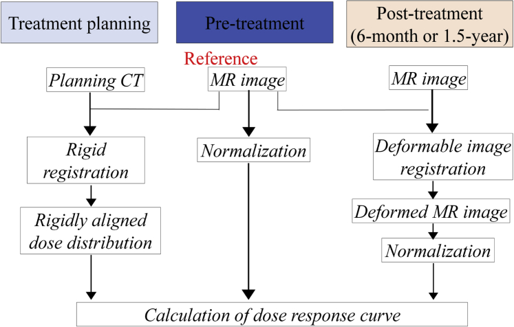
We enrolled 19 patients with esophageal cancer who were expected to have long-term survival by definitive treatment. They underwent delayed contrast-enhanced MRI (19 patients before treatment, 19 patients 6 months after treatment, and 12 patients 1.5 years after treatment). Dose distribution of the left ventricle was made using computed tomography, and the dose volume histogram of the left ventricle was calculated. Myocardial signal intensities in individual MRIs were normalized by the mean values in regions receiving low doses (<5 Gy). Changes in the normalized signal intensities after mediastinal radiation therapy were compared among regions where irradiation doses were 0 to 10 Gy, 10 to 20 Gy, 20 to 30 Gy, 30 to 40 Gy, 40 to 50 Gy, and 50 to 60 Gy, and we investigated whether intensity change was detected in a dose-dependent manner.

RESULTS

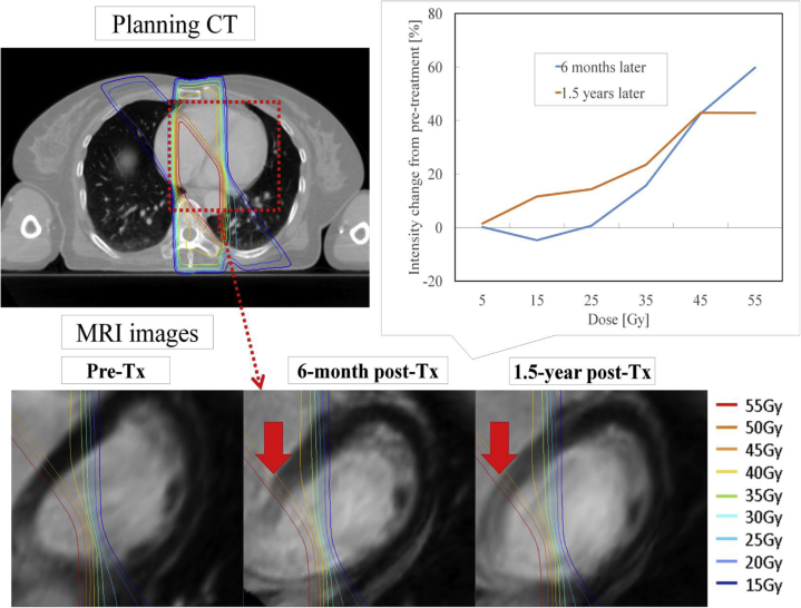
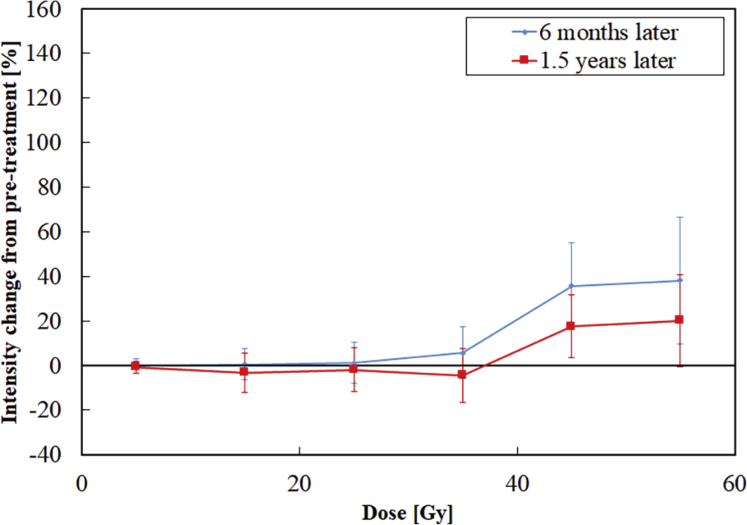
The registered patients were treated with concurrent chemoradiotherapy with a median total dose of 60 Gy (50.4-66 Gy). Chemotherapy consisting of cisplatin and 5-fluorouracil was administered. In the population-based dose-response curve, dose-dependent intensity changes progressively increased in regions receiving more than 30 Gy. The averages of relative intensity change at 6 months and 1.5 years after treatment were 1.1% and -1.9% at 20 to 30 Gy and 37.5% and 17.5% at 40 to 50 Gy, respectively. LGE in regions receiving more than 30 Gy was detected in 68% (13/19) of the patients.

CONCLUSIONS

A dose-dependent relationship for myocardial signal intensity change was found by using LGE MRI. It may be necessary to reduce the volume of the myocardium receiving more than 30 Gy.

摘要

目的

本前瞻性研究的目的是使用延迟钆增强(LGE)磁共振成像(MRI)评估纵隔放射治疗(RT)后辐射诱发的心肌损伤。

方法和材料

我们纳入了19例经确定性治疗有望长期存活的食管癌患者。他们接受了延迟对比增强MRI检查(19例患者在治疗前、19例患者在治疗后6个月、12例患者在治疗后1.5年)。使用计算机断层扫描绘制左心室的剂量分布,并计算左心室的剂量体积直方图。通过低剂量(<5 Gy)区域的平均值对各个MRI中的心肌信号强度进行归一化。比较纵隔放射治疗后归一化信号强度在照射剂量为0至10 Gy、10至20 Gy、20至30 Gy、30至40 Gy、40至50 Gy和50至60 Gy的区域中的变化,并且我们调查强度变化是否以剂量依赖性方式被检测到。

结果

登记的患者接受了同步放化疗,中位总剂量为60 Gy(50.4 - 66 Gy)。给予由顺铂和5-氟尿嘧啶组成的化疗。在基于人群的剂量反应曲线中,接受超过30 Gy的区域中剂量依赖性强度变化逐渐增加。治疗后6个月和1.5年时,20至30 Gy区域的相对强度变化平均值分别为1.1%和 -1.9%,40至50 Gy区域分别为37.5%和17.5%。68%(13/19)的患者在接受超过30 Gy的区域中检测到LGE。

结论

通过使用LGE MRI发现了心肌信号强度变化的剂量依赖性关系。可能有必要减少接受超过30 Gy的心肌体积。

https://cdn.ncbi.nlm.nih.gov/pmc/blobs/67f8/7718544/f433a69b95ed/gr3.jpg
https://cdn.ncbi.nlm.nih.gov/pmc/blobs/67f8/7718544/ff579970bb0a/gr1.jpg
https://cdn.ncbi.nlm.nih.gov/pmc/blobs/67f8/7718544/be55bdeff8fa/gr2.jpg
https://cdn.ncbi.nlm.nih.gov/pmc/blobs/67f8/7718544/f433a69b95ed/gr3.jpg
https://cdn.ncbi.nlm.nih.gov/pmc/blobs/67f8/7718544/ff579970bb0a/gr1.jpg
https://cdn.ncbi.nlm.nih.gov/pmc/blobs/67f8/7718544/be55bdeff8fa/gr2.jpg
https://cdn.ncbi.nlm.nih.gov/pmc/blobs/67f8/7718544/f433a69b95ed/gr3.jpg

相似文献

1
Dose-Dependent Radiation-Induced Myocardial Damage in Esophageal Cancer Treated With Chemoradiotherapy: A Prospective Cardiac Magnetic Resonance Imaging Study.同步放化疗治疗食管癌中剂量依赖性辐射诱导的心肌损伤:一项前瞻性心脏磁共振成像研究
Adv Radiat Oncol. 2020 Aug 5;5(6):1170-1178. doi: 10.1016/j.adro.2020.07.012. eCollection 2020 Nov-Dec.
2
Clinical impact of radiation-induced myocardial damage detected by cardiac magnetic resonance imaging and dose-volume histogram parameters of the left ventricle as prognostic factors of cardiac events after chemoradiotherapy for esophageal cancer.心脏磁共振成像检测到的放射性心肌损伤及左心室剂量-体积直方图参数对食管癌化放化疗后心脏事件的预后价值。
J Radiat Res. 2023 Jul 18;64(4):702-710. doi: 10.1093/jrr/rrad040.
3
Assessment of myocardial metabolic disorder associated with mediastinal radiotherapy for esophageal cancer -a pilot study.食管癌纵隔放疗相关心肌代谢紊乱的评估——一项初步研究。
Radiat Oncol. 2015 Apr 21;10:96. doi: 10.1186/s13014-015-0410-z.
4
Quantitative assessment of radiotherapy-induced myocardial damage using MRI: a systematic review.使用磁共振成像对放疗引起的心肌损伤进行定量评估:一项系统综述
Cardiooncology. 2023 May 18;9(1):24. doi: 10.1186/s40959-023-00175-0.
5
MRI findings of radiation-induced myocardial damage in patients with oesophageal cancer.食管癌患者放射性心肌损伤的磁共振成像表现
Clin Radiol. 2014 Dec;69(12):1273-9. doi: 10.1016/j.crad.2014.08.010. Epub 2014 Sep 20.
6
Quantitative Analysis of Radiation Therapy-Induced Cardiac and Aortic Sequelae in Patients With Lung Cancer via Magnetic Resonance Imaging: A Pilot Study.基于磁共振成像的肺癌患者放疗后心脏和主动脉不良事件的定量分析:一项初步研究。
Int J Radiat Oncol Biol Phys. 2024 May 1;119(1):281-291. doi: 10.1016/j.ijrobp.2023.10.037. Epub 2023 Nov 10.
7
Cardiac MRI utilizing late gadolinium enhancement (LGE) and T1 mapping in the detection of radiation induced heart disease.利用延迟钆增强(LGE)和T1映射的心脏磁共振成像在放射性心脏病检测中的应用
Cardiooncology. 2020 Jul 1;6:6. doi: 10.1186/s40959-020-00061-z. eCollection 2020.
8
Detection of late radiation damage on left atrial fibrosis using cardiac late gadolinium enhancement magnetic resonance imaging.使用心脏延迟钆增强磁共振成像检测左心房纤维化的晚期辐射损伤
Adv Radiat Oncol. 2016 Apr 26;1(2):106-114. doi: 10.1016/j.adro.2016.04.002. eCollection 2016 Apr-Jun.
9
Induction chemotherapy improved outcomes of patients with resectable esophageal cancer who received chemoradiotherapy followed by surgery.诱导化疗改善了接受放化疗后手术的可切除食管癌患者的预后。
Int J Radiat Oncol Biol Phys. 2004 Oct 1;60(2):427-36. doi: 10.1016/j.ijrobp.2004.03.033.
10
Dose-response relationship in locoregional control for patients with stage II-III esophageal cancer treated with concurrent chemotherapy and radiotherapy.同步放化疗治疗II-III期食管癌患者局部区域控制中的剂量反应关系。
Int J Radiat Oncol Biol Phys. 2005 Mar 1;61(3):656-64. doi: 10.1016/j.ijrobp.2004.06.022.

引用本文的文献

1
Clinical outcomes of transcatheter aortic valve replacement in patients with radiation-induced aortic stenosis: a systematic review and meta-analysis.放射诱导的主动脉瓣狭窄患者经导管主动脉瓣置换术的临床结局:一项系统评价和荟萃分析。
Front Cardiovasc Med. 2025 Aug 12;12:1537220. doi: 10.3389/fcvm.2025.1537220. eCollection 2025.
2
Acute Myocarditis and Inflammatory Cardiomyopathies: Insights From Cardiac Magnetic Resonance Findings.急性心肌炎与炎症性心肌病:心脏磁共振成像结果的见解
Echocardiography. 2025 Feb;42(2):e70099. doi: 10.1111/echo.70099.
3
Cardiac volume reduction during radiotherapy in patients with esophageal carcinoma.

本文引用的文献

1
Long-Term Results of NRG Oncology RTOG 0617: Standard- Versus High-Dose Chemoradiotherapy With or Without Cetuximab for Unresectable Stage III Non-Small-Cell Lung Cancer.NRG肿瘤学RTOG 0617的长期结果:不可切除的III期非小细胞肺癌采用标准剂量与高剂量放化疗联合或不联合西妥昔单抗的疗效对比
J Clin Oncol. 2020 Mar 1;38(7):706-714. doi: 10.1200/JCO.19.01162. Epub 2019 Dec 16.
2
Cardiac Events After Radiation Therapy: Combined Analysis of Prospective Multicenter Trials for Locally Advanced Non-Small-Cell Lung Cancer.放射治疗后的心脏事件:局部晚期非小细胞肺癌前瞻性多中心试验的联合分析
J Clin Oncol. 2017 May 1;35(13):1395-1402. doi: 10.1200/JCO.2016.71.6142. Epub 2017 Mar 16.
3
食管癌患者放疗期间的心腔容积减小
Mol Clin Oncol. 2024 Jan 16;20(3):19. doi: 10.3892/mco.2024.2717. eCollection 2024 Mar.
4
The potential overdose of heart and left anterior descending coronary artery region during intensity-modulated radiation therapy in patients with esophageal cancer.食管癌调强放疗中心脏和左前降支区域潜在的超量照射。
J Radiat Res. 2024 Mar 22;65(2):238-243. doi: 10.1093/jrr/rrad100.
5
Identification of key genes in sepsis-induced cardiomyopathy based on integrated bioinformatical analysis and experiments and .基于整合生物信息学分析和实验鉴定脓毒症诱导性心肌病的关键基因
PeerJ. 2023 Nov 21;11:e16222. doi: 10.7717/peerj.16222. eCollection 2023.
6
Radiotherapy-induced diffuse myocardial fibrosis in early-stage breast cancer patients - multimodality imaging study with six-year follow-up.早期乳腺癌患者放疗后弥漫性心肌纤维化的多模态影像学研究-六年随访。
Radiat Oncol. 2023 Jul 26;18(1):124. doi: 10.1186/s13014-023-02319-z.
7
Clinical impact of radiation-induced myocardial damage detected by cardiac magnetic resonance imaging and dose-volume histogram parameters of the left ventricle as prognostic factors of cardiac events after chemoradiotherapy for esophageal cancer.心脏磁共振成像检测到的放射性心肌损伤及左心室剂量-体积直方图参数对食管癌化放化疗后心脏事件的预后价值。
J Radiat Res. 2023 Jul 18;64(4):702-710. doi: 10.1093/jrr/rrad040.
8
Quantitative assessment of radiotherapy-induced myocardial damage using MRI: a systematic review.使用磁共振成像对放疗引起的心肌损伤进行定量评估:一项系统综述
Cardiooncology. 2023 May 18;9(1):24. doi: 10.1186/s40959-023-00175-0.
9
Quantitative assessment of intra- and inter-modality deformable image registration of the heart, left ventricle, and thoracic aorta on longitudinal 4D-CT and MR images.基于纵向 4D-CT 和 MR 图像的心脏、左心室和胸主动脉的内模态和间模态可变形图像配准的定量评估。
J Appl Clin Med Phys. 2022 Feb;23(2):e13500. doi: 10.1002/acm2.13500. Epub 2021 Dec 27.
Cardiac Toxicity After Radiotherapy for Stage III Non-Small-Cell Lung Cancer: Pooled Analysis of Dose-Escalation Trials Delivering 70 to 90 Gy.
Ⅲ期非小细胞肺癌放疗后的心脏毒性:70至90 Gy剂量递增试验的汇总分析
J Clin Oncol. 2017 May 1;35(13):1387-1394. doi: 10.1200/JCO.2016.70.0229. Epub 2017 Jan 23.
4
Association between radiation dose to the heart and myocardial fatty acid metabolic impairment due to chemoradiation-therapy: Prospective study using I-123 BMIPP SPECT/CT.心脏辐射剂量与放化疗导致的心肌脂肪酸代谢障碍的相关性:使用 I-123 BMIPP SPECT/CT 的前瞻性研究。
Radiother Oncol. 2016 Apr;119(1):77-83. doi: 10.1016/j.radonc.2016.01.024. Epub 2016 Feb 9.
5
Radiation modality use and cardiopulmonary mortality risk in elderly patients with esophageal cancer.老年食管癌患者的放疗方式使用与心肺死亡风险
Cancer. 2016 Mar 15;122(6):917-28. doi: 10.1002/cncr.29857. Epub 2015 Dec 30.
6
Cardiac Function After Multimodal Breast Cancer Therapy Assessed With Functional Magnetic Resonance Imaging and Echocardiography Imaging.采用功能磁共振成像和超声心动图成像评估多模式乳腺癌治疗后的心脏功能
Int J Radiat Oncol Biol Phys. 2015 Nov 15;93(4):836-44. doi: 10.1016/j.ijrobp.2015.07.2287. Epub 2015 Aug 3.
7
Long-term results of chemoradiotherapy for stage II-III thoracic esophageal cancer in a single institution after 2000 -with a focus on comparison of three protocols.2000年后某单一机构中Ⅱ-Ⅲ期胸段食管癌放化疗的长期结果——以三种方案的比较为重点
BMC Cancer. 2015 Oct 27;15:813. doi: 10.1186/s12885-015-1836-2.
8
Increased risk of death due to heart disease after radiotherapy for esophageal cancer.食管癌放疗后因心脏病导致的死亡风险增加。
J Gastrointest Oncol. 2015 Oct;6(5):516-23. doi: 10.3978/j.issn.2078-6891.2015.040.
9
Assessment of myocardial metabolic disorder associated with mediastinal radiotherapy for esophageal cancer -a pilot study.食管癌纵隔放疗相关心肌代谢紊乱的评估——一项初步研究。
Radiat Oncol. 2015 Apr 21;10:96. doi: 10.1186/s13014-015-0410-z.
10
ACE inhibition attenuates radiation-induced cardiopulmonary damage.血管紧张素转换酶抑制可减轻辐射诱导的心肺损伤。
Radiother Oncol. 2015 Jan;114(1):96-103. doi: 10.1016/j.radonc.2014.11.017. Epub 2014 Nov 25.