• 文献检索
  • 文档翻译
  • 深度研究
  • 学术资讯
  • Suppr Zotero 插件Zotero 插件
  • 邀请有礼
  • 套餐&价格
  • 历史记录
应用&插件
Suppr Zotero 插件Zotero 插件浏览器插件Mac 客户端Windows 客户端微信小程序
定价
高级版会员购买积分包购买API积分包
服务
文献检索文档翻译深度研究API 文档MCP 服务
关于我们
关于 Suppr公司介绍联系我们用户协议隐私条款
关注我们

Suppr 超能文献

核心技术专利:CN118964589B侵权必究
粤ICP备2023148730 号-1Suppr @ 2026

文献检索

告别复杂PubMed语法,用中文像聊天一样搜索,搜遍4000万医学文献。AI智能推荐,让科研检索更轻松。

立即免费搜索

文件翻译

保留排版,准确专业,支持PDF/Word/PPT等文件格式,支持 12+语言互译。

免费翻译文档

深度研究

AI帮你快速写综述,25分钟生成高质量综述,智能提取关键信息,辅助科研写作。

立即免费体验

基于活性的荧光探针用于亮氨酸氨肽酶成像的最新进展。

Recent Progress of Activity-Based Fluorescent Probes for Imaging Leucine Aminopeptidase.

机构信息

Key Laboratory of Biomedical Engineering of Hainan Province, School of Biomedical Engineering, Hainan University, Haikou 570228, China.

One Health Institute, Hainan University, Haikou 570228, China.

出版信息

Biosensors (Basel). 2023 Jul 21;13(7):752. doi: 10.3390/bios13070752.

DOI:10.3390/bios13070752
PMID:37504150
原文链接:https://pmc.ncbi.nlm.nih.gov/articles/PMC10377407/
Abstract

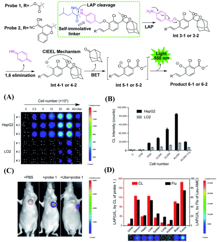
Leucine aminopeptidase (LAP) is an important protease that can specifically hydrolyze Leucine residues. LAP occurs in microorganisms, plants, animals, and humans and is involved in a variety of physiological processes in the human body. In the physiological system, abnormal levels of LAP are associated with a variety of diseases and pathological processes, such as cancer and drug-induced liver injury; thus, LAP was chosen as the early biochemical marker for many physiological processes, including cancer. Considering the importance of LAP in physiological and pathological processes, it is critical that high-efficiency and dependable technology be developed to monitor LAP levels. Herein, we summarize the organic small molecule fluorescence/chemiluminescence probes used for LAP detection in recent years, which can image LAP in cancer, drug-induced liver injury (DILI), and bacteria. It can also reveal the role of LAP in tumors and differentiate the serum of cirrhotic, drug-induced liver injury and normal models.

摘要

亮氨酸氨基肽酶(LAP)是一种重要的蛋白酶,能够特异性水解亮氨酸残基。LAP 存在于微生物、植物、动物和人类中,参与人体的多种生理过程。在生理系统中,LAP 水平异常与多种疾病和病理过程有关,如癌症和药物性肝损伤;因此,LAP 被选为许多生理过程(包括癌症)的早期生化标志物。鉴于 LAP 在生理和病理过程中的重要性,开发高效、可靠的技术来监测 LAP 水平至关重要。在此,我们总结了近年来用于 LAP 检测的有机小分子荧光/化学发光探针,这些探针可用于癌症、药物性肝损伤(DILI)和细菌的 LAP 成像,还可以揭示 LAP 在肿瘤中的作用,并区分肝硬化、药物性肝损伤和正常模型的血清。

https://cdn.ncbi.nlm.nih.gov/pmc/blobs/669f/10377407/812b770acc1f/biosensors-13-00752-g033.jpg
https://cdn.ncbi.nlm.nih.gov/pmc/blobs/669f/10377407/63a4d39b0b2f/biosensors-13-00752-g001.jpg
https://cdn.ncbi.nlm.nih.gov/pmc/blobs/669f/10377407/0b92dee1928e/biosensors-13-00752-g002.jpg
https://cdn.ncbi.nlm.nih.gov/pmc/blobs/669f/10377407/f3f7d44c1b75/biosensors-13-00752-g003.jpg
https://cdn.ncbi.nlm.nih.gov/pmc/blobs/669f/10377407/b141936d1bbd/biosensors-13-00752-g004.jpg
https://cdn.ncbi.nlm.nih.gov/pmc/blobs/669f/10377407/58ffa590aeab/biosensors-13-00752-g005.jpg
https://cdn.ncbi.nlm.nih.gov/pmc/blobs/669f/10377407/e67befccc00e/biosensors-13-00752-g006.jpg
https://cdn.ncbi.nlm.nih.gov/pmc/blobs/669f/10377407/1b0c0c2bcb02/biosensors-13-00752-g007.jpg
https://cdn.ncbi.nlm.nih.gov/pmc/blobs/669f/10377407/77034f808027/biosensors-13-00752-g008.jpg
https://cdn.ncbi.nlm.nih.gov/pmc/blobs/669f/10377407/782bc3e7191b/biosensors-13-00752-g009.jpg
https://cdn.ncbi.nlm.nih.gov/pmc/blobs/669f/10377407/6b7b2cfcf295/biosensors-13-00752-g010.jpg
https://cdn.ncbi.nlm.nih.gov/pmc/blobs/669f/10377407/4a30e5ad6bc1/biosensors-13-00752-g011.jpg
https://cdn.ncbi.nlm.nih.gov/pmc/blobs/669f/10377407/27e860bdab5d/biosensors-13-00752-g012.jpg
https://cdn.ncbi.nlm.nih.gov/pmc/blobs/669f/10377407/068458d01ae2/biosensors-13-00752-g013.jpg
https://cdn.ncbi.nlm.nih.gov/pmc/blobs/669f/10377407/8e6778a14cf5/biosensors-13-00752-g014.jpg
https://cdn.ncbi.nlm.nih.gov/pmc/blobs/669f/10377407/ccdafabe9262/biosensors-13-00752-g015.jpg
https://cdn.ncbi.nlm.nih.gov/pmc/blobs/669f/10377407/e70133be0fd4/biosensors-13-00752-g016.jpg
https://cdn.ncbi.nlm.nih.gov/pmc/blobs/669f/10377407/01a56e5b7e91/biosensors-13-00752-g017.jpg
https://cdn.ncbi.nlm.nih.gov/pmc/blobs/669f/10377407/86683f02d073/biosensors-13-00752-g018.jpg
https://cdn.ncbi.nlm.nih.gov/pmc/blobs/669f/10377407/24cd033be5e4/biosensors-13-00752-g019.jpg
https://cdn.ncbi.nlm.nih.gov/pmc/blobs/669f/10377407/fb8482b2fcb9/biosensors-13-00752-g020.jpg
https://cdn.ncbi.nlm.nih.gov/pmc/blobs/669f/10377407/1901c00573a0/biosensors-13-00752-g021.jpg
https://cdn.ncbi.nlm.nih.gov/pmc/blobs/669f/10377407/69ecedd6838c/biosensors-13-00752-g022.jpg
https://cdn.ncbi.nlm.nih.gov/pmc/blobs/669f/10377407/eb968b7a7c29/biosensors-13-00752-g023.jpg
https://cdn.ncbi.nlm.nih.gov/pmc/blobs/669f/10377407/c8990d876c36/biosensors-13-00752-g024.jpg
https://cdn.ncbi.nlm.nih.gov/pmc/blobs/669f/10377407/61f15a3a5997/biosensors-13-00752-g025.jpg
https://cdn.ncbi.nlm.nih.gov/pmc/blobs/669f/10377407/57f4033cea2c/biosensors-13-00752-g026.jpg
https://cdn.ncbi.nlm.nih.gov/pmc/blobs/669f/10377407/e450e3ecf0cc/biosensors-13-00752-g027.jpg
https://cdn.ncbi.nlm.nih.gov/pmc/blobs/669f/10377407/fd698ab4c5cc/biosensors-13-00752-g028.jpg
https://cdn.ncbi.nlm.nih.gov/pmc/blobs/669f/10377407/daf58ac200ad/biosensors-13-00752-g029.jpg
https://cdn.ncbi.nlm.nih.gov/pmc/blobs/669f/10377407/b0cc7328decf/biosensors-13-00752-g030.jpg
https://cdn.ncbi.nlm.nih.gov/pmc/blobs/669f/10377407/706ef40cb9b2/biosensors-13-00752-g031.jpg
https://cdn.ncbi.nlm.nih.gov/pmc/blobs/669f/10377407/530ca069795f/biosensors-13-00752-g032.jpg
https://cdn.ncbi.nlm.nih.gov/pmc/blobs/669f/10377407/812b770acc1f/biosensors-13-00752-g033.jpg

相似文献

1
Recent Progress of Activity-Based Fluorescent Probes for Imaging Leucine Aminopeptidase.基于活性的荧光探针用于亮氨酸氨肽酶成像的最新进展。
Biosensors (Basel). 2023 Jul 21;13(7):752. doi: 10.3390/bios13070752.
2
Rational design of a fluorescent probe for the detection of LAP and its application in drug-induced liver injury.用于检测亮氨酸氨基肽酶的荧光探针的合理设计及其在药物性肝损伤中的应用
Spectrochim Acta A Mol Biomol Spectrosc. 2021 Apr 15;251:119362. doi: 10.1016/j.saa.2020.119362. Epub 2021 Jan 22.
3
Diagnosing Drug-Induced Liver Injury by Multispectral Optoacoustic Tomography and Fluorescence Imaging Using a Leucine-Aminopeptidase-Activated Probe.基于亮氨酸氨肽酶激活探针的多光谱光声断层成像和荧光成像技术诊断药物性肝损伤。
Anal Chem. 2019 Jul 2;91(13):8085-8092. doi: 10.1021/acs.analchem.9b00107. Epub 2019 Jun 10.
4
A turn-on fluorescence probe based on aggregation-induced emission for leucine aminopeptidase in living cells and tumor tissue.基于聚集诱导发光的亮氨酸氨基肽酶荧光探针用于活细胞和肿瘤组织。
Anal Chim Acta. 2018 Nov 15;1031:169-177. doi: 10.1016/j.aca.2018.05.032. Epub 2018 May 12.
5
Enhancing the Selectivity of Leucine Aminopeptidase Near-Infrared Fluorescent Probes for Assisting in Surgical Tumor Resection.提高亮氨酸氨肽酶近红外荧光探针的选择性以辅助手术肿瘤切除
Anal Chem. 2023 Jan 31;95(4):2428-2435. doi: 10.1021/acs.analchem.2c04587. Epub 2023 Jan 17.
6
Near-Infrared Fluorescent Probe with Remarkable Large Stokes Shift and Favorable Water Solubility for Real-Time Tracking Leucine Aminopeptidase in Living Cells and In Vivo.近红外荧光探针具有显著的大斯托克斯位移和良好的水溶性,可用于实时跟踪活细胞和体内亮氨酸氨肽酶。
Anal Chem. 2017 Nov 21;89(22):12319-12326. doi: 10.1021/acs.analchem.7b03332. Epub 2017 Oct 30.
7
A ratiometric fluorescent probe based on quinoline for monitoring and imaging of Leucine aminopeptidase in liver tumor cells.一种基于喹啉的比率荧光探针,用于监测和成像肝肿瘤细胞中的亮氨酸氨肽酶。
Spectrochim Acta A Mol Biomol Spectrosc. 2021 Mar 15;249:119328. doi: 10.1016/j.saa.2020.119328. Epub 2020 Dec 15.
8
A Ratiometric Fluorescent Probe for Monitoring Leucine Aminopeptidase in Living Cells and Zebrafish Model.用于监测活细胞和斑马鱼模型中亮氨酸氨肽酶的比率荧光探针。
Anal Chem. 2017 Nov 7;89(21):11576-11582. doi: 10.1021/acs.analchem.7b02910. Epub 2017 Oct 26.
9
Leucine Aminopeptidase-Mediated Multifunctional Molecular Imaging Tool for Diagnosis, Drug Evaluation, and Surgical Guidance of Liver-Related Diseases.亮氨酸氨肽酶介导的多功能分子成像工具用于肝脏相关疾病的诊断、药物评估和手术指导
Anal Chem. 2023 Aug 15;95(32):12089-12096. doi: 10.1021/acs.analchem.3c02130. Epub 2023 Jul 31.
10
A diketopyrrolopyrrole-based ratiometric fluorescent probe for endogenous leucine aminopeptidase detecting and imaging with specific phototoxicity in tumor cells.基于二酮吡咯并吡咯的比率型荧光探针用于内源性亮氨酸氨肽酶的检测和成像,并具有肿瘤细胞特异性光毒性。
J Mater Chem B. 2021 Nov 3;9(42):8842-8850. doi: 10.1039/d1tb01480c.

本文引用的文献

1
Recent progress in the development of fluorescent probes for imaging pathological oxidative stress.近年来,用于成像病理性氧化应激的荧光探针的研究进展。
Chem Soc Rev. 2023 Jun 6;52(11):3873-3926. doi: 10.1039/d2cs00172a.
2
The design of small-molecule prodrugs and activatable phototherapeutics for cancer therapy.用于癌症治疗的小分子前药和可激活光疗药物的设计。
Chem Soc Rev. 2023 Feb 6;52(3):879-920. doi: 10.1039/d2cs00673a.
3
Fluorescent Probes for Lipid Membranes: From the Cell Surface to Organelles.荧光探针用于脂质膜:从细胞膜到细胞器。
Acc Chem Res. 2023 Jan 3;56(1):1-12. doi: 10.1021/acs.accounts.2c00586. Epub 2022 Dec 19.
4
A preliminary study for the development of cleavable linkers using activatable fluorescent probes targeting leucine aminopeptidase.使用针对亮氨酸氨肽酶的活化荧光探针开发可裂解连接子的初步研究。
Analyst. 2022 Nov 21;147(23):5386-5394. doi: 10.1039/d2an01145j.
5
State-of-the-art self-luminescence: a win-win situation.最先进的自发光:双赢局面。
Chem Soc Rev. 2022 Oct 31;51(21):8815-8831. doi: 10.1039/d2cs00228k.
6
Near-infrared (NIR) fluorescence-emitting small organic molecules for cancer imaging and therapy.近红外(NIR)荧光小分子用于癌症成像和治疗。
Chem Soc Rev. 2022 Oct 31;51(21):8957-9008. doi: 10.1039/d2cs00722c.
7
Chameleon-Like Fluorescent Probe for Monitoring Interplays between Three Organelles and Reporting Cell Damage Processes through Dramatic Color Change.用于监测三种细胞器之间相互作用并通过显著颜色变化报告细胞损伤过程的变色龙状荧光探针。
Small. 2022 Nov;18(45):e2205026. doi: 10.1002/smll.202205026. Epub 2022 Sep 26.
8
A ratiometric fluorescence probe for selective and sensitive detection of leucine aminopeptidase in lysosome.用于溶酶体中亮氨酸氨肽酶选择性和灵敏检测的比率荧光探针。
Chem Commun (Camb). 2022 Jul 26;58(60):8364-8367. doi: 10.1039/d2cc02214a.
9
A Molecular Logic Gate for Developing "AND" Logic Probes and the Application in Hepatopathy Differentiation.一种用于开发“与”逻辑探针的分子逻辑门及其在肝病鉴别中的应用。
ACS Cent Sci. 2022 Jun 22;8(6):837-844. doi: 10.1021/acscentsci.2c00387. Epub 2022 Jun 6.
10
Permeability-Controlled Probe for Directly Visualizing the Opening of Mitochondrial Permeability Transition Pore in Native Status.通透性控制探针直接可视化天然状态下线粒体通透性转换孔的开放。
Anal Chem. 2022 Apr 5;94(13):5255-5264. doi: 10.1021/acs.analchem.1c04751. Epub 2022 Mar 23.