Cardile Alessia, Passarini Carlotta, Zanrè Valentina, Fiore Alessandra, Menegazzi Marta
Section of Biochemistry, Department of Neuroscience, Biomedicine and Movement Sciences, School of Medicine, University of Verona, Strada Le Grazie, 8, 37134 Verona, Italy.
Antioxidants (Basel). 2023 Jun 30;12(7):1369. doi: 10.3390/antiox12071369.
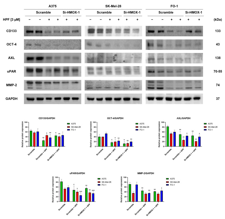
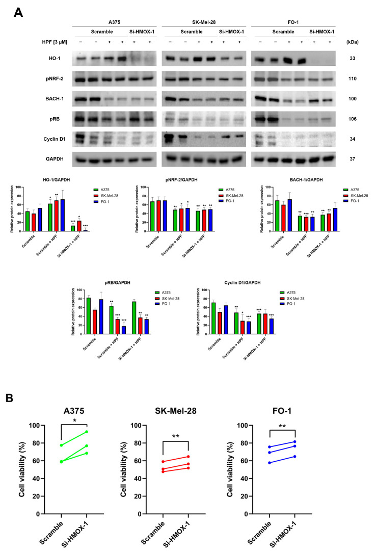
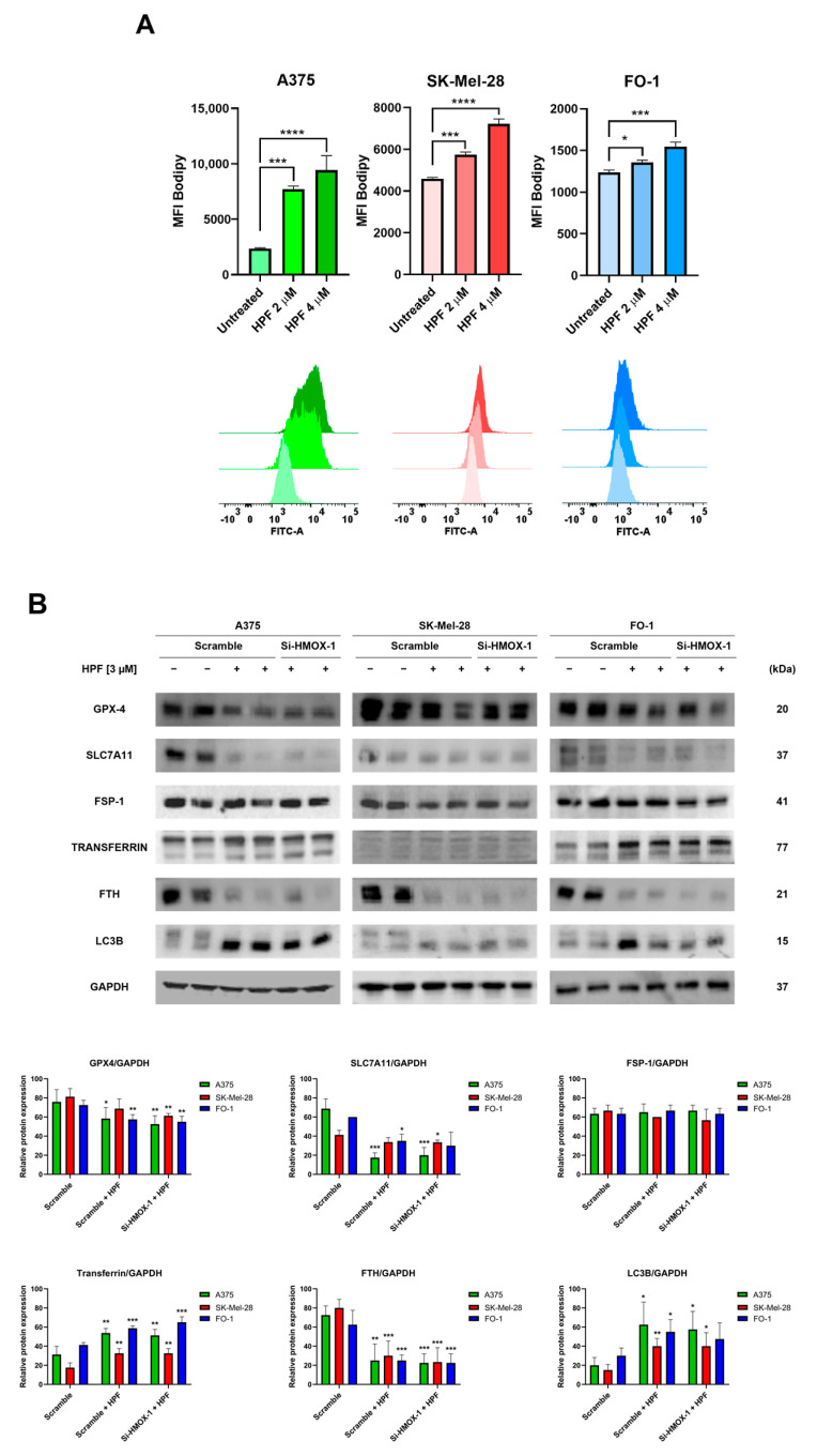
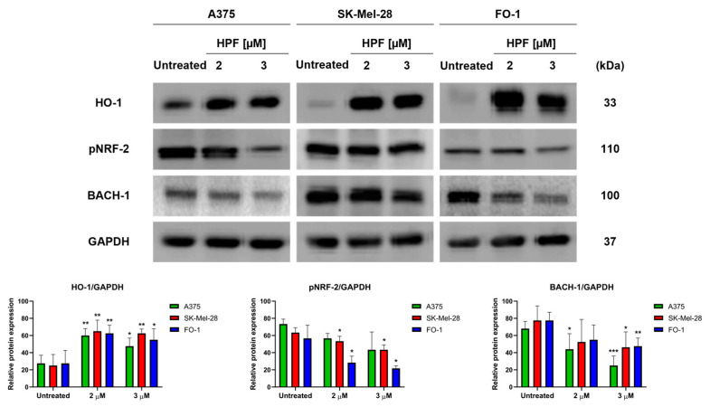
Hyperforin (HPF) is an acylphloroglucinol compound found abundantly in extract which exhibits antidepressant, anti-inflammatory, antimicrobial, and antitumor activities. Our recent study revealed a potent antimelanoma effect of HPF, which hinders melanoma cell proliferation, motility, colony formation, and induces apoptosis. Furthermore, we have identified glutathione peroxidase-4 (GPX-4), a key enzyme involved in cellular protection against iron-induced lipid peroxidation, as one of the molecular targets of HPF. Thus, in three BRAF-mutated melanoma cell lines, we investigated whether iron unbalance and lipid peroxidation may be a part of the molecular mechanisms underlying the antimelanoma activity of HPF. Initially, we focused on heme oxygenase-1 (HO-1), which catalyzes the heme group into CO, biliverdin, and free iron, and observed that HPF treatment triggered the expression of this inducible enzyme. In order to investigate the mechanism involved in HO-1 induction, we verified that HPF downregulates the BTB and CNC homology 1 (BACH-1) transcription factor, an inhibitor of the heme oxygenase 1 (HMOX-1) gene transcription. Remarkably, we observed a partial recovery of cell viability and an increase in the expression of the phosphorylated and active form of retinoblastoma protein when we suppressed the HMOX-1 gene using HMOX-1 siRNA while HPF was present. This suggests that the HO-1 pathway is involved in the cytostatic effect of HPF in melanoma cells. To explore whether lipid peroxidation is induced, we conducted cytofluorimetric analysis and observed a significant increase in the fluorescence of the BODIPY C-11 probe 48 h after HPF administration in all tested melanoma cell lines. To discover the mechanism by which HPF triggers lipid peroxidation, along with the induction of HO-1, we examined the expression of additional proteins associated with iron homeostasis and lipid peroxidation. After HPF administration, we confirmed the downregulation of GPX-4 and observed low expression levels of SLC7A11, a cystine transporter crucial for the glutathione production, and ferritin, able to sequester free iron. A decreased expression level of these proteins can sensitize cells to lipid peroxidation. On the other hand, HPF treatment resulted in increased expression levels of transferrin, which facilitates iron uptake, and LC3B proteins, a molecular marker of autophagy induction. Indeed, ferritin and GPX-4 have been reported to be digested during autophagy. Altogether, these findings suggest that HPF induced lipid peroxidation likely through iron overloading and decreasing the expression of proteins that protect cells from lipid peroxidation. Finally, we examined the expression levels of proteins associated with melanoma cell invasion and metastatic potential. We observed the decreased expression of CD133, octamer-4, tyrosine-kinase receptor AXL, urokinase plasminogen activator receptor, and metalloproteinase-2 following HPF treatment. These findings provide further support for our previous observations, demonstrating the inhibitory effects of HPF on cell motility and colony formation in soft agar, which are both metastasis-related processes in tumor cells.
金丝桃素(HPF)是一种酰基间苯三酚化合物,在提取物中含量丰富,具有抗抑郁、抗炎、抗菌和抗肿瘤活性。我们最近的研究揭示了HPF具有强大的抗黑色素瘤作用,它能阻碍黑色素瘤细胞的增殖、运动、集落形成并诱导细胞凋亡。此外,我们已确定谷胱甘肽过氧化物酶-4(GPX-4)是参与细胞抵御铁诱导的脂质过氧化的关键酶,也是HPF的分子靶点之一。因此,在三种BRAF突变的黑色素瘤细胞系中,我们研究了铁失衡和脂质过氧化是否可能是HPF抗黑色素瘤活性潜在分子机制的一部分。最初,我们聚焦于血红素加氧酶-1(HO-1),它催化血红素基团生成CO、胆绿素和游离铁,并观察到HPF处理可触发这种诱导酶的表达。为了研究HO-1诱导所涉及的机制,我们证实HPF下调了BTB和CNC同源蛋白1(BACH-1)转录因子,该因子是血红素加氧酶1(HMOX-1)基因转录的抑制剂。值得注意的是,当在存在HPF的情况下使用HMOX-1 siRNA抑制HMOX-1基因时,我们观察到细胞活力部分恢复,并且视网膜母细胞瘤蛋白的磷酸化和活性形式的表达增加。这表明HO-1途径参与了HPF对黑色素瘤细胞的细胞生长抑制作用。为了探究是否诱导了脂质过氧化,我们进行了细胞荧光分析,并观察到在所有测试的黑色素瘤细胞系中,HPF给药48小时后BODIPY C-11探针的荧光显著增加。为了发现HPF触发脂质过氧化的机制,连同HO-1的诱导,我们检测了与铁稳态和脂质过氧化相关的其他蛋白质的表达。HPF给药后,我们证实了GPX-4的下调,并观察到SLC7A11(一种对谷胱甘肽产生至关重要的胱氨酸转运蛋白)和铁蛋白(能够螯合游离铁)的低表达水平。这些蛋白质表达水平的降低会使细胞对脂质过氧化敏感。另一方面HPF处理导致转铁蛋白(促进铁摄取)和LC3B蛋白(自噬诱导的分子标志物)的表达水平增加。事实上,据报道铁蛋白和GPX-4在自噬过程中会被消化。总之,这些发现表明HPF可能通过铁过载和降低保护细胞免受脂质过氧化的蛋白质表达来诱导脂质过氧化。最后,我们检测了与黑色素瘤细胞侵袭和转移潜能相关的蛋白质表达水平。我们观察到HPF处理后CD133、八聚体-4、酪氨酸激酶受体AXL、尿激酶型纤溶酶原激活剂受体和金属蛋白酶-2的表达降低。这些发现为我们之前的观察提供了进一步支持,证明了HPF对细胞运动和软琼脂中集落形成的抑制作用,而这两个过程都是肿瘤细胞中与转移相关进程。