Department of Biochemistry, Faculty of Medicine, University of Medicine and Pharmacy of Craiova, Romania;
Rom J Morphol Embryol. 2023 Apr-Jun;64(2):173-180. doi: 10.47162/RJME.64.2.07.
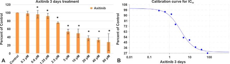
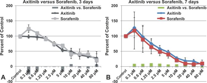
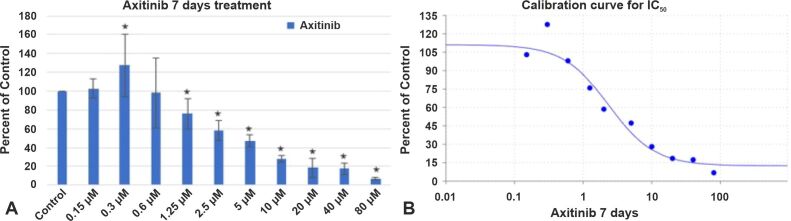
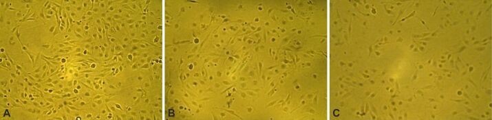
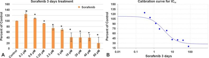
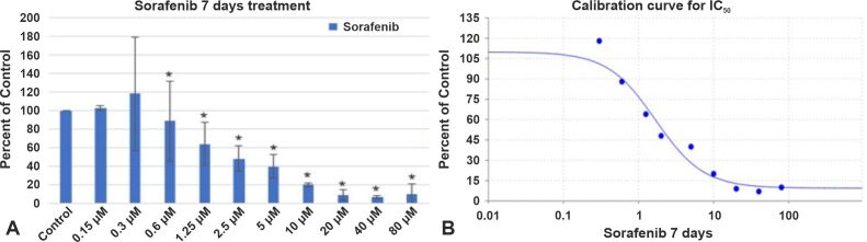
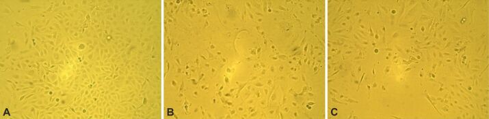
The formation, proliferation, and evolution of glioblastoma (GB) are significantly influenced by pathological angiogenesis. This is supported by several growth factor receptors, such as the vascular endothelial growth factor receptor (VEGFR). In this experiment, we examined how the Food and Drug Administration (FDA) approved VEGFR blockers Sorafenib and Axitinib affect the viability of GB cells in vitro. Cells were cultivated in 96-well culture plates for the experiments, afterwards Sorafenib and Axitinib were administered at doses ranging from 0.3 μM to 80 μM. 2,5-Diphenyl-2H-tetrazolium bromide (MTT) assay was used to assess the impact of VEGFR inhibition on high-grade glioma (HGG) cell lines. To observe the morphological changes in cell shape, we used a 10× magnification microscopy. Our results showed that both Axitinib and Sorafenib retarded GB1B culture proliferation in a dose- and time-dependent manner in comparison to control cohorts that had not received any treatment. The half maximal inhibitory concentration (IC50) value for Axitinib was 3.5839 μM after three days of drug administration and 2.2133 μM after seven days of drug administration. The IC50 value for Sorafenib was 3.5152 μM after three days of drug administration and 1.6846 μM after seven days of drug administration. After the treatment with Axitinib or Sorafenib, very few cells became rounded and detached from the support, others remained adherent to the culture substrate, but acquired a larger, flatter shape. Our results indicate that VEGFR might serve as a key target in the treatment of GB. Although it is known that in vitro some drugs block the VEGFR more potently, clinical evidence is required to show whether this actually translates to better clinical outcomes.
胶质母细胞瘤(GB)的形成、增殖和演变受到病理性血管生成的显著影响。这得到了几种生长因子受体的支持,例如血管内皮生长因子受体(VEGFR)。在这项实验中,我们研究了美国食品和药物管理局(FDA)批准的 VEGFR 阻滞剂索拉非尼(Sorafenib)和阿昔替尼(Axitinib)如何影响体外 GB 细胞的活力。将细胞在 96 孔培养板中进行培养,然后将索拉非尼和阿昔替尼以 0.3 μM 至 80 μM 的剂量给药。使用 2,5-二苯基-2H-四唑溴盐(MTT)测定法来评估 VEGFR 抑制对高级别神经胶质瘤(HGG)细胞系的影响。为了观察细胞形态的变化,我们使用了 10×放大倍数的显微镜。结果表明,与未接受任何治疗的对照组相比,阿昔替尼和索拉非尼均以剂量和时间依赖的方式延迟了 GB1B 培养物的增殖。在给药三天后,阿昔替尼的半最大抑制浓度(IC50)值为 3.5839 μM,在给药七天后为 2.2133 μM。在给药三天后,索拉非尼的 IC50 值为 3.5152 μM,在给药七天后为 1.6846 μM。用阿昔替尼或索拉非尼处理后,很少有细胞变成圆形并从支持物上脱落,其他细胞仍然附着在培养底物上,但获得了更大、更平坦的形状。我们的结果表明,VEGFR 可能是治疗 GB 的关键靶标。虽然已知在体外一些药物更有效地阻断 VEGFR,但需要临床证据表明这是否实际上转化为更好的临床结果。