Qu Kai, Wang Caihong, Huang Lu, Qin Xian, Zhang Kun, Qiu Juhui, Wang Guixue
Key Laboratory for Biorheological Science and Technology of Ministry of Education, State and Local Joint Engineering Laboratory for Vascular Implants, Bioengineering College of Chongqing University, Chongqing, China.
APL Bioeng. 2023 Jul 31;7(3):036104. doi: 10.1063/5.0141289. eCollection 2023 Sep.
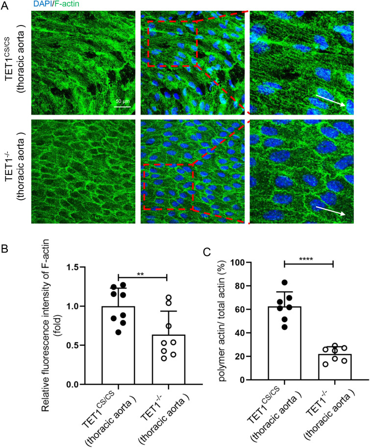
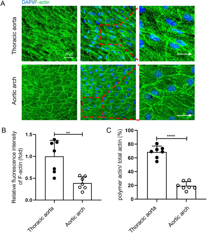
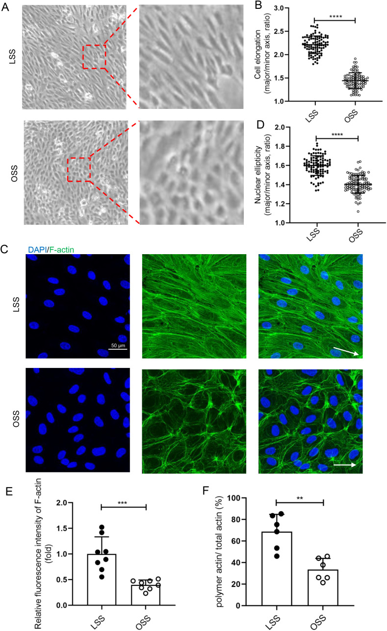
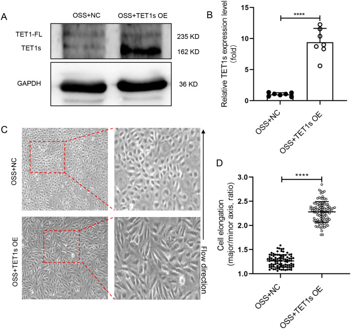
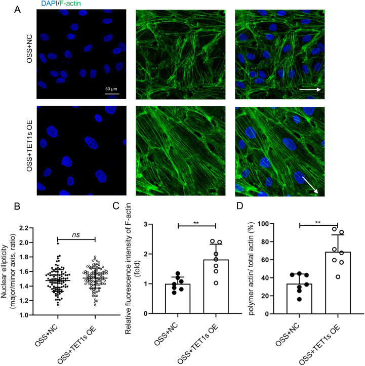
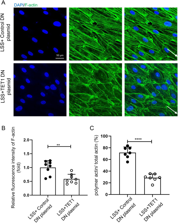
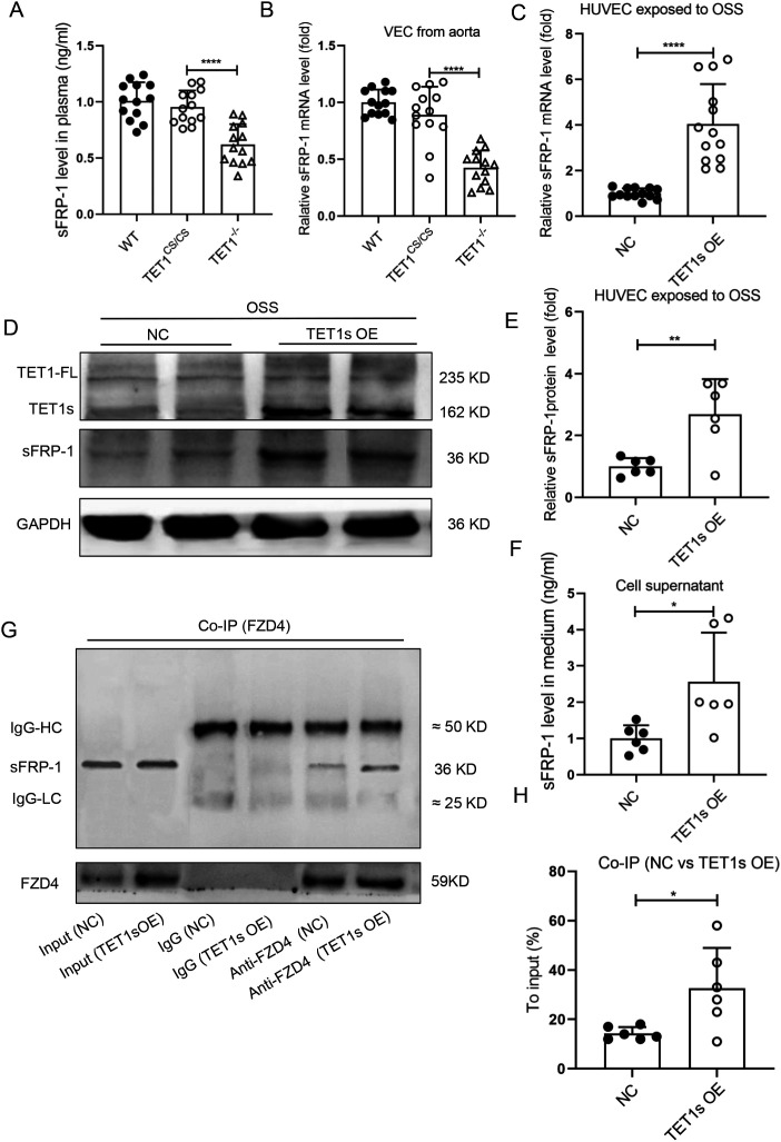
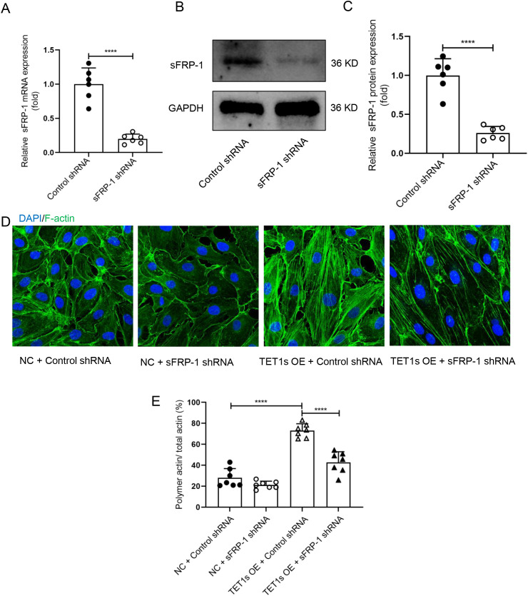
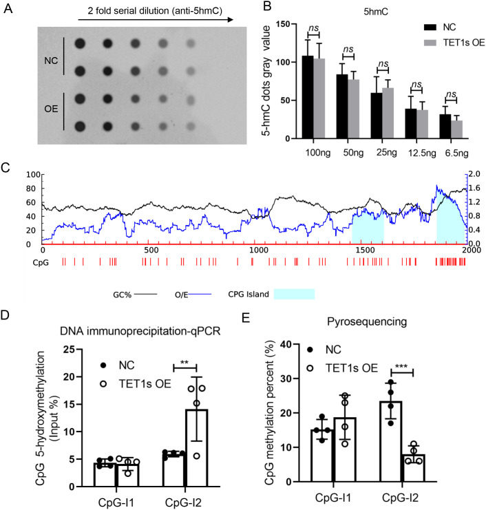
Vascular endothelial polarity induced by blood flow plays crucial roles in the development of atherosclerosis. Loss of endothelial polarity leads to an increase in permeability and leukocyte recruitment, which are crucial hallmarks of atherosclerotic initiation. Endothelial cells exhibit a morphological adaptation to hemodynamic shear stress and possesses planar cell polarity to the direction of blood flow. However, the mechanism of how hemodynamic shear stress regulates endothelial planar cell polarity has not been firmly established. Here, we found that TET1s, a short isoform of Tet methylcytosine dioxygenase 1, was a mediator in the regulation of the planar cell polarity in endothelial cells in response to hemodynamic shear stress. In the process, low expression of TET1s induced by oscillatory shear stress led to the endothelial planar polarity damage through inhibition of F-actin polymerization. TET1s can regulate demethylation level of the sFRP-1 promoter to alter the expression of sFRP-1, which affects the interaction of sFRP-1/Fzd4 and F-actin polymerization. Our study revealed the mechanism of how TET1s mediates endothelial planar cell polarity in response to hemodynamic shear stress and provides a new insight for the prevention of atherosclerosis.
血流诱导的血管内皮极性在动脉粥样硬化的发展中起关键作用。内皮极性丧失导致通透性增加和白细胞募集,这是动脉粥样硬化起始的关键标志。内皮细胞对血流动力学剪切应力表现出形态学适应,并在血流方向上具有平面细胞极性。然而,血流动力学剪切应力如何调节内皮平面细胞极性的机制尚未完全明确。在此,我们发现TET1s(Tet甲基胞嘧啶双加氧酶1的一种短异构体)是响应血流动力学剪切应力调节内皮细胞平面细胞极性的一种介质。在此过程中,振荡剪切应力诱导的TET1s低表达通过抑制F-肌动蛋白聚合导致内皮平面极性损伤。TET1s可调节sFRP-1启动子的去甲基化水平以改变sFRP-1的表达,从而影响sFRP-1/Fzd4的相互作用和F-肌动蛋白聚合。我们的研究揭示了TET1s如何介导内皮细胞响应血流动力学剪切应力的平面细胞极性的机制,并为动脉粥样硬化的预防提供了新的见解。