• 文献检索
  • 文档翻译
  • 深度研究
  • 学术资讯
  • Suppr Zotero 插件Zotero 插件
  • 邀请有礼
  • 套餐&价格
  • 历史记录
应用&插件
Suppr Zotero 插件Zotero 插件浏览器插件Mac 客户端Windows 客户端微信小程序
定价
高级版会员购买积分包购买API积分包
服务
文献检索文档翻译深度研究API 文档MCP 服务
关于我们
关于 Suppr公司介绍联系我们用户协议隐私条款
关注我们

Suppr 超能文献

核心技术专利:CN118964589B侵权必究
粤ICP备2023148730 号-1Suppr @ 2026

文献检索

告别复杂PubMed语法,用中文像聊天一样搜索,搜遍4000万医学文献。AI智能推荐,让科研检索更轻松。

立即免费搜索

文件翻译

保留排版,准确专业,支持PDF/Word/PPT等文件格式,支持 12+语言互译。

免费翻译文档

深度研究

AI帮你快速写综述,25分钟生成高质量综述,智能提取关键信息,辅助科研写作。

立即免费体验

评估口腔放射摄影中矩形准直器中的锥形束切割:ALADA 的应用和辐射管理。

Evaluating cone cut in rectangular collimation in intraoral radiography: application of ALADA and radiation stewardship.

机构信息

Department of Periodontology, Academic Centre for Dentistry, Amsterdam (ACTA), University of Amsterdam and Vrije Universiteit Amsterdam, Gustav Mahlerlaan 3004, 1081 LA, Amsterdam, The Netherlands.

Department of Oral Radiology, Academic Centre for Dentistry, Amsterdam (ACTA), University of Amsterdam and Vrije Universiteit Amsterdam, Amsterdam, The Netherlands.

出版信息

Clin Oral Investig. 2023 Sep;27(9):5391-5402. doi: 10.1007/s00784-023-05158-0. Epub 2023 Aug 4.

DOI:10.1007/s00784-023-05158-0
PMID:37537518
原文链接:https://pmc.ncbi.nlm.nih.gov/articles/PMC10492766/
Abstract

OBJECTIVE

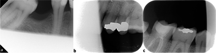
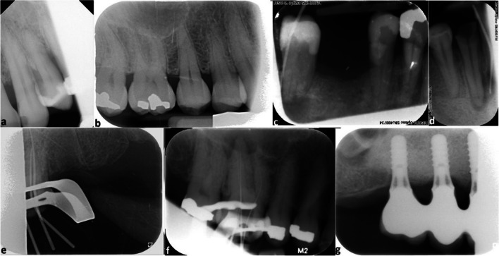
Rectangular collimation is a popular method used in intraoral radiography to reduce patient exposure to ionizing radiation. One of the perceived drawbacks of rectangular collimation is the possibility of an increase in cone cut errors ultimately impacting the diagnostic value of the radiographs. Thus, the aim of this study was to explore the frequency of cone cut errors in radiographs taken using a rectangular collimator.

MATERIALS AND METHODS

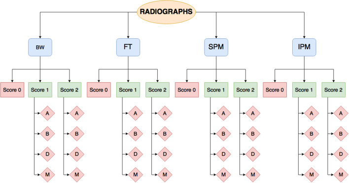
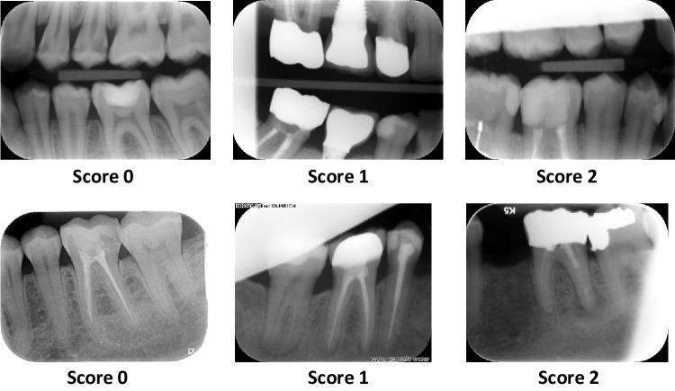
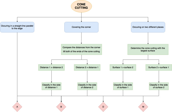
Radiographs taken using PSP plates at Academic Center for Dentistry Amsterdam in the Netherlands by staff and students from January to December 2015 were assessed for cone cut errors. The radiographs were grouped as bitewings, front teeth, inferior premolars and molars, and superior premolars and molars and categorized as no cone cut, cone cut but diagnostically usable, and cone cut but diagnostically not usable. The results were entered into Microsoft Excel and analyzed thereafter.

RESULTS

A total of 53,684 radiographs were assessed, 79% had no cone cut errors and consequently 21% had some degree of cone cut. However, the diagnostic value was unaffected in 18% of the radiographs with cone cut. Only 3% of the radiographs were deemed diagnostically unusable due to cone cut. The most common area of cone cut was in the premolar and molar areas while cone cut in the front teeth was least likely to be diagnostically unusable.

CONCLUSION

Cone cut from the use of a rectangular collimator does not seem to result in an increase of diagnostically unusable radiographs. Thus, rectangular collimation should be preferred as it decreases the amount of radiation exposure to the patient while producing diagnostically usable radiographs and thus allowing the dental professional to adhere to the ALADA principle and practice radiation stewardship.

CLINICAL RELEVANCE

Scientific rationale for the study: rectangular collimation is a method used to reduce patient exposure to ionizing radiation; however, this benefit is negligible if radiographs must be retaken due to cone cut errors that make the radiograph diagnostically unusable. Therefore, the aim of this study was to explore the frequency of cone cut in radiographs taken using a rectangular collimator.

PRINCIPAL FINDINGS

cone cut was observed in 21% of the radiographs; however, only 3% of the radiographs were considered diagnostically unusable.

PRACTICAL IMPLICATIONS

rectangular collimation does not result in a high number of diagnostically unusable radiographs and should be used to reduce patient exposure to ionizing radiation.

摘要

目的

在口腔放射摄影中,矩形准直器是一种常用的方法,可减少患者接受电离辐射的剂量。使用矩形准直器的一个被认为的缺点是可能会增加锥形切割误差,最终影响射线照片的诊断价值。因此,本研究的目的是探讨使用矩形准直器拍摄的射线照片中锥形切割误差的频率。

材料和方法

对 2015 年 1 月至 12 月期间,荷兰阿姆斯特丹牙科学院的工作人员和学生使用 PSP 板拍摄的射线照片进行锥形切割误差评估。将射线照片分为咬合片、前牙、下前磨牙和磨牙以及上前磨牙和磨牙,并分为无锥形切割、锥形切割但可诊断使用和锥形切割但不可诊断使用。结果输入 Microsoft Excel 并进行分析。

结果

共评估了 53684 张射线照片,79%无锥形切割误差,因此 21%存在一定程度的锥形切割。然而,18%的带有锥形切割的射线照片的诊断价值未受影响。只有 3%的射线照片因锥形切割而被认为不可诊断使用。最常见的锥形切割区域是在后磨牙区域,而前牙的锥形切割最不可能不可诊断使用。

结论

使用矩形准直器产生的锥形切割似乎不会导致不可诊断使用的射线照片数量增加。因此,应首选矩形准直器,因为它可以减少患者的辐射暴露量,同时生成可诊断使用的射线照片,从而使牙科专业人员能够遵守 ALADA 原则并进行辐射管理。

临床相关性

研究的科学依据:矩形准直是一种用于减少患者接受电离辐射的方法;然而,如果由于锥形切割错误导致射线照片必须重拍,从而使射线照片不可诊断使用,那么这种益处就微不足道了。因此,本研究的目的是探讨使用矩形准直器拍摄的射线照片中锥形切割的频率。

主要发现

在 21%的射线照片中观察到锥形切割;然而,只有 3%的射线照片被认为不可诊断使用。

实际意义

矩形准直不会导致大量不可诊断使用的射线照片,应使用它来减少患者接受电离辐射的剂量。

https://cdn.ncbi.nlm.nih.gov/pmc/blobs/dae4/10492766/97d3fd87b8a2/784_2023_5158_Fig7_HTML.jpg
https://cdn.ncbi.nlm.nih.gov/pmc/blobs/dae4/10492766/33bf3ace8a40/784_2023_5158_Fig1_HTML.jpg
https://cdn.ncbi.nlm.nih.gov/pmc/blobs/dae4/10492766/6954f4cf60fd/784_2023_5158_Fig2_HTML.jpg
https://cdn.ncbi.nlm.nih.gov/pmc/blobs/dae4/10492766/8dceca73a568/784_2023_5158_Fig3_HTML.jpg
https://cdn.ncbi.nlm.nih.gov/pmc/blobs/dae4/10492766/c0c5eed58e8d/784_2023_5158_Fig4_HTML.jpg
https://cdn.ncbi.nlm.nih.gov/pmc/blobs/dae4/10492766/4fffa8a460df/784_2023_5158_Fig5_HTML.jpg
https://cdn.ncbi.nlm.nih.gov/pmc/blobs/dae4/10492766/8146c0822452/784_2023_5158_Fig6_HTML.jpg
https://cdn.ncbi.nlm.nih.gov/pmc/blobs/dae4/10492766/97d3fd87b8a2/784_2023_5158_Fig7_HTML.jpg

相似文献

1
Evaluating cone cut in rectangular collimation in intraoral radiography: application of ALADA and radiation stewardship.评估口腔放射摄影中矩形准直器中的锥形束切割:ALADA 的应用和辐射管理。
Clin Oral Investig. 2023 Sep;27(9):5391-5402. doi: 10.1007/s00784-023-05158-0. Epub 2023 Aug 4.
2
A comparison between bitewing radiographs taken with rectangular and circular collimators in UK military dental practices: a retrospective study.英国军人口腔实践中使用矩形和圆形遮线器拍摄的咬翼片的比较:一项回顾性研究。
Dentomaxillofac Radiol. 2011 Feb;40(2):102-9. doi: 10.1259/dmfr/86968802.
3
Digital intraoral imaging: analysis of retakes and reduction in radiation exposure using rectangular collimation.数字化口腔内成像:使用矩形准直器分析重拍和减少辐射暴露。
Gen Dent. 2022 Nov-Dec;70(6):66-70.
4
[Intraoral radiography: qualitative study].[口腔内放射摄影术:定性研究]
Swiss Dent J. 2023 Feb 6;133(2):89-96. doi: 10.61872/sdj-2023-02-02.
5
A comparison of rectangular and cylindrical collimation for intraoral radiographs.口腔内X光片矩形准直和圆柱形准直的比较。
J Dent Educ. 1983 Dec;47(12):771-3.
6
Errors generated with the use of rectangular collimation.使用矩形准直器产生的误差。
Oral Surg Oral Med Oral Pathol. 1991 Apr;71(4):509-13. doi: 10.1016/0030-4220(91)90442-f.
7
A survey on radiation exposure reduction methods including rectangular collimation for intraoral radiography by pediatric dentists in the United States.美国儿童牙医采用矩形准直器降低口腔内放射摄影辐射暴露的方法调查。
J Am Dent Assoc. 2020 Apr;151(4):287-296. doi: 10.1016/j.adaj.2020.01.014.
8
Intraoral Imaging in Dental Private Practice - A Rectangular Collimator Study.牙科私人诊所中的口腔内成像——一项矩形准直器研究。
J Can Dent Assoc. 2020 Oct;86:k16.
9
Evidence on radiation dose reduction using rectangular collimation: a systematic review.使用矩形准直器降低放射剂量的证据:系统评价。
Int Dent J. 2019 Apr;69(2):84-97. doi: 10.1111/idj.12411. Epub 2018 Jun 29.
10
Technical Performance of Universal and Enhanced Intraoral Imaging Rectangular Collimators.通用型和增强型口腔内成像矩形准直器的技术性能
J Dent Hyg. 2015 Aug;89(4):238-46.

引用本文的文献

1
Impact of Rectangular Collimation on Quality of Intraoral Radiographs: Findings from a Clinical Audit at a Dental Practice.矩形准直对口腔内X光片质量的影响:牙科诊所临床审计结果
Diagnostics (Basel). 2025 Apr 2;15(7):911. doi: 10.3390/diagnostics15070911.

本文引用的文献

1
[Intraoral radiography: qualitative study].[口腔内放射摄影术:定性研究]
Swiss Dent J. 2023 Feb 6;133(2):89-96. doi: 10.61872/sdj-2023-02-02.
2
Dental X-Rays and the Risk of Thyroid Cancer and Meningioma: A Systematic Review and Meta-Analysis of Current Epidemiological Evidence.牙科 X 射线与甲状腺癌和脑膜瘤风险:当前流行病学证据的系统评价和荟萃分析。
Thyroid. 2019 Nov;29(11):1572-1593. doi: 10.1089/thy.2019.0105. Epub 2019 Oct 14.
3
Evidence on radiation dose reduction using rectangular collimation: a systematic review.使用矩形准直器降低放射剂量的证据:系统评价。
Int Dent J. 2019 Apr;69(2):84-97. doi: 10.1111/idj.12411. Epub 2018 Jun 29.
4
The REporting of studies Conducted using Observational Routinely-collected health Data (RECORD) statement.使用常规收集的健康数据进行研究的报告(RECORD)声明
PLoS Med. 2015 Oct 6;12(10):e1001885. doi: 10.1371/journal.pmed.1001885. eCollection 2015 Oct.
5
Exposure to ionizing radiation during dental X-rays is not associated with risk of developing meningioma: a meta-analysis based on seven case-control studies.牙科X线检查时接触电离辐射与患脑膜瘤的风险无关:一项基于七项病例对照研究的荟萃分析。
PLoS One. 2015 Feb 6;10(2):e0113210. doi: 10.1371/journal.pone.0113210. eCollection 2015.
6
A comparison between bitewing radiographs taken with rectangular and circular collimators in UK military dental practices: a retrospective study.英国军人口腔实践中使用矩形和圆形遮线器拍摄的咬翼片的比较:一项回顾性研究。
Dentomaxillofac Radiol. 2011 Feb;40(2):102-9. doi: 10.1259/dmfr/86968802.
7
Patient risk related to common dental radiographic examinations: the impact of 2007 International Commission on Radiological Protection recommendations regarding dose calculation.与常见牙科X线检查相关的患者风险:2007年国际放射防护委员会关于剂量计算建议的影响
J Am Dent Assoc. 2008 Sep;139(9):1237-43. doi: 10.14219/jada.archive.2008.0339.
8
Dental X-rays and the risk of intracranial meningioma: a population-based case-control study.牙科X线检查与颅内脑膜瘤风险:一项基于人群的病例对照研究。
Cancer. 2004 Mar 1;100(5):1026-34. doi: 10.1002/cncr.20036.
9
Diagnostic X-ray exposure and female papillary thyroid cancer: a pooled analysis of two Swedish studies.诊断性X射线暴露与女性甲状腺乳头状癌:两项瑞典研究的汇总分析
Eur J Cancer Prev. 1997 Dec;6(6):550-6. doi: 10.1097/00008469-199712000-00010.
10
Environmental factors and the risk of salivary gland cancer.环境因素与唾液腺癌风险
Epidemiology. 1997 Jul;8(4):414-9. doi: 10.1097/00001648-199707000-00011.