Madaki Saudatu Umar, Habib Saudat Garba, Yero Musa Zainab
Department of Ophthalmology, Gombe State University/Federal Teaching Hospital, Gombe, Gombe State, Nigeria.
Department of Ophthalmology, Aminu Kano Teaching Hospital/Faculty of Clinical Sciences-College of Health Sciences, Bayero University, Kano, Kano State, Nigeria.
J West Afr Coll Surg. 2023 Jul-Sep;13(3):111-115. doi: 10.4103/jwas.jwas_13_23. Epub 2023 Jun 27.
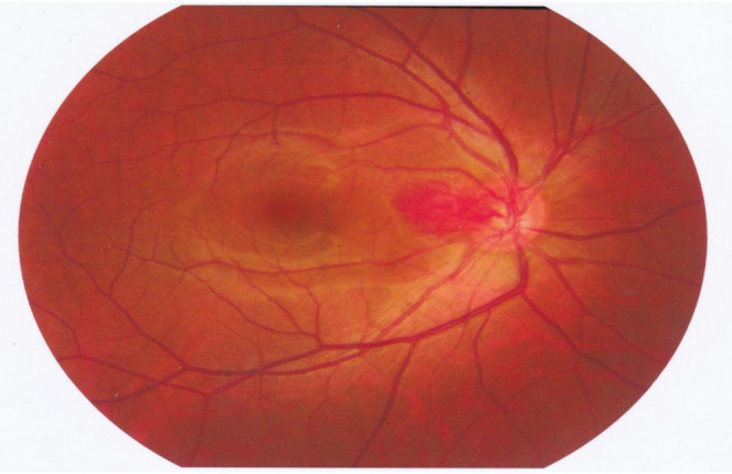
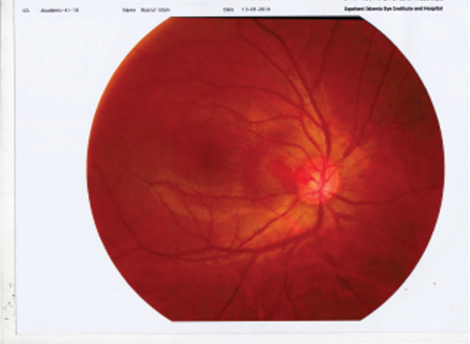
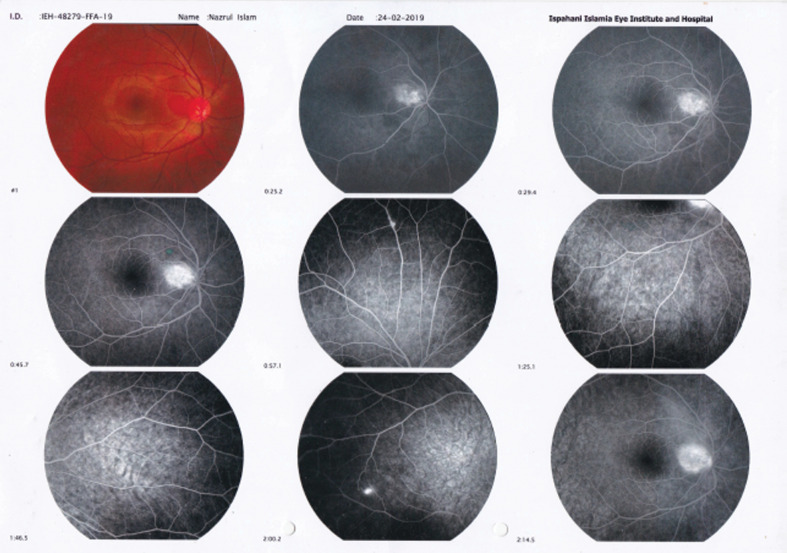
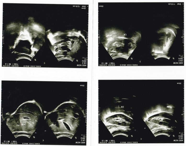
Retinal hemangioma has posed a therapeutic problem for ophthalmologists for almost a decade. The location of hemangioma in the retina is an important factor in determining the treatment options as well as the size of the tumor, clarity of media, and secondary features of the mass. A 22-year-old male presented to vitreoretina clinic at a tertiary eye hospital, with a history of sudden decrease of vision in the left eye for 1 year associated with mild ocular pains. Physical and systemic examination revealed no positive findings. On ocular examination, the visual acuity was 6/6 in the right eye and perception of light in the left eye, and there was no improvement with best correction. Anterior segment examination in both eyes was essentially normal. Dilated funduscopy revealed a well-circumscribed and elevated vascular orange reddish mass in the juxtapapillary area, temporal to the disc with dilated tortuous feeder blood vessels measuring 6.4 × 3.0 mm in the right eye and vitreous hemorrhage with tractional retinal detachment in the left eye. The small solitary lesion in the right eye was treated with focal laser. This was followed by intravitreal bevacizumab avastin 1.25 mg in 0.05 mL in the same eye, monthly three doses, with the aim of targeting the bigger lesion at the juxtapapillary area to reduce the risk of visual loss. Patient also had pars plana vitrectomy, endolaser photocoagulation, and silicon oil in the left eye. There was complete resolution of the tumor, which was not measurable after third dose of intravitreal avastin. Patient vision remained 6/6, and there was no recurrence at the last follow-up visit 3 years after treatment. Antivascular endothelial growth factors like avastin are effective in the treatment of retinal capillary hemangioma, thereby inducing complete tumor resolution as well as maintaining good vision in the early stages of the disease before complication occurs, as it is evident in this case study.
近十年来,视网膜血管瘤一直给眼科医生带来治疗难题。血管瘤在视网膜中的位置是决定治疗方案的重要因素,此外还包括肿瘤大小、屈光间质清晰度以及肿块的继发特征。一名22岁男性前往一家三级眼科医院的玻璃体视网膜诊所就诊,有左眼视力突然下降1年并伴有轻度眼痛的病史。体格检查和全身检查均未发现阳性体征。眼部检查显示,右眼视力为6/6,左眼仅存光感,最佳矫正后视力无改善。双眼眼前节检查基本正常。散瞳眼底检查发现,在视乳头旁区域有一个边界清晰、隆起的血管性橙红色肿块,位于视盘颞侧,右眼有迂曲扩张的供血血管,大小为6.4×3.0毫米,左眼有玻璃体出血并伴有牵拉性视网膜脱离。右眼的小孤立病灶接受了局部激光治疗。随后,在同一只眼中每月注射3次0.05毫升含1.25毫克阿瓦斯汀的玻璃体腔内抗血管内皮生长因子,目的是针对视乳头旁区域较大的病灶,以降低视力丧失的风险。患者左眼还接受了玻璃体切割术、眼内激光光凝术并植入了硅油。肿瘤完全消退,在第三次玻璃体腔内注射阿瓦斯汀后已无法测量。患者视力仍为6/6,治疗后3年的最后一次随访未发现复发。正如本病例研究所示,像阿瓦斯汀这样的抗血管内皮生长因子在治疗视网膜毛细血管瘤方面是有效的,从而能在疾病早期并发症发生之前诱导肿瘤完全消退并保持良好视力。