Department of Urology, Renmin Hospital of Wuhan University, Wuhan, Hubei Province, China.
Medicine (Baltimore). 2023 Aug 4;102(31):e34443. doi: 10.1097/MD.0000000000034443.
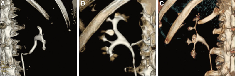
To study the anatomical orientation of the posterior group of calyces based on reconstructed images of computerized tomography urography (CTU) and provide a novel classification with its clinical significance. Clinical data of a total of 1321 patients, who underwent CTU examination in our hospital were retrospectively analyzed. Among these, a total of 2642 3-dimensional reconstructed images of CTU scans were considered in this study. Based on the morphology of the renal calyces and the influence on the establishment of surgical access, the posterior group renal calyces are classified into 3 major types including pot-belly type, classically branched and elongated branched. The classically branched type is further classified into 3 sub-types: a, b and c, based on the association of minor calyces of the posterior group to the major calyces. Type a is derived from 1 group of major calyces only, type b is derived from 2 groups of major calyces simultaneously, and type c is derived from 3 groups of major calyces simultaneously. Statistical findings revealed that all kidneys possess posterior group calyces. The percentage of occurrence of pot-belly type, classically branched and elongated branched is 8.06%, 73.13%, and 18.81%, respectively. The anatomical typing of the classical branching type occurred in 19.36%, 68.17%, and 12.47% for types a, b, and c, respectively. In this study, the posterior group calyces were found to be present across all patients. The posterior group calyces were highest in the classical branching type, of which anatomical typing was highest in type b. The typing of the posterior group of calyces could provide an anatomical basis for percutaneous nephrolithotomy (PCNL) puncture from the posterior group.
研究基于 CTU 重建图像的后组肾盏解剖方位,并提供一种新的分类及其临床意义。回顾性分析我院 1321 例行 CTU 检查的患者的临床资料。本研究共纳入 2642 例 3D 重建 CTU 扫描图像。根据肾盏形态和对建立手术入路的影响,将后组肾盏分为 3 大类,包括壶腹型、经典分支型和长分支型。经典分支型根据后组肾小盏与主肾盏的关系进一步分为 3 个亚型:a、b 和 c。a 型仅来源于 1 组主肾盏,b 型同时来源于 2 组主肾盏,c 型同时来源于 3 组主肾盏。统计发现所有肾脏均有后组肾盏。壶腹型、经典分支型和长分支型的发生率分别为 8.06%、73.13%和 18.81%。经典分支型的解剖分型发生率分别为 a 型 19.36%、b 型 68.17%和 c 型 12.47%。本研究发现所有患者均存在后组肾盏。后组肾盏以经典分支型最多,其中解剖分型以 b 型最多。后组肾盏的分型可为经皮肾镜取石术(PCNL)从后组穿刺提供解剖学依据。