• 文献检索
  • 文档翻译
  • 深度研究
  • 学术资讯
  • Suppr Zotero 插件Zotero 插件
  • 邀请有礼
  • 套餐&价格
  • 历史记录
应用&插件
Suppr Zotero 插件Zotero 插件浏览器插件Mac 客户端Windows 客户端微信小程序
定价
高级版会员购买积分包购买API积分包
服务
文献检索文档翻译深度研究API 文档MCP 服务
关于我们
关于 Suppr公司介绍联系我们用户协议隐私条款
关注我们

Suppr 超能文献

核心技术专利:CN118964589B侵权必究
粤ICP备2023148730 号-1Suppr @ 2026

文献检索

告别复杂PubMed语法,用中文像聊天一样搜索,搜遍4000万医学文献。AI智能推荐,让科研检索更轻松。

立即免费搜索

文件翻译

保留排版,准确专业,支持PDF/Word/PPT等文件格式,支持 12+语言互译。

免费翻译文档

深度研究

AI帮你快速写综述,25分钟生成高质量综述,智能提取关键信息,辅助科研写作。

立即免费体验

用于高效分析3D病理学样本的弱监督人工智能

Weakly Supervised AI for Efficient Analysis of 3D Pathology Samples.

作者信息

Song Andrew H, Williams Mane, Williamson Drew F K, Jaume Guillaume, Zhang Andrew, Chen Bowen, Serafin Robert, Liu Jonathan T C, Baras Alex, Parwani Anil V, Mahmood Faisal

机构信息

Department of Pathology, Brigham and Women's Hospital, Harvard Medical School, Boston, MA, USA.

Department of Pathology, Massachusetts General Hospital, Harvard Medical School, Boston, MA, USA.

出版信息

ArXiv. 2023 Jul 27:arXiv:2307.14907v1.

PMID:37547660
原文链接:https://pmc.ncbi.nlm.nih.gov/articles/PMC10402184/
Abstract

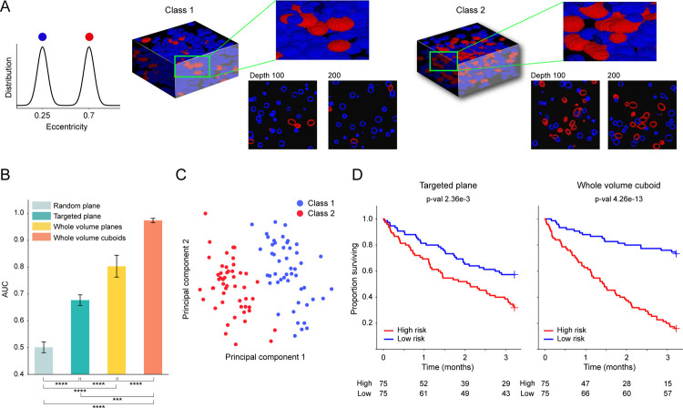
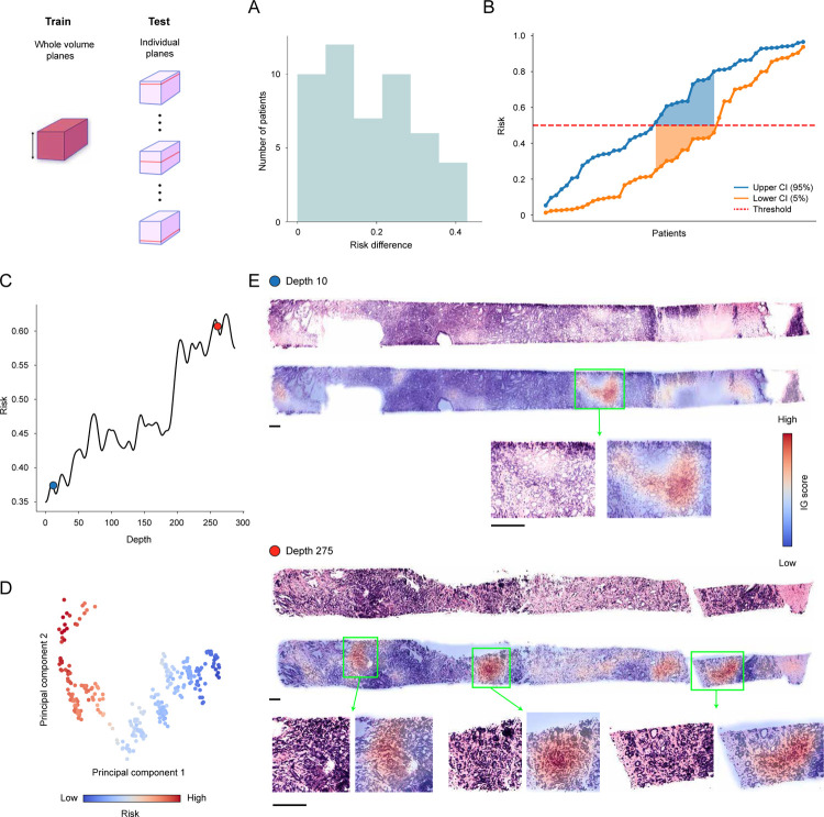
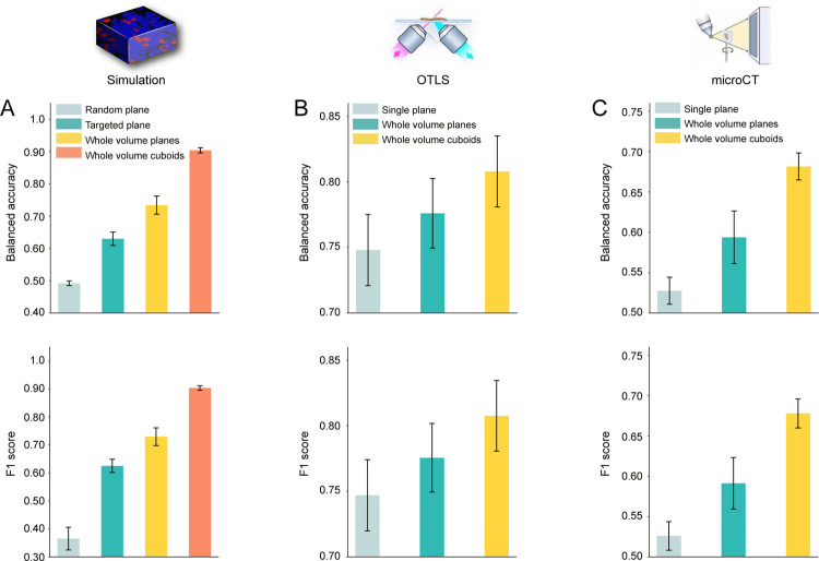
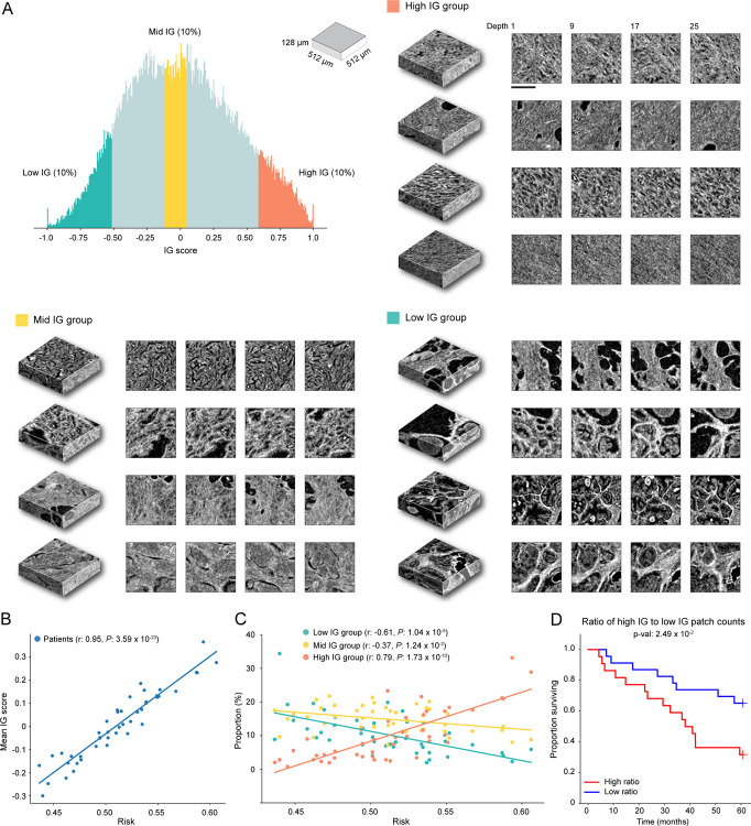
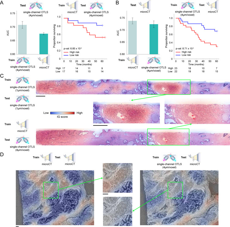
Human tissue consists of complex structures that display a diversity of morphologies, forming a tissue microenvironment that is, by nature, three-dimensional (3D). However, the current standard-of-care involves slicing 3D tissue specimens into two-dimensional (2D) sections and selecting a few for microscopic evaluation, with concomitant risks of sampling bias and misdiagnosis. To this end, there have been intense efforts to capture 3D tissue morphology and transition to 3D pathology, with the development of multiple high-resolution 3D imaging modalities. However, these tools have had little translation to clinical practice as manual evaluation of such large data by pathologists is impractical and there is a lack of computational platforms that can efficiently process the 3D images and provide patient-level clinical insights. Here we present Modality-Agnostic Multiple instance learning for volumetric Block Analysis (MAMBA), a deep-learning-based platform for processing 3D tissue images from diverse imaging modalities and predicting patient outcomes. Archived prostate cancer specimens were imaged with open-top light-sheet microscopy or microcomputed tomography and the resulting 3D datasets were used to train risk-stratification networks based on 5-year biochemical recurrence outcomes via MAMBA. With the 3D block-based approach, MAMBA achieves an area under the receiver operating characteristic curve (AUC) of 0.86 and 0.74, superior to 2D traditional single-slice-based prognostication (AUC of 0.79 and 0.57), suggesting superior prognostication with 3D morphological features. Further analyses reveal that the incorporation of greater tissue volume improves prognostic performance and mitigates risk prediction variability from sampling bias, suggesting that there is value in capturing larger extents of spatially heterogeneous 3D morphology. With the rapid growth and adoption of 3D spatial biology and pathology techniques by researchers and clinicians, MAMBA provides a general and efficient framework for 3D weakly supervised learning for clinical decision support and can help to reveal novel 3D morphological biomarkers for prognosis and therapeutic response.

摘要

人体组织由复杂结构组成,呈现出多样的形态,形成了本质上为三维(3D)的组织微环境。然而,当前的标准治疗方法是将3D组织标本切成二维(2D)切片,并选择少数切片进行显微镜评估,这伴随着抽样偏差和误诊的风险。为此,人们付出了巨大努力来捕捉3D组织形态并向3D病理学转变,开发了多种高分辨率3D成像模式。然而,这些工具在临床实践中的应用很少,因为病理学家对手动评估如此大量的数据不切实际,而且缺乏能够有效处理3D图像并提供患者水平临床见解的计算平台。在此,我们展示了用于体积块分析的模态无关多实例学习(MAMBA),这是一个基于深度学习的平台,用于处理来自不同成像模式的3D组织图像并预测患者预后。对存档的前列腺癌标本进行了开放式光片显微镜或微型计算机断层扫描成像,并通过MAMBA将所得的3D数据集用于基于5年生化复发结果训练风险分层网络。通过基于3D块的方法,MAMBA在受试者工作特征曲线(AUC)下的面积分别达到0.86和0.74,优于基于2D传统单切片的预测(AUC分别为0.79和0.57),表明具有3D形态特征的预测更优。进一步分析表明,纳入更大的组织体积可提高预后性能并减轻抽样偏差导致的风险预测变异性,这表明捕捉更大范围的空间异质性3D形态具有价值。随着研究人员和临床医生对3D空间生物学和病理学技术的快速发展和采用,MAMBA为用于临床决策支持的3D弱监督学习提供了一个通用且高效的框架,并有助于揭示用于预后和治疗反应的新型3D形态生物标志物。

https://cdn.ncbi.nlm.nih.gov/pmc/blobs/b612/10402184/963f1e58cecf/nihpp-2307.14907v1-f0012.jpg
https://cdn.ncbi.nlm.nih.gov/pmc/blobs/b612/10402184/de004782dcf6/nihpp-2307.14907v1-f0005.jpg
https://cdn.ncbi.nlm.nih.gov/pmc/blobs/b612/10402184/1a3f17f476ea/nihpp-2307.14907v1-f0006.jpg
https://cdn.ncbi.nlm.nih.gov/pmc/blobs/b612/10402184/1bc2edf1c31b/nihpp-2307.14907v1-f0010.jpg
https://cdn.ncbi.nlm.nih.gov/pmc/blobs/b612/10402184/f4f2fd1fb0d2/nihpp-2307.14907v1-f0011.jpg
https://cdn.ncbi.nlm.nih.gov/pmc/blobs/b612/10402184/963f1e58cecf/nihpp-2307.14907v1-f0012.jpg

相似文献

1
Weakly Supervised AI for Efficient Analysis of 3D Pathology Samples.用于高效分析3D病理学样本的弱监督人工智能
ArXiv. 2023 Jul 27:arXiv:2307.14907v1.
2
Analysis of 3D pathology samples using weakly supervised AI.使用弱监督 AI 分析 3D 病理样本。
Cell. 2024 May 9;187(10):2502-2520.e17. doi: 10.1016/j.cell.2024.03.035.
3
Nondestructive 3D pathology with analysis of nuclear features for prostate cancer risk assessment.用于前列腺癌风险评估的具有核特征分析的无损 3D 病理学。
J Pathol. 2023 Aug;260(4):390-401. doi: 10.1002/path.6090. Epub 2023 May 26.
4
Prostate Cancer Risk Stratification via Nondestructive 3D Pathology with Deep Learning-Assisted Gland Analysis.基于深度学习辅助腺体分析的非破坏性 3D 病理学前列腺癌风险分层。
Cancer Res. 2022 Jan 15;82(2):334-345. doi: 10.1158/0008-5472.CAN-21-2843. Epub 2021 Dec 1.
5
Direct three-dimensional segmentation of prostate glands with nnU-Net.使用nnU-Net对前列腺进行直接三维分割。
J Biomed Opt. 2024 Mar;29(3):036001. doi: 10.1117/1.JBO.29.3.036001. Epub 2024 Mar 1.
6
Bi-channel image registration and deep-learning segmentation (BIRDS) for efficient, versatile 3D mapping of mouse brain.双通道图像配准和深度学习分割(BIRDS)用于高效、通用的小鼠大脑 3D 映射。
Elife. 2021 Jan 18;10:e63455. doi: 10.7554/eLife.63455.
7
Erratum: Eyestalk Ablation to Increase Ovarian Maturation in Mud Crabs.勘误:切除眼柄以增加泥蟹的卵巢成熟度。
J Vis Exp. 2023 May 26(195). doi: 10.3791/6561.
8
Automatic prostate segmentation using deep learning on clinically diverse 3D transrectal ultrasound images.基于临床多样的三维经直肠超声图像,利用深度学习进行前列腺自动分割。
Med Phys. 2020 Jun;47(6):2413-2426. doi: 10.1002/mp.14134. Epub 2020 Apr 8.
9
Weakly Supervised Learning of 3D Deep Network for Neuron Reconstruction.用于神经元重建的3D深度网络的弱监督学习
Front Neuroanat. 2020 Jul 28;14:38. doi: 10.3389/fnana.2020.00038. eCollection 2020.
10
Digital Volumetric Biopsy Cores Improve Gleason Grading of Prostate Cancer Using Deep Learning.数字体积活检芯利用深度学习改善前列腺癌的 Gleason 分级
ArXiv. 2024 Sep 12:arXiv:2409.08331v1.