• 文献检索
  • 文档翻译
  • 深度研究
  • 学术资讯
  • Suppr Zotero 插件Zotero 插件
  • 邀请有礼
  • 套餐&价格
  • 历史记录
应用&插件
Suppr Zotero 插件Zotero 插件浏览器插件Mac 客户端Windows 客户端微信小程序
定价
高级版会员购买积分包购买API积分包
服务
文献检索文档翻译深度研究API 文档MCP 服务
关于我们
关于 Suppr公司介绍联系我们用户协议隐私条款
关注我们

Suppr 超能文献

核心技术专利:CN118964589B侵权必究
粤ICP备2023148730 号-1Suppr @ 2026

文献检索

告别复杂PubMed语法,用中文像聊天一样搜索,搜遍4000万医学文献。AI智能推荐,让科研检索更轻松。

立即免费搜索

文件翻译

保留排版,准确专业,支持PDF/Word/PPT等文件格式,支持 12+语言互译。

免费翻译文档

深度研究

AI帮你快速写综述,25分钟生成高质量综述,智能提取关键信息,辅助科研写作。

立即免费体验

使用nnU-Net对前列腺进行直接三维分割。

Direct three-dimensional segmentation of prostate glands with nnU-Net.

作者信息

Wang Rui, Chow Sarah S L, Serafin Robert B, Xie Weisi, Han Qinghua, Baraznenok Elena, Lan Lydia, Bishop Kevin W, Liu Jonathan T C

机构信息

University of Washington, Department of Mechanical Engineering, Seattle, Washington, United States.

University of Washington, Department of Bioengineering, Seattle, Washington, United States.

出版信息

J Biomed Opt. 2024 Mar;29(3):036001. doi: 10.1117/1.JBO.29.3.036001. Epub 2024 Mar 1.

DOI:10.1117/1.JBO.29.3.036001
PMID:38434772
原文链接:https://pmc.ncbi.nlm.nih.gov/articles/PMC10905031/
Abstract

SIGNIFICANCE

In recent years, we and others have developed non-destructive methods to obtain three-dimensional (3D) pathology datasets of clinical biopsies and surgical specimens. For prostate cancer risk stratification (prognostication), standard-of-care Gleason grading is based on examining the morphology of prostate glands in thin 2D sections. This motivates us to perform 3D segmentation of prostate glands in our 3D pathology datasets for the purposes of computational analysis of 3D glandular features that could offer improved prognostic performance.

AIM

To facilitate prostate cancer risk assessment, we developed a computationally efficient and accurate deep learning model for 3D gland segmentation based on open-top light-sheet microscopy datasets of human prostate biopsies stained with a fluorescent analog of hematoxylin and eosin (H&E).

APPROACH

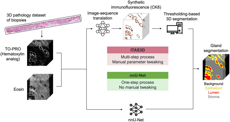
For 3D gland segmentation based on our H&E-analog 3D pathology datasets, we previously developed a hybrid deep learning and computer vision-based pipeline, called image translation-assisted segmentation in 3D (ITAS3D), which required a complex two-stage procedure and tedious manual optimization of parameters. To simplify this procedure, we use the 3D gland-segmentation masks previously generated by ITAS3D as training datasets for a direct end-to-end deep learning-based segmentation model, nnU-Net. The inputs to this model are 3D pathology datasets of prostate biopsies rapidly stained with an inexpensive fluorescent analog of H&E and the outputs are 3D semantic segmentation masks of the gland epithelium, gland lumen, and surrounding stromal compartments within the tissue.

RESULTS

nnU-Net demonstrates remarkable accuracy in 3D gland segmentations even with limited training data. Moreover, compared with the previous ITAS3D pipeline, nnU-Net operation is simpler and faster, and it can maintain good accuracy even with lower-resolution inputs.

CONCLUSIONS

Our trained DL-based 3D segmentation model will facilitate future studies to demonstrate the value of computational 3D pathology for guiding critical treatment decisions for patients with prostate cancer.

摘要

意义

近年来,我们和其他研究团队开发了非破坏性方法来获取临床活检和手术标本的三维(3D)病理学数据集。对于前列腺癌风险分层(预后评估),标准的Gleason分级是基于在薄二维切片中检查前列腺腺体的形态。这促使我们在3D病理学数据集中对前列腺腺体进行3D分割,以便对3D腺体特征进行计算分析,从而提高预后评估性能。

目的

为便于进行前列腺癌风险评估,我们基于苏木精和伊红(H&E)荧光类似物染色的人类前列腺活检的开放式光片显微镜数据集,开发了一种计算效率高且准确的用于3D腺体分割的深度学习模型。

方法

对于基于我们的H&E类似物3D病理学数据集的3D腺体分割,我们之前开发了一种基于深度学习和计算机视觉的混合流程,称为三维图像翻译辅助分割(ITAS3D),该流程需要复杂的两阶段程序和繁琐的参数手动优化。为简化此程序,我们将之前由ITAS3D生成的3D腺体分割掩码用作基于深度学习的直接端到端分割模型nnU-Net的训练数据集。该模型的输入是用廉价的H&E荧光类似物快速染色的前列腺活检的3D病理学数据集,输出是组织内腺体上皮、腺腔和周围基质区域的3D语义分割掩码。

结果

即使训练数据有限,nnU-Net在3D腺体分割中也表现出显著的准确性。此外,与之前的ITAS3D流程相比,nnU-Net操作更简单、更快,并且即使输入分辨率较低也能保持良好的准确性。

结论

我们训练的基于深度学习的3D分割模型将有助于未来的研究,以证明计算3D病理学在指导前列腺癌患者关键治疗决策方面的价值。

https://cdn.ncbi.nlm.nih.gov/pmc/blobs/d5b1/10905031/5798ef0b0bac/JBO-029-036001-g005.jpg
https://cdn.ncbi.nlm.nih.gov/pmc/blobs/d5b1/10905031/3ca2172d520e/JBO-029-036001-g001.jpg
https://cdn.ncbi.nlm.nih.gov/pmc/blobs/d5b1/10905031/862e8d94541f/JBO-029-036001-g002.jpg
https://cdn.ncbi.nlm.nih.gov/pmc/blobs/d5b1/10905031/016548525d55/JBO-029-036001-g003.jpg
https://cdn.ncbi.nlm.nih.gov/pmc/blobs/d5b1/10905031/364758dd51c8/JBO-029-036001-g004.jpg
https://cdn.ncbi.nlm.nih.gov/pmc/blobs/d5b1/10905031/5798ef0b0bac/JBO-029-036001-g005.jpg

相似文献

1
Direct three-dimensional segmentation of prostate glands with nnU-Net.使用nnU-Net对前列腺进行直接三维分割。
J Biomed Opt. 2024 Mar;29(3):036001. doi: 10.1117/1.JBO.29.3.036001. Epub 2024 Mar 1.
2
Prostate Cancer Risk Stratification via Nondestructive 3D Pathology with Deep Learning-Assisted Gland Analysis.基于深度学习辅助腺体分析的非破坏性 3D 病理学前列腺癌风险分层。
Cancer Res. 2022 Jan 15;82(2):334-345. doi: 10.1158/0008-5472.CAN-21-2843. Epub 2021 Dec 1.
3
Effect of dataset size, image quality, and image type on deep learning-based automatic prostate segmentation in 3D ultrasound.基于深度学习的三维超声自动前列腺分割中数据集大小、图像质量和图像类型的影响。
Phys Med Biol. 2022 Mar 29;67(7). doi: 10.1088/1361-6560/ac5a93.
4
Automatic prostate segmentation using deep learning on clinically diverse 3D transrectal ultrasound images.基于临床多样的三维经直肠超声图像,利用深度学习进行前列腺自动分割。
Med Phys. 2020 Jun;47(6):2413-2426. doi: 10.1002/mp.14134. Epub 2020 Apr 8.
5
Nondestructive 3D pathology with analysis of nuclear features for prostate cancer risk assessment.用于前列腺癌风险评估的具有核特征分析的无损 3D 病理学。
J Pathol. 2023 Aug;260(4):390-401. doi: 10.1002/path.6090. Epub 2023 May 26.
6
Use of Deep Learning to Develop and Analyze Computational Hematoxylin and Eosin Staining of Prostate Core Biopsy Images for Tumor Diagnosis.使用深度学习开发和分析前列腺核心活检图像的计算苏木精和伊红染色,用于肿瘤诊断。
JAMA Netw Open. 2020 May 1;3(5):e205111. doi: 10.1001/jamanetworkopen.2020.5111.
7
Deep learning-based ultrasound auto-segmentation of the prostate with brachytherapy implanted needles.基于深度学习的放射性治疗植入针前列腺超声自动分割。
Med Phys. 2024 Apr;51(4):2665-2677. doi: 10.1002/mp.16811. Epub 2023 Oct 27.
8
Automatic Segmentation of Type A Aortic Dissection on Computed Tomography Images Using Deep Learning Approach.基于深度学习方法的计算机断层扫描图像上A型主动脉夹层自动分割
Diagnostics (Basel). 2024 Jun 23;14(13):1332. doi: 10.3390/diagnostics14131332.
9
Epithelium segmentation using deep learning in H&E-stained prostate specimens with immunohistochemistry as reference standard.使用深度学习对 H&E 染色前列腺标本进行上皮分割,以免疫组织化学作为参考标准。
Sci Rep. 2019 Jan 29;9(1):864. doi: 10.1038/s41598-018-37257-4.
10
Open-Top Light-Sheet Microscopy Image Atlas of Prostate Core Needle Biopsies.前列腺核心针活检的开放式光片显微镜图像图谱。
Arch Pathol Lab Med. 2019 Sep;143(9):1069-1075. doi: 10.5858/arpa.2018-0466-OA. Epub 2019 Mar 20.

引用本文的文献

1
Automated detection of small hepatocellular carcinoma in cirrhotic livers: applying deep learning to Gd-EOB-DTPA-enhanced MRI.肝硬化肝脏中微小肝细胞癌的自动检测:将深度学习应用于钆塞酸二钠增强磁共振成像
Abdom Radiol (NY). 2025 Mar 10. doi: 10.1007/s00261-025-04853-8.
2
Identification of Gingival Inflammation Surface Image Features Using Intraoral Scanning and Deep Learning.使用口腔内扫描和深度学习识别牙龈炎症表面图像特征
Int Dent J. 2025 Jun;75(3):2104-2114. doi: 10.1016/j.identj.2025.01.002. Epub 2025 Jan 27.
3
Adrenal Volume Quantitative Visualization Tool by Multiple Parameters and an nnU-Net Deep Learning Automatic Segmentation Model.

本文引用的文献

1
Nondestructive 3D pathology with analysis of nuclear features for prostate cancer risk assessment.用于前列腺癌风险评估的具有核特征分析的无损 3D 病理学。
J Pathol. 2023 Aug;260(4):390-401. doi: 10.1002/path.6090. Epub 2023 May 26.
2
Prostate Cancer Risk Stratification via Nondestructive 3D Pathology with Deep Learning-Assisted Gland Analysis.基于深度学习辅助腺体分析的非破坏性 3D 病理学前列腺癌风险分层。
Cancer Res. 2022 Jan 15;82(2):334-345. doi: 10.1158/0008-5472.CAN-21-2843. Epub 2021 Dec 1.
3
Deep learning in histopathology: the path to the clinic.
基于多参数的肾上腺体积定量可视化工具及 nnU-Net 深度学习自动分割模型
J Imaging Inform Med. 2025 Feb;38(1):47-59. doi: 10.1007/s10278-024-01158-y. Epub 2024 Jul 2.
深度学习在组织病理学中的应用:通往临床的道路。
Nat Med. 2021 May;27(5):775-784. doi: 10.1038/s41591-021-01343-4. Epub 2021 May 14.
4
Harnessing non-destructive 3D pathology.利用非破坏性三维病理学
Nat Biomed Eng. 2021 Mar;5(3):203-218. doi: 10.1038/s41551-020-00681-x. Epub 2021 Feb 15.
5
Cancer Statistics, 2021.癌症统计数据,2021.
CA Cancer J Clin. 2021 Jan;71(1):7-33. doi: 10.3322/caac.21654. Epub 2021 Jan 12.
6
nnU-Net: a self-configuring method for deep learning-based biomedical image segmentation.nnU-Net:一种基于深度学习的生物医学图像分割的自配置方法。
Nat Methods. 2021 Feb;18(2):203-211. doi: 10.1038/s41592-020-01008-z. Epub 2020 Dec 7.
7
FalseColor-Python: A rapid intensity-leveling and digital-staining package for fluorescence-based slide-free digital pathology.FalseColor-Python:用于基于荧光的无载玻片数字病理学的快速强度均衡和数字染色包。
PLoS One. 2020 Oct 1;15(10):e0233198. doi: 10.1371/journal.pone.0233198. eCollection 2020.
8
Multi-immersion open-top light-sheet microscope for high-throughput imaging of cleared tissues.多浸开放式光片显微镜,用于高通量成像透明化组织。
Nat Commun. 2019 Jul 4;10(1):2781. doi: 10.1038/s41467-019-10534-0.
9
Open-Top Light-Sheet Microscopy Image Atlas of Prostate Core Needle Biopsies.前列腺核心针活检的开放式光片显微镜图像图谱。
Arch Pathol Lab Med. 2019 Sep;143(9):1069-1075. doi: 10.5858/arpa.2018-0466-OA. Epub 2019 Mar 20.
10
Combining fluorescence-based image segmentation and automated microfluidics for ultrafast cell-by-cell assessment of biomarkers for HER2-type breast carcinoma.结合基于荧光的图像分割和自动化微流控技术,实现了对 HER2 型乳腺癌生物标志物的超快速单细胞评估。
J Biomed Opt. 2018 Nov;24(2):1-8. doi: 10.1117/1.JBO.24.2.021204.