• 文献检索
  • 文档翻译
  • 深度研究
  • 学术资讯
  • Suppr Zotero 插件Zotero 插件
  • 邀请有礼
  • 套餐&价格
  • 历史记录
应用&插件
Suppr Zotero 插件Zotero 插件浏览器插件Mac 客户端Windows 客户端微信小程序
定价
高级版会员购买积分包购买API积分包
服务
文献检索文档翻译深度研究API 文档MCP 服务
关于我们
关于 Suppr公司介绍联系我们用户协议隐私条款
关注我们

Suppr 超能文献

核心技术专利:CN118964589B侵权必究
粤ICP备2023148730 号-1Suppr @ 2026

文献检索

告别复杂PubMed语法,用中文像聊天一样搜索,搜遍4000万医学文献。AI智能推荐,让科研检索更轻松。

立即免费搜索

文件翻译

保留排版,准确专业,支持PDF/Word/PPT等文件格式,支持 12+语言互译。

免费翻译文档

深度研究

AI帮你快速写综述,25分钟生成高质量综述,智能提取关键信息,辅助科研写作。

立即免费体验

认知障碍患者对比增强脑磁共振成像的检出率。

Detection rate of contrast-enhanced brain magnetic resonance imaging in patients with cognitive impairment.

机构信息

Department of Radiology, Korea University Guro Hospital, Seoul, Republic of Korea.

Department of Radiology and Research Institute of Radiology, Asan Medical Center, University of Ulsan College of Medicine, Seoul, Republic of Korea.

出版信息

PLoS One. 2023 Aug 7;18(8):e0289638. doi: 10.1371/journal.pone.0289638. eCollection 2023.

DOI:10.1371/journal.pone.0289638
PMID:37549181
原文链接:https://pmc.ncbi.nlm.nih.gov/articles/PMC10406288/
Abstract

INTRODUCTION

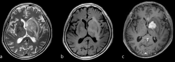
The number of brain MRI with contrast media performed in patients with cognitive impairment has increased without universal agreement. We aimed to evaluate the detection rate of contrast-enhanced brain MRI in patients with cognitive impairment.

MATERIALS AND METHODS

This single-institution, retrospective study included 4,838 patients who attended outpatient clinics for cognitive impairment evaluation and underwent brain MRI with or without contrast enhancement from December 2015 to February 2020. Patients who tested positive for cognitive impairment were followed-up to confirm whether the result was true-positive and provide follow-up management. Detection rate was defined as the proportion of patients with true-positive results and was compared between groups with and without contrast enhancement. Individual matching in a 1:2 ratio according to age, sex, and year of test was performed.

RESULTS

The overall detection rates of brain MRI with and without contrast media were 4.7% (57/1,203; 95% CI: 3.6%-6.1%) and 1.8% (65/3,635; 95% CI: 1.4%-2.3%), respectively (P<0.001); individual matching demonstrated similar results (4.7% and 1.9%). Among 1,203 patients with contrast media, 3.6% was only detectable with the aid of contrast media. The proportion of patients who underwent follow-up imaging or treatment for the detected lesions were significantly higher in the group with contrast media (2.0% and 0.6%, P < .001).

CONCLUSIONS

Detection rate of brain MRI for lesions only detectable with contrast media in patients with cognitive impairment was not high enough and further study is needed to identify whom would truly benefit with contrast media.

摘要

简介

在认知障碍患者中进行增强对比剂脑 MRI 的数量有所增加,但尚未达成普遍共识。我们旨在评估认知障碍患者增强对比剂脑 MRI 的检出率。

材料与方法

这是一项单中心、回顾性研究,纳入了 2015 年 12 月至 2020 年 2 月在门诊就诊评估认知障碍并接受 MRI 检查(有无增强对比剂)的 4838 例患者。对认知障碍检测结果阳性的患者进行随访,以确认结果是否为真阳性,并提供随访管理。检出率定义为真阳性结果患者的比例,并比较有无增强对比剂的两组间的检出率。按照年龄、性别和检查年份进行 1:2 个体匹配。

结果

有和无增强对比剂的脑 MRI 的总体检出率分别为 4.7%(57/1203;95%CI:3.6%-6.1%)和 1.8%(65/3635;95%CI:1.4%-2.3%)(P<0.001);个体匹配也得到了相似的结果(4.7%和 1.9%)。在 1203 例使用增强对比剂的患者中,仅 3.6%的病灶可通过增强对比剂检出。在有增强对比剂组中,接受检出病灶的随访影像学或治疗的患者比例显著更高(2.0%和 0.6%,P<.001)。

结论

认知障碍患者中仅能通过增强对比剂检出的病灶的脑 MRI 检出率不高,需要进一步研究以确定哪些患者真正受益于增强对比剂。

https://cdn.ncbi.nlm.nih.gov/pmc/blobs/3975/10406288/036788aba53f/pone.0289638.g004.jpg
https://cdn.ncbi.nlm.nih.gov/pmc/blobs/3975/10406288/7cfc45a38cc1/pone.0289638.g001.jpg
https://cdn.ncbi.nlm.nih.gov/pmc/blobs/3975/10406288/9d863cff86a4/pone.0289638.g002.jpg
https://cdn.ncbi.nlm.nih.gov/pmc/blobs/3975/10406288/7f7d900b9e70/pone.0289638.g003.jpg
https://cdn.ncbi.nlm.nih.gov/pmc/blobs/3975/10406288/036788aba53f/pone.0289638.g004.jpg

相似文献

1
Detection rate of contrast-enhanced brain magnetic resonance imaging in patients with cognitive impairment.认知障碍患者对比增强脑磁共振成像的检出率。
PLoS One. 2023 Aug 7;18(8):e0289638. doi: 10.1371/journal.pone.0289638. eCollection 2023.
2
From Dose Reduction to Contrast Maximization: Can Deep Learning Amplify the Impact of Contrast Media on Brain Magnetic Resonance Image Quality? A Reader Study.从降低剂量到最大化对比:深度学习能否增强对比剂对脑磁共振图像质量的影响?一项读者研究。
Invest Radiol. 2022 Aug 1;57(8):527-535. doi: 10.1097/RLI.0000000000000867. Epub 2022 Apr 20.
3
Identification of multiple sclerosis patients at highest risk of cognitive impairment using an integrated brain magnetic resonance imaging assessment approach.采用综合脑磁共振成像评估方法识别认知障碍风险最高的多发性硬化症患者。
Eur J Neurol. 2017 Feb;24(2):292-301. doi: 10.1111/ene.13200. Epub 2016 Nov 22.
4
Contrast-enhanced ultrasound using SonoVue® (sulphur hexafluoride microbubbles) compared with contrast-enhanced computed tomography and contrast-enhanced magnetic resonance imaging for the characterisation of focal liver lesions and detection of liver metastases: a systematic review and cost-effectiveness analysis.超声造影使用声诺维®(六氟化硫微泡)与对比增强计算机断层扫描和对比增强磁共振成像在局灶性肝脏病变的特征描述和肝转移检测中的比较:系统评价和成本效益分析。
Health Technol Assess. 2013 Apr;17(16):1-243. doi: 10.3310/hta17160.
5
Does focal inflammation have an impact on cognition in multiple sclerosis? An MRI study.局灶性炎症对多发性硬化症患者的认知功能有影响吗?一项 MRI 研究。
Mult Scler Relat Disord. 2018 Jul;23:83-87. doi: 10.1016/j.msard.2018.05.012. Epub 2018 May 16.
6
Deep-learning-based synthesis of post-contrast T1-weighted MRI for tumour response assessment in neuro-oncology: a multicentre, retrospective cohort study.基于深度学习的对比后 T1 加权 MRI 合成用于神经肿瘤学中的肿瘤反应评估:一项多中心、回顾性队列研究。
Lancet Digit Health. 2021 Dec;3(12):e784-e794. doi: 10.1016/S2589-7500(21)00205-3. Epub 2021 Oct 20.
7
Accuracy of Unenhanced MRI in the Detection of New Brain Lesions in Multiple Sclerosis.磁共振非增强扫描对多发性硬化症中新发脑损害的检测准确性。
Radiology. 2019 May;291(2):429-435. doi: 10.1148/radiol.2019181568. Epub 2019 Mar 12.
8
Structural magnetic resonance imaging for the early diagnosis of dementia due to Alzheimer's disease in people with mild cognitive impairment.用于轻度认知障碍患者阿尔茨海默病所致痴呆早期诊断的结构磁共振成像
Cochrane Database Syst Rev. 2020 Mar 2;3(3):CD009628. doi: 10.1002/14651858.CD009628.pub2.
9
Comparison between MRI and CEUS in the follow-up of patients with blunt abdominal trauma managed conservatively.磁共振成像(MRI)与超声造影(CEUS)在保守治疗的钝性腹部创伤患者随访中的比较。
Radiol Med. 2016 Jan;121(1):27-37. doi: 10.1007/s11547-015-0578-1. Epub 2015 Aug 8.
10
Pigmented villonodular synovitis (PVNS) of the knee joint: magnetic resonance imaging (MRI) using standard and dynamic paramagnetic contrast media. Report of 52 cases surgically and histologically controlled.膝关节色素沉着绒毛结节性滑膜炎(PVNS):使用标准和动态顺磁性造影剂的磁共振成像(MRI)。52例手术及组织学对照报告。
Radiol Med. 2004 Apr;107(4):356-66.

本文引用的文献

1
Use of "Diagnostic Yield" in Imaging Research Reports: Results from Articles Published in Two General Radiology Journals.影像学研究报告中“诊断收益”的使用:两篇普通放射学期刊文章的结果。
Korean J Radiol. 2022 Dec;23(12):1290-1300. doi: 10.3348/kjr.2022.0741.
2
Gadolinium Deposition and Retention in the Brain: Should We Be Concerned?钆在大脑中的沉积与滞留:我们应该担心吗?
Radiol Cardiothorac Imaging. 2019 Aug 29;1(3):e190104. doi: 10.1148/ryct.2019190104. eCollection 2019 Aug.
3
ACR Appropriateness Criteria® Dementia.美国放射学会适宜性标准® 痴呆症
J Am Coll Radiol. 2020 May;17(5S):S100-S112. doi: 10.1016/j.jacr.2020.01.040.
4
Incidental findings on brain imaging and blood tests: results from the first phase of Insight 46, a prospective observational substudy of the 1946 British birth cohort.脑成像和血液检查的偶然发现:来自 Insight 46 第一阶段的结果,这是对 1946 年英国出生队列进行的前瞻性观察性子研究。
BMJ Open. 2019 Jul 31;9(7):e029502. doi: 10.1136/bmjopen-2019-029502.
5
Acute Adverse Events Following Gadolinium-based Contrast Agent Administration: A Single-Center Retrospective Study of 281 945 Injections.基于钆造影剂的急性不良反应:281945 次注射的单中心回顾性研究。
Radiology. 2019 Sep;292(3):620-627. doi: 10.1148/radiol.2019182834. Epub 2019 Jul 2.
6
Potentially serious incidental findings on brain and body magnetic resonance imaging of apparently asymptomatic adults: systematic review and meta-analysis.脑和身体磁共振成像在貌似无症状成人中发现的潜在严重偶发结果:系统评价和荟萃分析。
BMJ. 2018 Nov 22;363:k4577. doi: 10.1136/bmj.k4577.
7
Impact of detecting potentially serious incidental findings during multi-modal imaging.多模态成像期间检测到潜在严重偶然发现的影响。
Wellcome Open Res. 2017 Nov 30;2:114. doi: 10.12688/wellcomeopenres.13181.3. eCollection 2017.
8
Prevalence and outcomes of incidental imaging findings: umbrella review.偶然影像学发现的患病率和结局:伞式综述。
BMJ. 2018 Jun 18;361:k2387. doi: 10.1136/bmj.k2387.
9
Incidental findings in research: A focus group study about the perspective of the research participant.研究中的偶发发现:研究参与者视角的焦点小组研究。
J Magn Reson Imaging. 2018 Jan;47(1):230-237. doi: 10.1002/jmri.25739. Epub 2017 May 4.
10
Prevalence, Clinical Management, and Natural Course of Incidental Findings on Brain MR Images: The Population-based Rotterdam Scan Study.脑磁共振成像偶然发现的患病率、临床管理和自然病程:基于人群的鹿特丹扫描研究。
Radiology. 2016 Nov;281(2):507-515. doi: 10.1148/radiol.2016160218. Epub 2016 Jun 23.