School of Biomedical Sciences and Pharmacy, College of Health, Medicine and Wellbeing, The University of Newcastle, Callaghan, NSW, Australia.
Hunter Medical Research Institute, New Lambton, NSW, Australia.
Cell Death Dis. 2023 Aug 8;14(8):509. doi: 10.1038/s41419-023-06031-4.
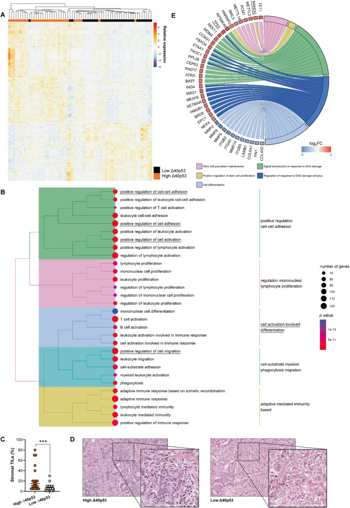
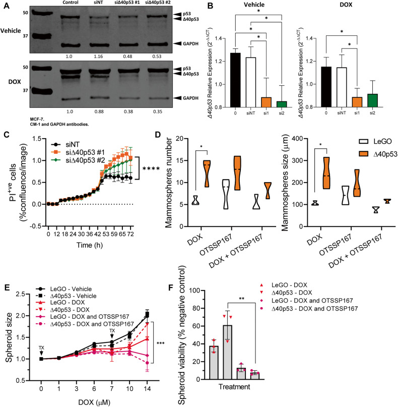
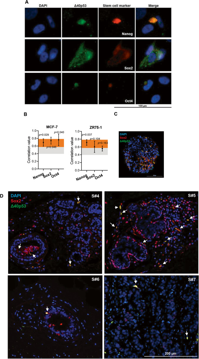
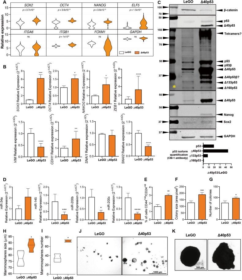
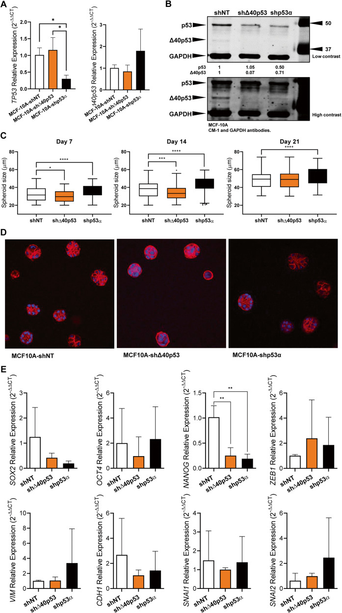
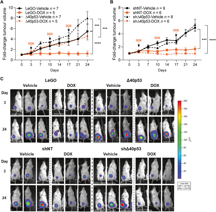
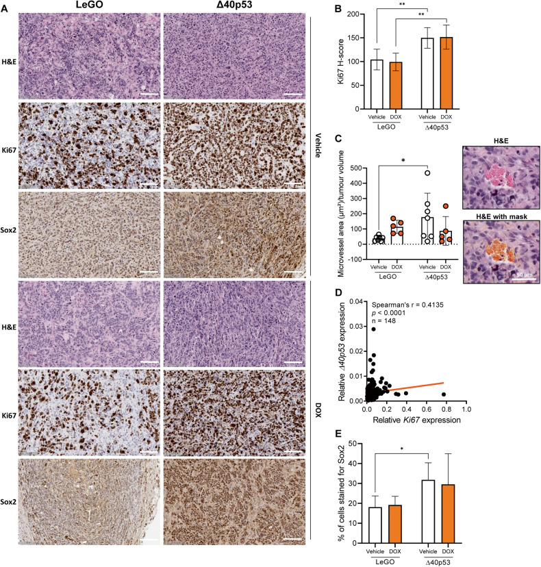
In breast cancer, dysregulated TP53 expression signatures are a better predictor of chemotherapy response and survival outcomes than TP53 mutations. Our previous studies have shown that high levels of Δ40p53 are associated with worse disease-free survival and disruption of p53-induced DNA damage response in breast cancers. Here, we further investigated the in vitro and in vivo implications of Δ40p53 expression in breast cancer. We have shown that genes associated with cell differentiation are downregulated while those associated with stem cell regulation are upregulated in invasive ductal carcinomas expressing high levels of Δ40p53. In contrast to p53, endogenous ∆40p53 co-localised with the stem cell markers Sox2, Oct4, and Nanog in MCF-7 and ZR75-1 cell lines. ∆40p53 and Sox2 co-localisation was also detected in breast cancer specimens. Further, in cells expressing a high ∆40p53:p53 ratio, increased expression of stem cell markers, greater mammosphere and colony formation capacities, and downregulation of miR-145 and miR-200 (p53-target microRNAs that repress stemness) were observed compared to the control subline. In vivo, a high ∆40p53:p53 ratio led to increased tumour growth, Ki67 and Sox2 expression, and blood microvessel areas in the vehicle-treated mice. High expression of ∆40p53 also reduced tumour sensitivity to doxorubicin compared to control tumours. Enhanced therapeutic efficacy of doxorubicin was observed when transiently targeting Δ40p53 or when treating cells with OTSSP167 with concomitant chemotherapy. Taken together, high Δ40p53 levels induce tumour growth and may promote chemoresistance by inducing a stemness phenotype in breast cancer; thus, targeting Δ40p53 in tumours that have a high Δ40p53:p53 ratio could enhance the efficacy of standard-of-care therapies such as doxorubicin.
在乳腺癌中,失调的 TP53 表达谱比 TP53 突变更能预测化疗反应和生存结果。我们之前的研究表明,高水平的 Δ40p53 与乳腺癌无病生存率较差和 p53 诱导的 DNA 损伤反应破坏有关。在这里,我们进一步研究了 Δ40p53 在乳腺癌中的体外和体内意义。我们已经表明,在表达高水平 Δ40p53 的浸润性导管癌中,与细胞分化相关的基因下调,而与干细胞调节相关的基因上调。与 p53 不同,内源性 ∆40p53 与 MCF-7 和 ZR75-1 细胞系中的干细胞标记物 Sox2、Oct4 和 Nanog 共定位。在乳腺癌标本中也检测到 ∆40p53 和 Sox2 的共定位。此外,在表达高 ∆40p53:p53 比值的细胞中,与对照亚系相比,观察到干细胞标记物的表达增加、更大的乳腺球体和集落形成能力、miR-145 和 miR-200(抑制干性的 p53 靶标 microRNAs)的下调。在体内,高 ∆40p53:p53 比值导致荷瘤小鼠肿瘤生长、Ki67 和 Sox2 表达以及血液微血管面积增加。与对照肿瘤相比,Δ40p53 高表达还降低了肿瘤对阿霉素的敏感性。与对照肿瘤相比,当瞬时靶向 Δ40p53 或用 OTSSP167 联合化疗治疗细胞时,观察到阿霉素的治疗效果增强。综上所述,高水平的 Δ40p53 诱导肿瘤生长,并可能通过在乳腺癌中诱导干性表型来促进化疗耐药性;因此,在具有高 Δ40p53:p53 比值的肿瘤中靶向 Δ40p53 可能增强阿霉素等标准治疗方法的疗效。