M Sandra C, Peethambar Breman A
Diagnostic Radiology, Muslim Educational Society (MES) Medical College, Perinthalmanna, IND.
Medicine, Madras Medical College, Chennai, IND.
Cureus. 2023 Jul 8;15(7):e41550. doi: 10.7759/cureus.41550. eCollection 2023 Jul.
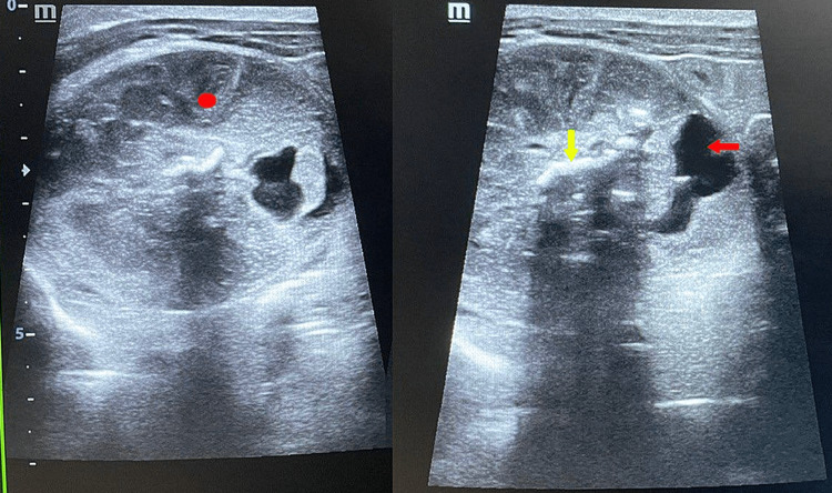
Fetus in fetu (FIF) is a rare congenital anomaly with two controversial theories regarding its embryogenesis. Although it is an extremely rare condition, it should be kept in mind as a differential diagnosis while evaluating children with abdominal calcification. Radiological findings on postnatal days 2 and 5 of a neonate with an antenatal scan showing an abdominal mass in the fetus are described here. Ultrasonography and magnetic resonance imaging (MRI) revealed the mass in which the contents favored a diagnosis of the FIF. Characteristic features of FIF on MRI have been less explored and knowledge regarding the same will be of immense help to the radiologist. Complete surgical excision followed by histopathology confirmed the diagnosis.
胎内胎(FIF)是一种罕见的先天性异常,关于其胚胎发生有两种存在争议的理论。尽管这是一种极其罕见的病症,但在评估有腹部钙化的儿童时,应将其作为鉴别诊断予以考虑。本文描述了一名产前扫描显示胎儿腹部有肿块的新生儿出生后第2天和第5天的影像学表现。超声检查和磁共振成像(MRI)显示了一个肿块,其内容物支持胎内胎的诊断。MRI上胎内胎的特征性表现研究较少,了解这些知识将对放射科医生有极大帮助。完整的手术切除并经组织病理学检查确诊。