Phan Alexander T, Hu Janie, Oganesian Buzand, Williams Shammah O
Department of Internal Medicine, Arrowhead Regional Medical Center, Colton, CA 92324, USA.
Department of Cardiology, Arrowhead Regional Medical Center, Colton, CA 92324, USA.
Cardiol Res. 2023 Aug;14(4):315-318. doi: 10.14740/cr1511. Epub 2023 Jul 12.
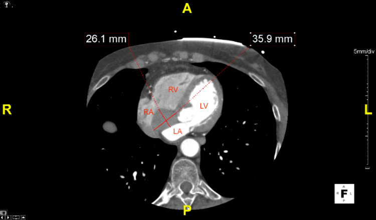
Intracardiac bronchogenic cysts are extremely rare congenital anomalies that arise during foregut development when the embryologic heart tube and ventral foregut are in close proximity to one another. We report a case of an interatrial septal bronchogenic cyst found on non-contrast enhanced computed tomography (CT) in a 66-year-old female who presented to the emergency department with chest pain. Further cardiac investigations, including contrast-enhanced CT angiogram of the heart, transthoracic echocardiogram, and transesophageal echocardiogram, revealed a cystic mass in the lipomatous interatrial septum. The patient was subsequently diagnosed with a bronchogenic cyst of the interatrial septum. No surgical intervention was pursued, as the mass remained stable, and the cardiothoracic surgeon did not recommend excision. This case highlights a rare case of a symptomatic bronchogenic cyst arising in the interatrial septum diagnosed by imaging modalities. Bronchogenic cysts should be included in the differential diagnosis of intracardiac tumors.
心内支气管源性囊肿是极其罕见的先天性异常,在前肠发育过程中,当胚胎心脏管和腹侧前肠彼此紧邻时出现。我们报告一例66岁女性,因胸痛就诊于急诊科,在非增强计算机断层扫描(CT)上发现房间隔支气管源性囊肿。进一步的心脏检查,包括心脏增强CT血管造影、经胸超声心动图和经食管超声心动图,显示脂肪性房间隔内有一个囊性肿块。该患者随后被诊断为房间隔支气管源性囊肿。由于肿块保持稳定,心胸外科医生不建议切除,因此未进行手术干预。本病例突出了通过影像学检查诊断出的一例罕见的有症状的房间隔支气管源性囊肿。支气管源性囊肿应纳入心内肿瘤的鉴别诊断。