• 文献检索
  • 文档翻译
  • 深度研究
  • 学术资讯
  • Suppr Zotero 插件Zotero 插件
  • 邀请有礼
  • 套餐&价格
  • 历史记录
应用&插件
Suppr Zotero 插件Zotero 插件浏览器插件Mac 客户端Windows 客户端微信小程序
定价
高级版会员购买积分包购买API积分包
服务
文献检索文档翻译深度研究API 文档MCP 服务
关于我们
关于 Suppr公司介绍联系我们用户协议隐私条款
关注我们

Suppr 超能文献

核心技术专利:CN118964589B侵权必究
粤ICP备2023148730 号-1Suppr @ 2026

文献检索

告别复杂PubMed语法,用中文像聊天一样搜索,搜遍4000万医学文献。AI智能推荐,让科研检索更轻松。

立即免费搜索

文件翻译

保留排版,准确专业,支持PDF/Word/PPT等文件格式,支持 12+语言互译。

免费翻译文档

深度研究

AI帮你快速写综述,25分钟生成高质量综述,智能提取关键信息,辅助科研写作。

立即免费体验

猕猴额叶的细胞构筑、受体分布和功能连接分析。

Cytoarchitectonic, receptor distribution and functional connectivity analyses of the macaque frontal lobe.

机构信息

Institute of Neuroscience and Medicine INM-1, Research Centre Jülich, Jülich, Germany.

Center for Neural Science, New York University, New York, United States.

出版信息

Elife. 2023 Aug 14;12:e82850. doi: 10.7554/eLife.82850.

DOI:10.7554/eLife.82850
PMID:37578332
原文链接:https://pmc.ncbi.nlm.nih.gov/articles/PMC10425179/
Abstract

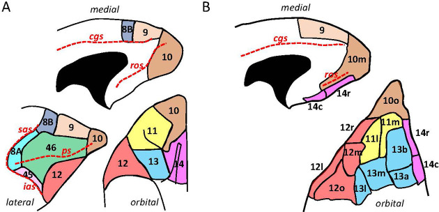
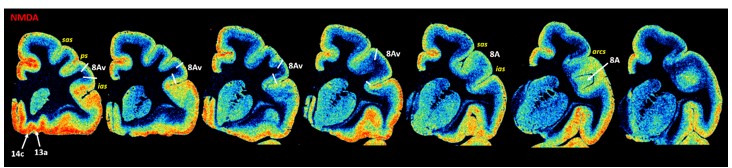
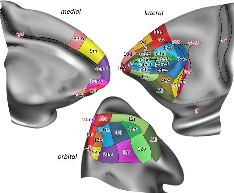
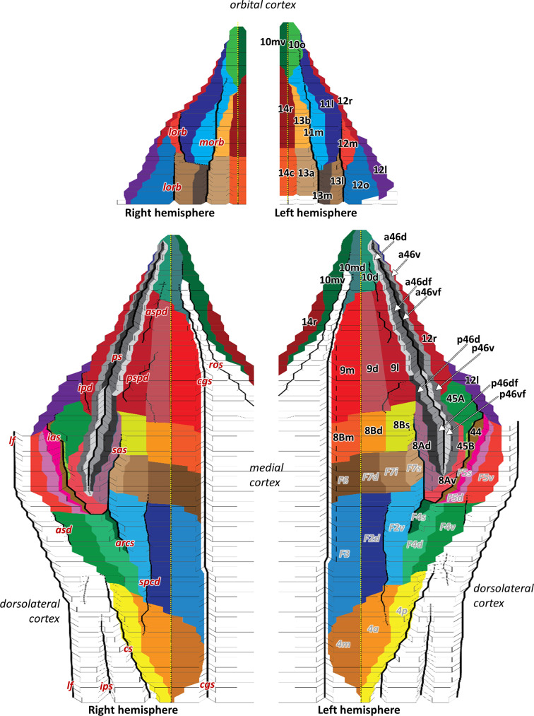
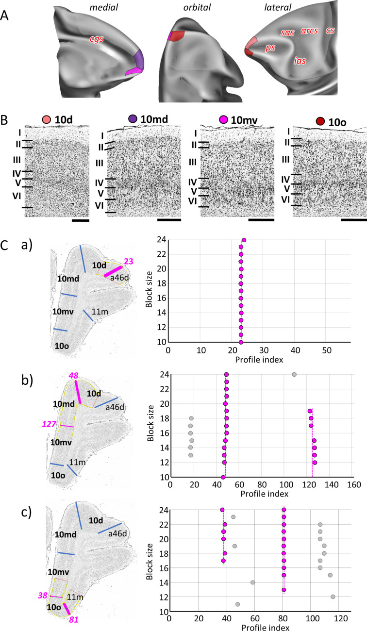
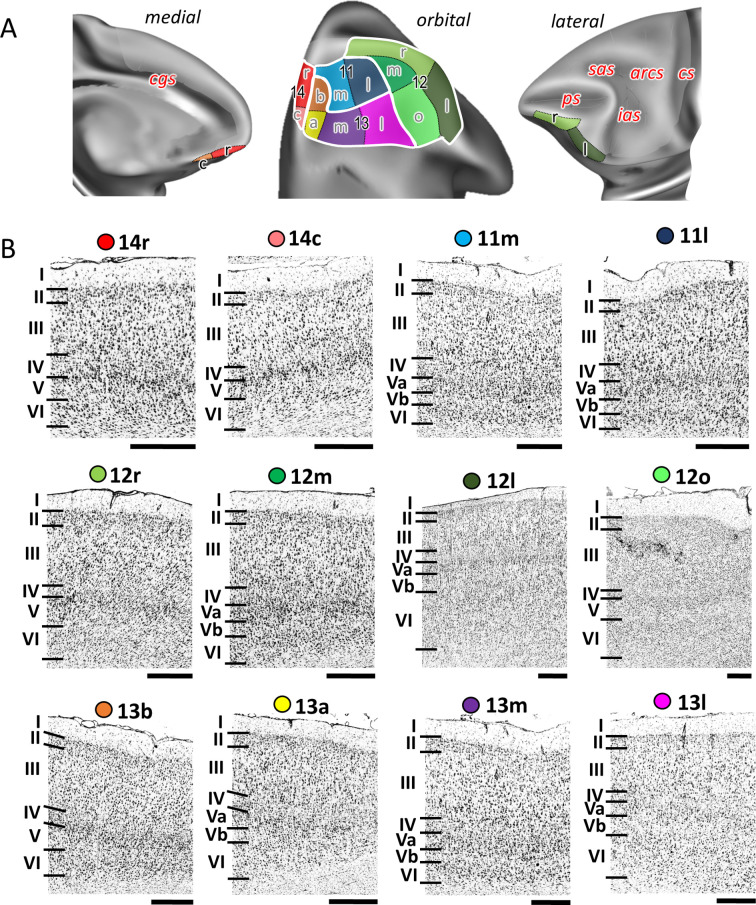
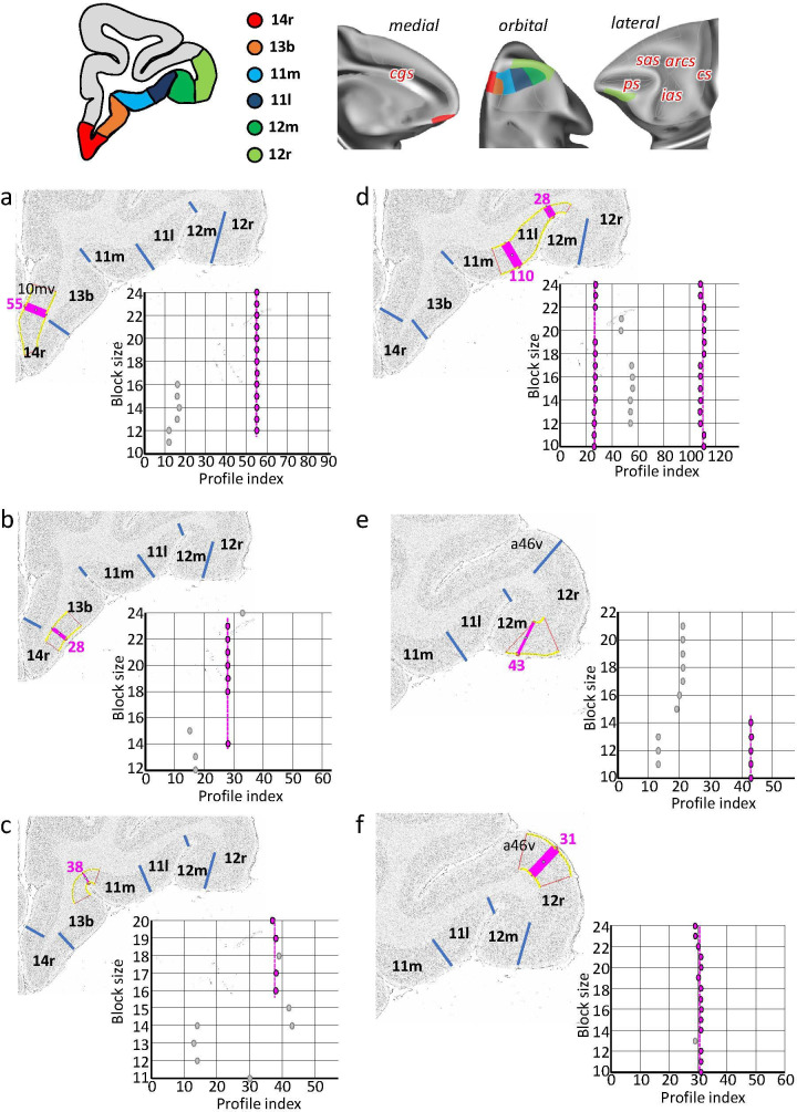
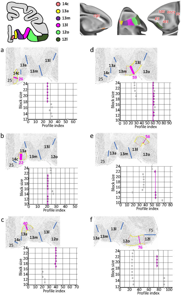
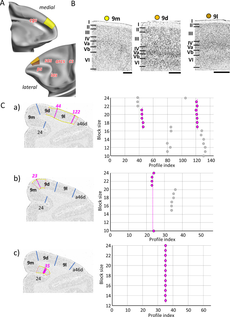
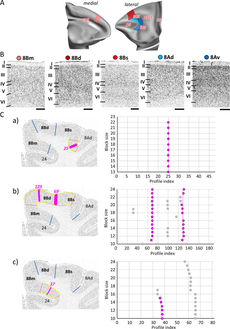
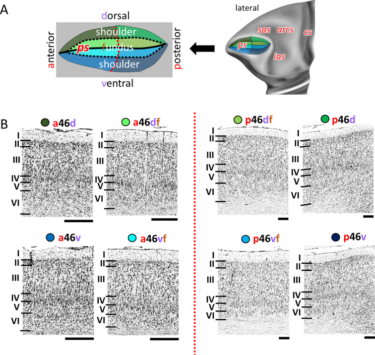
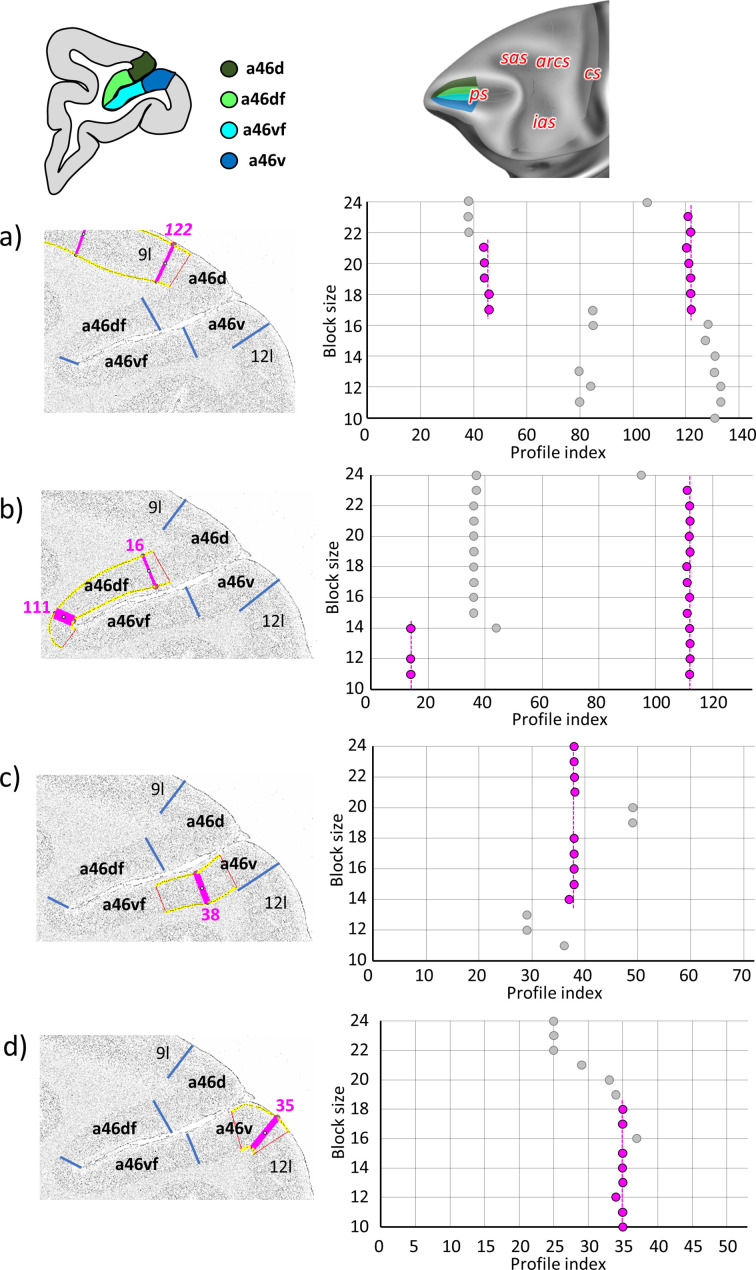
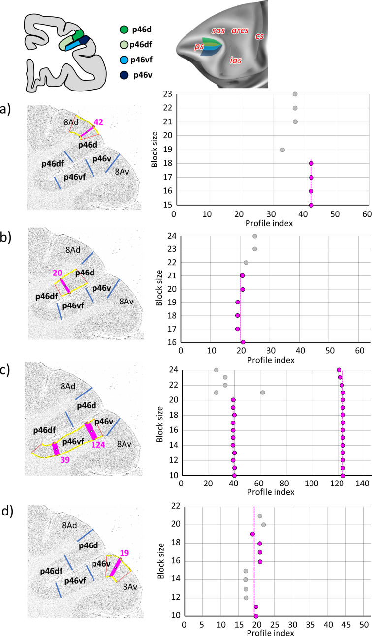
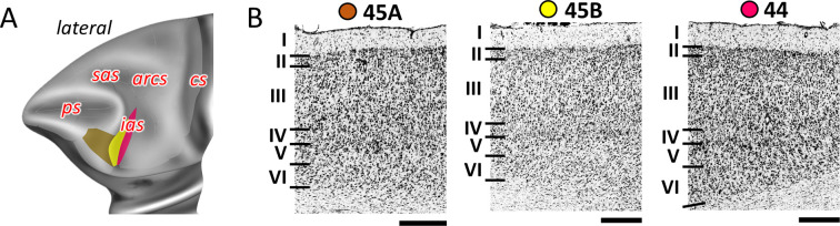
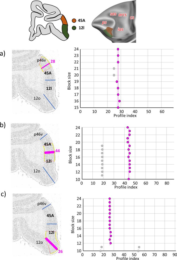
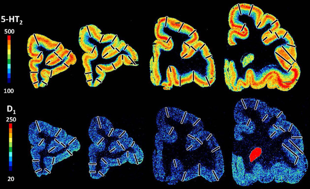
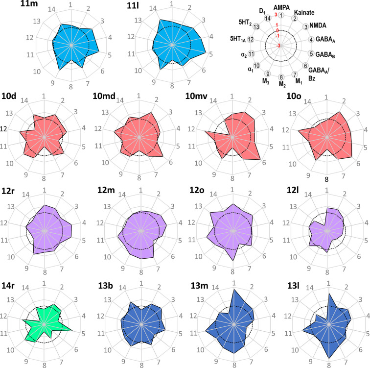
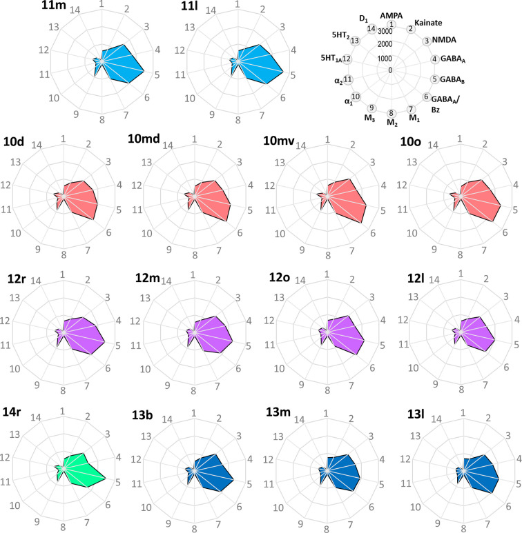
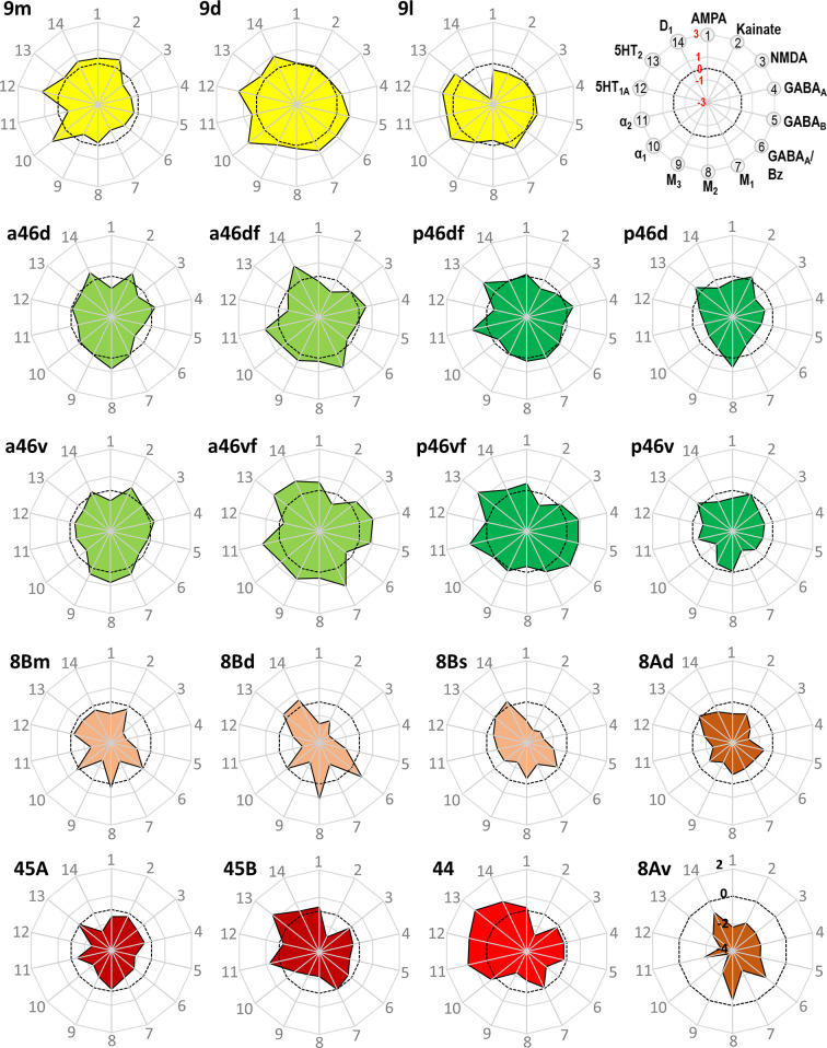
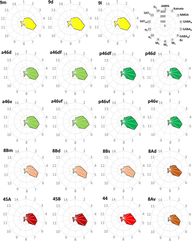
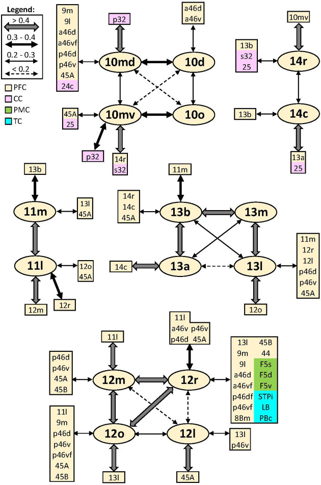
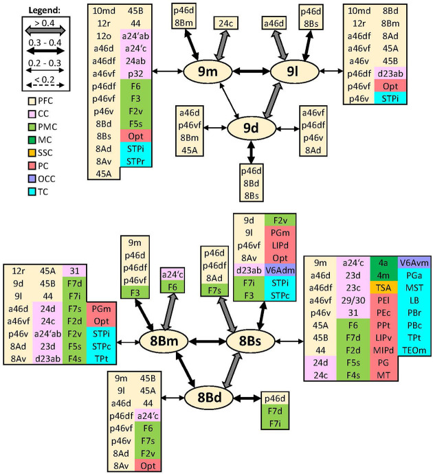
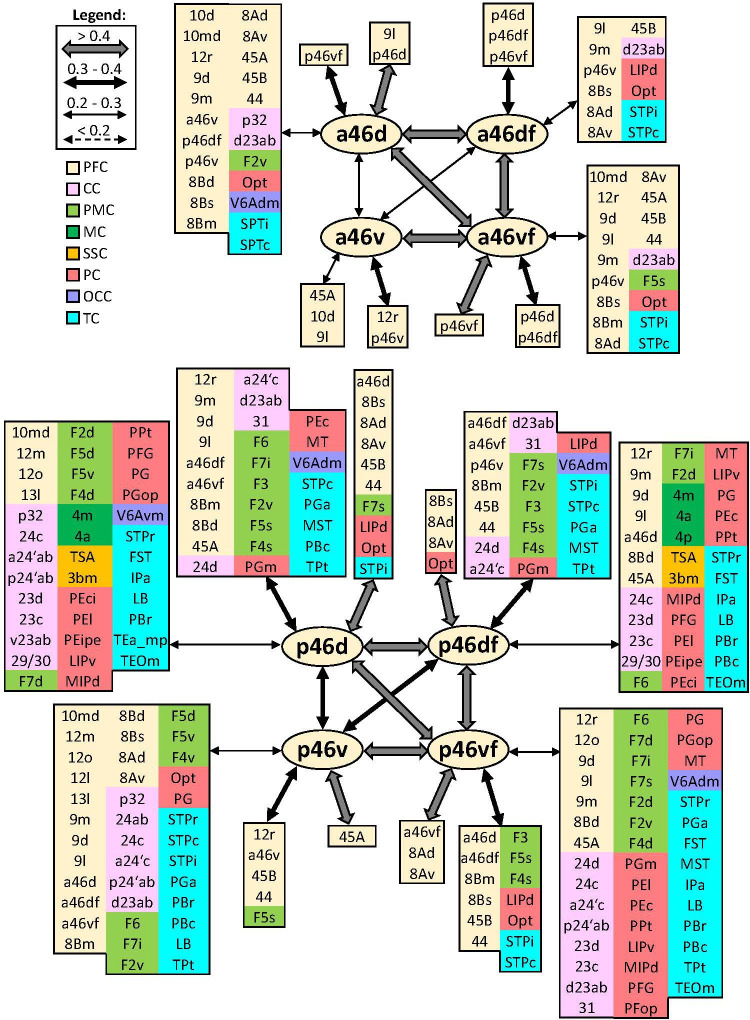
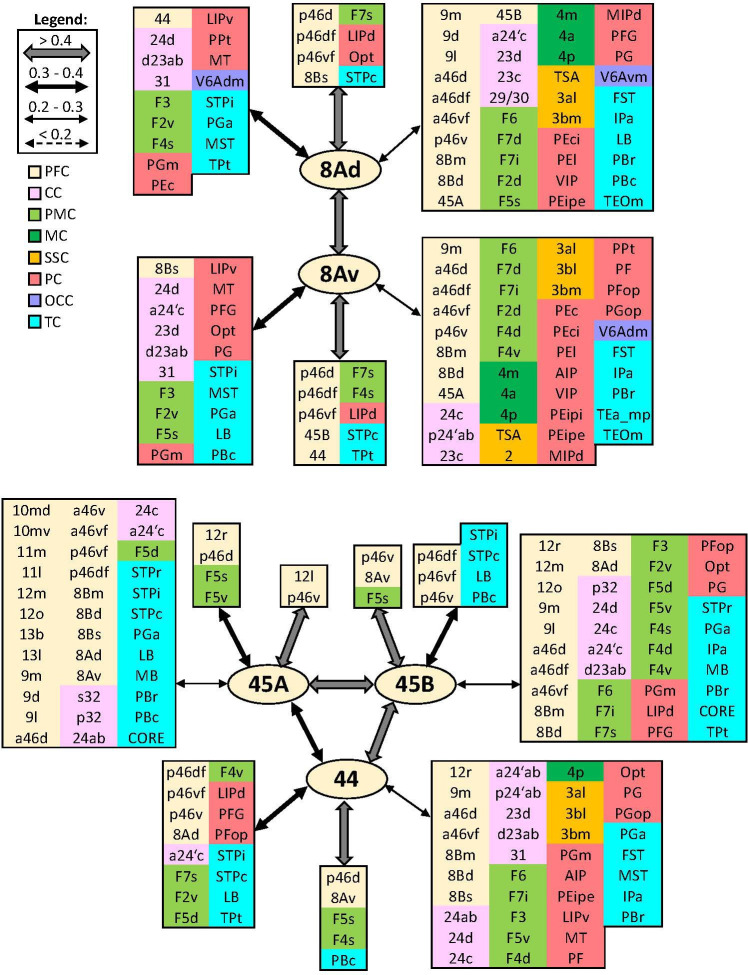
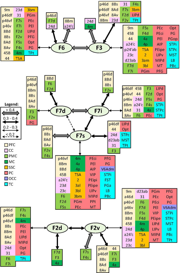
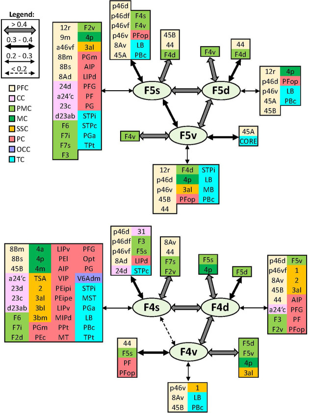
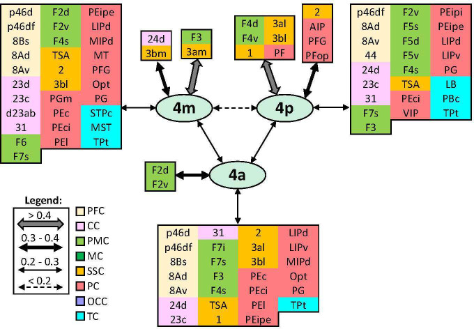
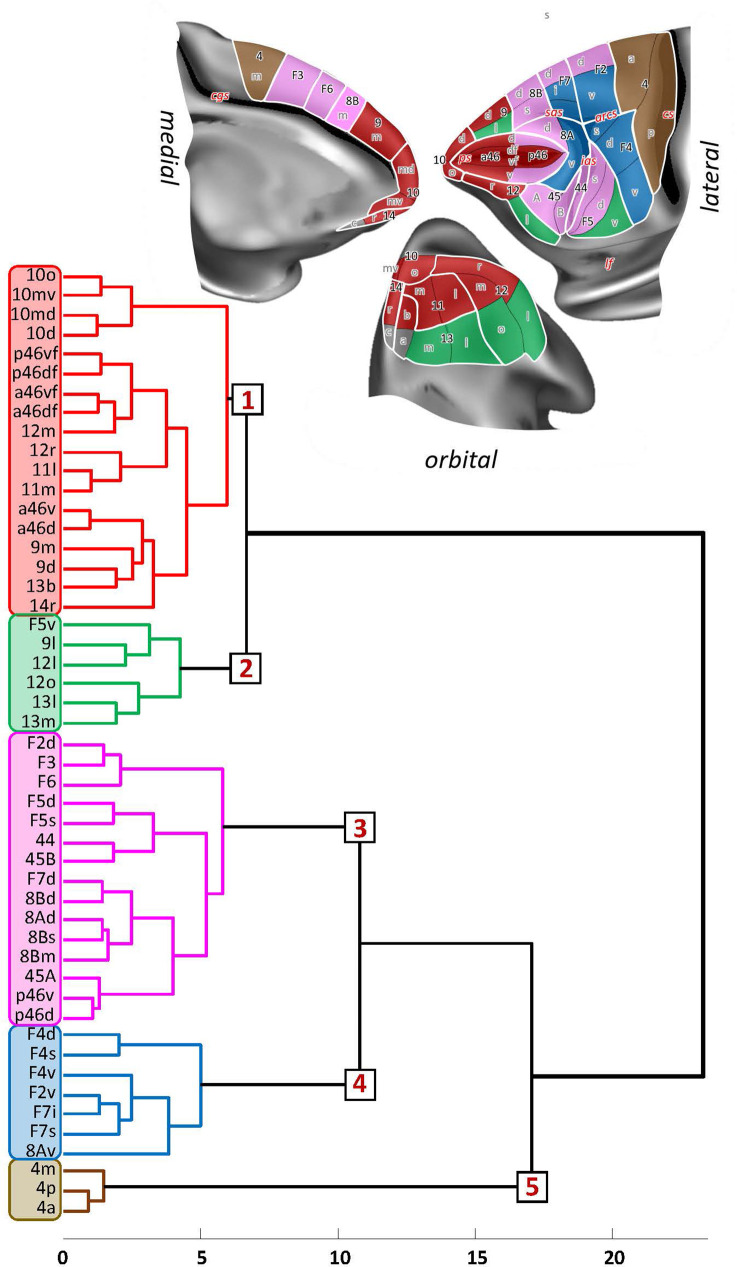
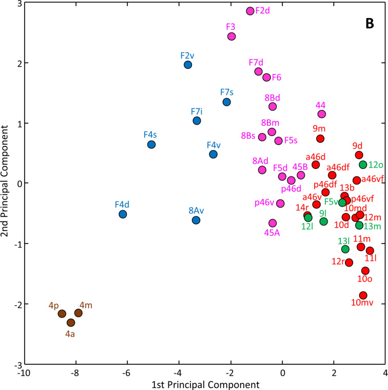
Based on quantitative cyto- and receptor architectonic analyses, we identified 35 prefrontal areas, including novel subdivisions of Walker's areas 10, 9, 8B, and 46. Statistical analysis of receptor densities revealed regional differences in lateral and ventrolateral prefrontal cortex. Indeed, structural and functional organization of subdivisions encompassing areas 46 and 12 demonstrated significant differences in the interareal levels of α receptors. Furthermore, multivariate analysis included receptor fingerprints of previously identified 16 motor areas in the same macaque brains and revealed 5 clusters encompassing frontal lobe areas. We used the MRI datasets from the non-human primate data sharing consortium PRIME-DE to perform functional connectivity analyses using the resulting frontal maps as seed regions. In general, rostrally located frontal areas were characterized by bigger fingerprints, that is, higher receptor densities, and stronger regional interconnections. Whereas more caudal areas had smaller fingerprints, but showed a widespread connectivity pattern with distant cortical regions. Taken together, this study provides a comprehensive insight into the molecular structure underlying the functional organization of the cortex and, thus, reconcile the discrepancies between the structural and functional hierarchical organization of the primate frontal lobe. Finally, our data are publicly available via the EBRAINS and BALSA repositories for the entire scientific community.

摘要

基于定量细胞和受体构筑分析,我们确定了 35 个前额叶区域,包括沃克氏区 10、9、8B 和 46 的新细分区域。受体密度的统计分析显示外侧和腹外侧前额叶皮层存在区域差异。事实上,包括区域 46 和 12 在内的细分区域的结构和功能组织在α受体的区域间水平上表现出显著差异。此外,多元分析包括同一猕猴大脑中先前确定的 16 个运动区域的受体指纹,并揭示了包含额叶区域的 5 个簇。我们使用非人类灵长类动物数据共享联盟 PRIME-DE 的 MRI 数据集,使用生成的额叶图谱作为种子区域进行功能连接分析。一般来说,位于前部的区域具有更大的指纹,即更高的受体密度和更强的区域内连接。而更靠后的区域具有较小的指纹,但与远处的皮质区域显示出广泛的连接模式。总的来说,这项研究提供了对皮层功能组织背后的分子结构的全面了解,从而调和了灵长类动物额叶的结构和功能层次组织之间的差异。最后,我们的数据通过 EBRAINS 和 BALSA 存储库向整个科学界公开。

https://cdn.ncbi.nlm.nih.gov/pmc/blobs/7a8a/10425179/a2dd9c8639c6/elife-82850-sa2-fig1.jpg
https://cdn.ncbi.nlm.nih.gov/pmc/blobs/7a8a/10425179/0bf0a1237d57/elife-82850-fig1.jpg
https://cdn.ncbi.nlm.nih.gov/pmc/blobs/7a8a/10425179/9d6ca79cafe3/elife-82850-fig2.jpg
https://cdn.ncbi.nlm.nih.gov/pmc/blobs/7a8a/10425179/aaad794bf243/elife-82850-fig2-figsupp1.jpg
https://cdn.ncbi.nlm.nih.gov/pmc/blobs/7a8a/10425179/662df40e5a99/elife-82850-fig2-figsupp2.jpg
https://cdn.ncbi.nlm.nih.gov/pmc/blobs/7a8a/10425179/9c9271303371/elife-82850-fig3.jpg
https://cdn.ncbi.nlm.nih.gov/pmc/blobs/7a8a/10425179/1af923cea7da/elife-82850-fig4.jpg
https://cdn.ncbi.nlm.nih.gov/pmc/blobs/7a8a/10425179/df38a8bedea5/elife-82850-fig4-figsupp1.jpg
https://cdn.ncbi.nlm.nih.gov/pmc/blobs/7a8a/10425179/7d66713fee65/elife-82850-fig4-figsupp2.jpg
https://cdn.ncbi.nlm.nih.gov/pmc/blobs/7a8a/10425179/7f9741ccabe9/elife-82850-fig5.jpg
https://cdn.ncbi.nlm.nih.gov/pmc/blobs/7a8a/10425179/1f76b32d7235/elife-82850-fig6.jpg
https://cdn.ncbi.nlm.nih.gov/pmc/blobs/7a8a/10425179/5b9b177345ed/elife-82850-fig7.jpg
https://cdn.ncbi.nlm.nih.gov/pmc/blobs/7a8a/10425179/e3a37cb0305f/elife-82850-fig7-figsupp1.jpg
https://cdn.ncbi.nlm.nih.gov/pmc/blobs/7a8a/10425179/695f4d0b5bef/elife-82850-fig7-figsupp2.jpg
https://cdn.ncbi.nlm.nih.gov/pmc/blobs/7a8a/10425179/930830669bf9/elife-82850-fig8.jpg
https://cdn.ncbi.nlm.nih.gov/pmc/blobs/7a8a/10425179/8bf7341d70df/elife-82850-fig8-figsupp1.jpg
https://cdn.ncbi.nlm.nih.gov/pmc/blobs/7a8a/10425179/4e20d8f7072c/elife-82850-fig8-figsupp2.jpg
https://cdn.ncbi.nlm.nih.gov/pmc/blobs/7a8a/10425179/2cfc394b6d96/elife-82850-fig9.jpg
https://cdn.ncbi.nlm.nih.gov/pmc/blobs/7a8a/10425179/307874654600/elife-82850-fig9-figsupp1.jpg
https://cdn.ncbi.nlm.nih.gov/pmc/blobs/7a8a/10425179/b925bce4725b/elife-82850-fig9-figsupp2.jpg
https://cdn.ncbi.nlm.nih.gov/pmc/blobs/7a8a/10425179/821d40ca92f8/elife-82850-fig9-figsupp3.jpg
https://cdn.ncbi.nlm.nih.gov/pmc/blobs/7a8a/10425179/04c448e34673/elife-82850-fig10.jpg
https://cdn.ncbi.nlm.nih.gov/pmc/blobs/7a8a/10425179/b7e56b0e7768/elife-82850-fig10-figsupp1.jpg
https://cdn.ncbi.nlm.nih.gov/pmc/blobs/7a8a/10425179/454a7e6be472/elife-82850-fig11.jpg
https://cdn.ncbi.nlm.nih.gov/pmc/blobs/7a8a/10425179/25c802328894/elife-82850-fig11-figsupp1.jpg
https://cdn.ncbi.nlm.nih.gov/pmc/blobs/7a8a/10425179/c12836265d04/elife-82850-fig12.jpg
https://cdn.ncbi.nlm.nih.gov/pmc/blobs/7a8a/10425179/3b9b47a9ad35/elife-82850-fig13.jpg
https://cdn.ncbi.nlm.nih.gov/pmc/blobs/7a8a/10425179/73a6abe7f63b/elife-82850-fig14.jpg
https://cdn.ncbi.nlm.nih.gov/pmc/blobs/7a8a/10425179/2ac86d94a2f4/elife-82850-fig15.jpg
https://cdn.ncbi.nlm.nih.gov/pmc/blobs/7a8a/10425179/f45410379bb6/elife-82850-fig16.jpg
https://cdn.ncbi.nlm.nih.gov/pmc/blobs/7a8a/10425179/b314e71ec29f/elife-82850-fig17.jpg
https://cdn.ncbi.nlm.nih.gov/pmc/blobs/7a8a/10425179/cae4cabeb496/elife-82850-fig18.jpg
https://cdn.ncbi.nlm.nih.gov/pmc/blobs/7a8a/10425179/5d14d5f655c3/elife-82850-fig19.jpg
https://cdn.ncbi.nlm.nih.gov/pmc/blobs/7a8a/10425179/f5f07fe8cc43/elife-82850-fig20.jpg
https://cdn.ncbi.nlm.nih.gov/pmc/blobs/7a8a/10425179/a2dd9c8639c6/elife-82850-sa2-fig1.jpg

相似文献

1
Cytoarchitectonic, receptor distribution and functional connectivity analyses of the macaque frontal lobe.猕猴额叶的细胞构筑、受体分布和功能连接分析。
Elife. 2023 Aug 14;12:e82850. doi: 10.7554/eLife.82850.
2
Multimodal 3D atlas of the macaque monkey motor and premotor cortex.猕猴运动和前运动皮层的多模态 3D 图谱。
Neuroimage. 2021 Feb 1;226:117574. doi: 10.1016/j.neuroimage.2020.117574. Epub 2020 Nov 20.
3
Connectivity-based parcellation of the macaque frontal cortex, and its relation with the cytoarchitectonic distribution described in current atlases.基于连接性的猕猴额叶皮层分区及其与当前图谱中细胞构筑分布的关系。
Brain Struct Funct. 2017 Apr;222(3):1331-1349. doi: 10.1007/s00429-016-1280-3. Epub 2016 Jul 28.
4
Medial to lateral frontal functional connectivity mapping reveals the organization of cingulate cortex.从中线到外侧额部的功能连接图揭示了扣带皮层的组织。
Cereb Cortex. 2024 Aug 1;34(8). doi: 10.1093/cercor/bhae322.
5
The prefrontal cortex: comparative architectonic organization in the human and the macaque monkey brains.前额皮质:人类和猕猴大脑的比较构筑组织。
Cortex. 2012 Jan;48(1):46-57. doi: 10.1016/j.cortex.2011.07.002. Epub 2011 Jul 29.
6
Mapping brain-wide excitatory projectome of primate prefrontal cortex at submicron resolution and comparison with diffusion tractography.以亚微米分辨率绘制灵长类前额叶皮层的全脑兴奋性投射组图谱,并与扩散轨迹进行比较。
Elife. 2022 May 20;11:e72534. doi: 10.7554/eLife.72534.
7
Divergence of rodent and primate medial frontal cortex functional connectivity.啮齿动物和灵长类动物内侧前额叶皮层功能连接的发散。
Proc Natl Acad Sci U S A. 2020 Sep 1;117(35):21681-21689. doi: 10.1073/pnas.2003181117. Epub 2020 Aug 17.
8
Comparative cytoarchitectonic analysis of the human and the macaque ventrolateral prefrontal cortex and corticocortical connection patterns in the monkey.人类与猕猴腹外侧前额叶皮层的比较细胞构筑分析以及猕猴的皮质-皮质连接模式
Eur J Neurosci. 2002 Jul;16(2):291-310. doi: 10.1046/j.1460-9568.2001.02090.x.
9
Broca's region: linking human brain functional connectivity data and non-human primate tracing anatomy studies.布罗卡区:连接人类大脑功能连接数据和非人类灵长类追踪解剖研究。
Eur J Neurosci. 2010 Aug;32(3):383-98. doi: 10.1111/j.1460-9568.2010.07279.x. Epub 2010 Jul 21.
10
The primate mediodorsal (MD) nucleus and its projection to the frontal lobe.灵长类动物的背内侧(MD)核及其向额叶的投射。
J Comp Neurol. 1985 Dec 22;242(4):535-60. doi: 10.1002/cne.902420406.

引用本文的文献

1
MEBRAINS 1.0: A new population-based macaque atlas.MEBRAINS 1.0:一种新的基于群体的猕猴脑图谱。
Imaging Neurosci (Camb). 2024 Feb 2;2. doi: 10.1162/imag_a_00077. eCollection 2024.
2
Consistent Hierarchies of Single-Neuron Timescales in Mice, Macaques, and Humans.小鼠、猕猴和人类中单个神经元时间尺度的一致层次结构。
J Neurosci. 2025 May 7;45(19):e2155242025. doi: 10.1523/JNEUROSCI.2155-24.2025.
3
Monkey Lateral Prefrontal Cortex Subregions Differentiate between Perceptual Exposure to Visual Stimuli.猕猴外侧前额叶皮质亚区域可区分对视觉刺激的感知暴露。

本文引用的文献

1
Receptor architecture of macaque and human early visual areas: not equal, but comparable.猕猴和人类早期视觉区域的受体结构:不平等,但可比较。
Brain Struct Funct. 2022 May;227(4):1247-1263. doi: 10.1007/s00429-021-02437-y. Epub 2021 Dec 20.
2
Organization of the macaque monkey inferior parietal lobule based on multimodal receptor architectonics.基于多模态受体构筑的猕猴下顶叶皮层组织。
Neuroimage. 2021 May 1;231:117843. doi: 10.1016/j.neuroimage.2021.117843. Epub 2021 Feb 10.
3
The genie in the bottle-magnified calcium signaling in dorsolateral prefrontal cortex.
J Cogn Neurosci. 2025 Apr 1;37(4):802-814. doi: 10.1162/jocn_a_02291.
4
Noradrenergic alpha-2a receptor stimulation enhances prediction error signaling and updating of attention sets in anterior cingulate cortex and striatum.去甲肾上腺素能α-2a 受体刺激增强了前扣带回皮层和纹状体中预测误差信号和注意力集的更新。
Nat Commun. 2024 Nov 15;15(1):9905. doi: 10.1038/s41467-024-54395-8.
5
Different Subregions of Monkey Lateral Prefrontal Cortex Respond to Abstract Sequences and Their Components.猴侧前额叶皮质的不同亚区对抽象序列及其成分作出反应。
J Neurosci. 2024 Nov 20;44(47):e1353242024. doi: 10.1523/JNEUROSCI.1353-24.2024.
6
Ventrolateral prefrontal cortex in macaques guides decisions in different learning contexts.猕猴的腹外侧前额叶皮层在不同学习情境中引导决策。
bioRxiv. 2024 Sep 19:2024.09.18.613767. doi: 10.1101/2024.09.18.613767.
7
Decision-making shapes dynamic inter-areal communication within macaque ventral frontal cortex.决策塑造猕猴腹侧前额叶皮层内的动态区域间通讯。
Curr Biol. 2024 Oct 7;34(19):4526-4538.e5. doi: 10.1016/j.cub.2024.08.034. Epub 2024 Sep 17.
8
Monkey lateral prefrontal cortex subregions differentiate between perceptual exposure to visual stimuli.猕猴外侧前额叶皮质亚区域可区分对视觉刺激的感知暴露。
bioRxiv. 2024 Aug 17:2024.07.28.605513. doi: 10.1101/2024.07.28.605513.
9
Decision-making shapes dynamic inter-areal communication within macaque ventral frontal cortex.决策塑造了猕猴腹侧前额叶皮质内动态的区域间交流。
bioRxiv. 2024 Jul 9:2024.07.05.602229. doi: 10.1101/2024.07.05.602229.
10
Dissociable Representations of Decision Variables within Subdivisions of the Macaque Orbital and Ventrolateral Frontal Cortex.猴脑眶额皮质和腹外侧前额皮质亚区对决策变量的可分离表示。
J Neurosci. 2024 Aug 28;44(35):e0464242024. doi: 10.1523/JNEUROSCI.0464-24.2024.
瓶中的精灵——放大背外侧前额叶皮层的钙信号。
Mol Psychiatry. 2021 Aug;26(8):3684-3700. doi: 10.1038/s41380-020-00973-3. Epub 2020 Dec 15.
4
Multimodal 3D atlas of the macaque monkey motor and premotor cortex.猕猴运动和前运动皮层的多模态 3D 图谱。
Neuroimage. 2021 Feb 1;226:117574. doi: 10.1016/j.neuroimage.2020.117574. Epub 2020 Nov 20.
5
The Ventral Part of Dorsolateral Frontal Area 8A Regulates Visual Attentional Selection and the Dorsal Part Auditory Attentional Selection.背外侧额叶8A区的腹侧部分调节视觉注意选择,而背侧部分调节听觉注意选择。
Neuroscience. 2020 Aug 10;441:209-216. doi: 10.1016/j.neuroscience.2020.05.057. Epub 2020 Jun 6.
6
Towards HCP-Style macaque connectomes: 24-Channel 3T multi-array coil, MRI sequences and preprocessing.迈向 HCP 风格的猕猴连接组学:24 通道 3T 多通道线圈、MRI 序列和预处理。
Neuroimage. 2020 Jul 15;215:116800. doi: 10.1016/j.neuroimage.2020.116800. Epub 2020 Apr 8.
7
Accelerating the Evolution of Nonhuman Primate Neuroimaging.加速非人类灵长类动物神经影像学的发展。
Neuron. 2020 Feb 19;105(4):600-603. doi: 10.1016/j.neuron.2019.12.023.
8
Receptor density pattern confirms and enhances the anatomic-functional features of the macaque superior parietal lobule areas.受体密度模式证实并增强了猕猴顶叶上小叶区域的解剖-功能特征。
Brain Struct Funct. 2019 Nov;224(8):2733-2756. doi: 10.1007/s00429-019-01930-9. Epub 2019 Aug 7.
9
Interindividual Variability of Functional Connectivity in Awake and Anesthetized Rhesus Macaque Monkeys.清醒和麻醉猕猴功能连接的个体间变异性。
Biol Psychiatry Cogn Neurosci Neuroimaging. 2019 Jun;4(6):543-553. doi: 10.1016/j.bpsc.2019.02.005. Epub 2019 Mar 11.
10
Rhesus Macaque Brain Atlas Regions Aligned to an MRI Template.恒河猴脑图谱区域与 MRI 模板对齐。
Neuroinformatics. 2019 Apr;17(2):295-306. doi: 10.1007/s12021-018-9400-2.