• 文献检索
  • 文档翻译
  • 深度研究
  • 学术资讯
  • Suppr Zotero 插件Zotero 插件
  • 邀请有礼
  • 套餐&价格
  • 历史记录
应用&插件
Suppr Zotero 插件Zotero 插件浏览器插件Mac 客户端Windows 客户端微信小程序
定价
高级版会员购买积分包购买API积分包
服务
文献检索文档翻译深度研究API 文档MCP 服务
关于我们
关于 Suppr公司介绍联系我们用户协议隐私条款
关注我们

Suppr 超能文献

核心技术专利:CN118964589B侵权必究
粤ICP备2023148730 号-1Suppr @ 2026

文献检索

告别复杂PubMed语法,用中文像聊天一样搜索,搜遍4000万医学文献。AI智能推荐,让科研检索更轻松。

立即免费搜索

文件翻译

保留排版,准确专业,支持PDF/Word/PPT等文件格式,支持 12+语言互译。

免费翻译文档

深度研究

AI帮你快速写综述,25分钟生成高质量综述,智能提取关键信息,辅助科研写作。

立即免费体验

抑制 ERK 通过减轻线粒体碎片化来下调自噬,从而保护 SH-SY5Y 细胞免受 OGD/R 损伤。

Inhibition of ERK downregulates autophagy via mitigating mitochondrial fragmentation to protect SH-SY5Y cells from OGD/R injury.

机构信息

Department of Emergency Medicine, The Second Affiliated Hospital of Guangxi Medical University, Nanning, 530007, Guangxi, People's Republic of China.

Intensive Care Unit, The Second Affiliated Hospital of Guangxi Medical University, 166 Daxuedong Road, Guangxi, 530007, Nanning, People's Republic of China.

出版信息

Cell Commun Signal. 2023 Aug 14;21(1):204. doi: 10.1186/s12964-023-01211-3.

DOI:10.1186/s12964-023-01211-3
PMID:37580749
原文链接:https://pmc.ncbi.nlm.nih.gov/articles/PMC10426156/
Abstract

BACKGROUND

Cerebral ischemia-reperfusion injury (CIRI) is the main cause leading to high mortality and neurological disability in patients with cardiac arrest/cardiopulmonary resuscitation (CA/CPR). Our previous study found that extracellular signal-regulated kinase (ERK) activation, dynamin-related protein1 (Drp1)/Mitofusin2 (Mfn2)-dependent mitochondrial dynamics imbalance, and excessive autophagy were involved in the mechanism of nerve injury after CA/CPR. However, the specific pathological signaling pathway is still unknown. This study aimed to explore the molecular function changes of ERK-Drp1/Mfn2-autophagy signaling pathway in SH-SY5Y cell oxygen-glucose deprivation/reoxygenation (OGD/R) model, to further clarify the pathophysiological mechanism of CIRI, and to provide a new strategy for cerebral protection after CIRI.

METHODS

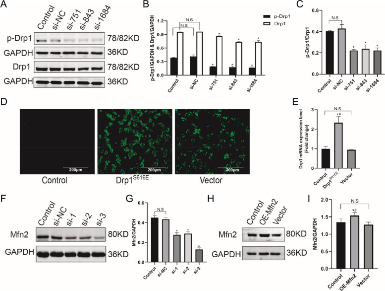
SH-SY5Y cells were pretreated with drugs 24 h before OGD/R. The Drp1 and Mfn2 knockdown were adopted small interfering RNAs. The overexpression of p-Drp1S616 and Mfn2 were used recombinant plasmids. The expression levels of mitochondrial dynamics proteins (p-Drp1, Drp1, Mfn2, Mfn1 and Opa1) and autophagy markers (LC3, Beclin1 and p62) were measured with the Western blotting. The mRNA levels after transfection were determined by PCR. Cell injury and viability were evaluated with released LDH activity and CCK8 assay kits. Mitochondria morphology and autophagosome were observed under transmission electron microscopy. Mitochondrial function was detected by the mitochondrial permeability transition pore assay kit. The co-expression of p-ERK, p-Drp1 and LC3 was assessed with multiple immunofluorescences. One-way analysis of variance followed by least significance difference post hoc analysis (for equal homogeneity) or Dunnett's T3 test (for unequal homogeneity) were used for statistical tests.

RESULTS

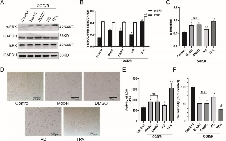
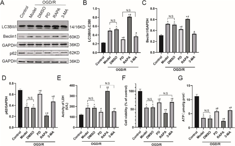
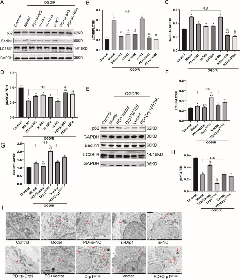
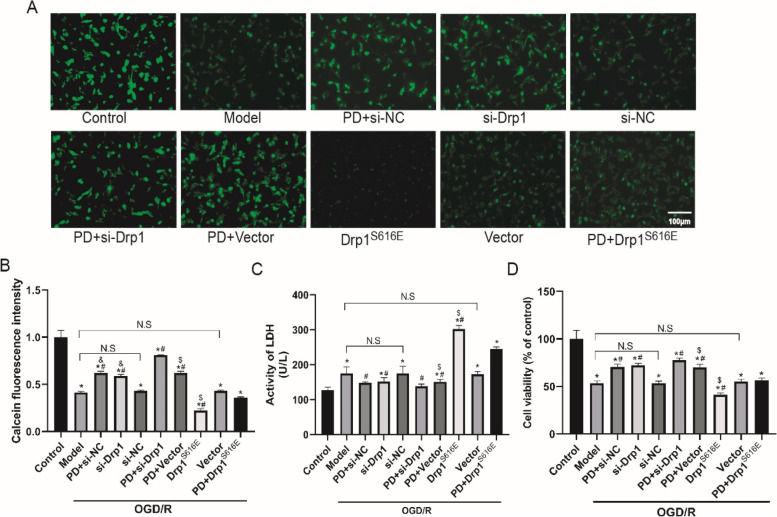
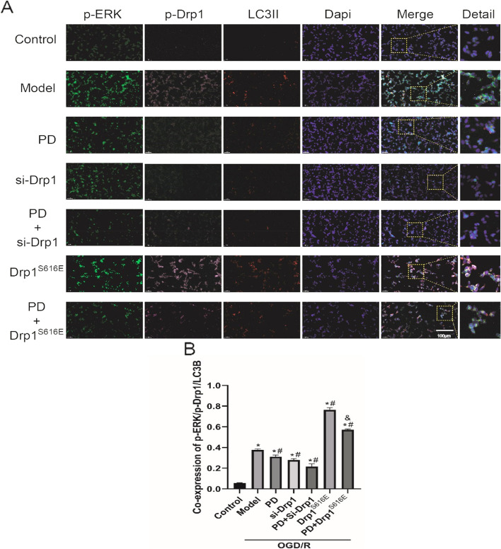
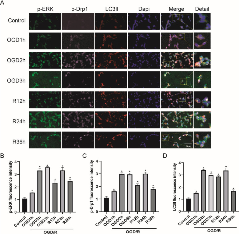
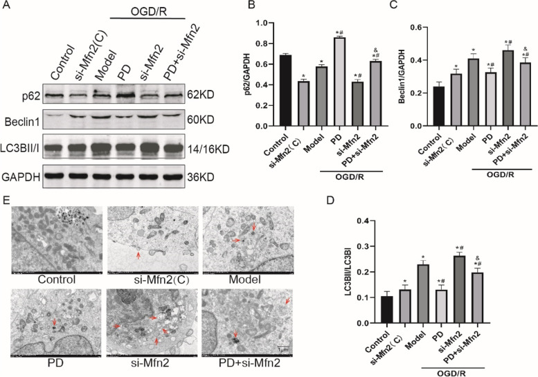
ERK inhibitor-PD98059 (PD) protects SH-SY5Y cells from OGD/R-induced injury; while ERK activator-TPA had the opposite effect. Similar to autophagy inhibitor 3-MA, PD downregulated autophagy to improve cell viability; while autophagy activator-rapamycin further aggravated cell death. PD and Drp1-knockdown synergistically attenuated OGD/R-induced Drp1 activation, mPTP opening and cell injury; overexpression of Drp1 or ablating Mfn2 partly abolished the protective effects of PD. Multiple immunofluorescences showed that p-ERK, p-Drp1 and LC3 were co-expressed.

CONCLUSION

Inhibition of ERK downregulates autophagy via reducing Drp1/Mfn2-dependent mitochondrial fragmentation to antagonize mitochondrial dysfunction and promotes cell survival in the SH-SY5Y cells OGD/R model. Video Abstract.

摘要

背景

脑缺血再灌注损伤(CIRI)是导致心搏骤停/心肺复苏(CA/CPR)患者高死亡率和神经功能障碍的主要原因。我们之前的研究发现细胞外信号调节激酶(ERK)的激活、动力相关蛋白 1(Drp1)/线粒体融合蛋白 2(Mfn2)依赖性线粒体动力学失衡以及过度自噬参与了 CA/CPR 后神经损伤的机制。然而,具体的病理信号通路仍不清楚。本研究旨在探讨 ERK-Drp1/Mfn2-自噬信号通路在 SH-SY5Y 细胞氧葡萄糖剥夺/复氧(OGD/R)模型中的分子功能变化,进一步阐明 CIRI 的病理生理机制,并为 CIRI 后脑保护提供新策略。

方法

SH-SY5Y 细胞在 OGD/R 前 24 小时用药物预处理。采用小干扰 RNA 转染沉默 Drp1 和 Mfn2。使用重组质粒过表达 p-Drp1S616 和 Mfn2。用 Western blot 检测线粒体动力学蛋白(p-Drp1、Drp1、Mfn2、Mfn1 和 Opa1)和自噬标志物(LC3、Beclin1 和 p62)的表达水平。转染后的 mRNA 水平通过 PCR 测定。通过释放 LDH 活性和 CCK8 试剂盒评估细胞损伤和活力。用透射电子显微镜观察线粒体形态和自噬体。用线粒体通透性转换孔测定试剂盒检测线粒体功能。用多重免疫荧光法评估 p-ERK、p-Drp1 和 LC3 的共表达。采用单因素方差分析,随后进行最小显著差异事后检验(对于同质性)或 Dunnett's T3 检验(对于非同质性)进行统计检验。

结果

ERK 抑制剂-PD98059(PD)可保护 SH-SY5Y 细胞免受 OGD/R 诱导的损伤;而 ERK 激活剂-TPA 则有相反的效果。类似于自噬抑制剂 3-MA,PD 通过下调自噬来提高细胞活力;而自噬激活剂-雷帕霉素则进一步加重细胞死亡。PD 和 Drp1 敲低协同减弱 OGD/R 诱导的 Drp1 激活、mPTP 开放和细胞损伤;过表达 Drp1 或破坏 Mfn2 部分消除了 PD 的保护作用。多重免疫荧光显示 p-ERK、p-Drp1 和 LC3 共表达。

结论

ERK 的抑制通过减少 Drp1/Mfn2 依赖性线粒体片段化来下调自噬,从而拮抗线粒体功能障碍,促进 SH-SY5Y 细胞 OGD/R 模型中的细胞存活。

https://cdn.ncbi.nlm.nih.gov/pmc/blobs/2ef6/10426156/c0c85f7b2be4/12964_2023_1211_Fig12_HTML.jpg
https://cdn.ncbi.nlm.nih.gov/pmc/blobs/2ef6/10426156/95f87db0f248/12964_2023_1211_Fig1_HTML.jpg
https://cdn.ncbi.nlm.nih.gov/pmc/blobs/2ef6/10426156/f42936904e76/12964_2023_1211_Fig2_HTML.jpg
https://cdn.ncbi.nlm.nih.gov/pmc/blobs/2ef6/10426156/a0a317401d2d/12964_2023_1211_Fig3_HTML.jpg
https://cdn.ncbi.nlm.nih.gov/pmc/blobs/2ef6/10426156/5696b5114cd3/12964_2023_1211_Fig4_HTML.jpg
https://cdn.ncbi.nlm.nih.gov/pmc/blobs/2ef6/10426156/6ef7208ab906/12964_2023_1211_Fig5_HTML.jpg
https://cdn.ncbi.nlm.nih.gov/pmc/blobs/2ef6/10426156/0db5903f32ae/12964_2023_1211_Fig6_HTML.jpg
https://cdn.ncbi.nlm.nih.gov/pmc/blobs/2ef6/10426156/dbeed24475d5/12964_2023_1211_Fig7_HTML.jpg
https://cdn.ncbi.nlm.nih.gov/pmc/blobs/2ef6/10426156/e567d8152eb7/12964_2023_1211_Fig8_HTML.jpg
https://cdn.ncbi.nlm.nih.gov/pmc/blobs/2ef6/10426156/0b559054e6ac/12964_2023_1211_Fig9_HTML.jpg
https://cdn.ncbi.nlm.nih.gov/pmc/blobs/2ef6/10426156/a8cb6519e20e/12964_2023_1211_Fig10_HTML.jpg
https://cdn.ncbi.nlm.nih.gov/pmc/blobs/2ef6/10426156/4be859d0e61f/12964_2023_1211_Fig11_HTML.jpg
https://cdn.ncbi.nlm.nih.gov/pmc/blobs/2ef6/10426156/c0c85f7b2be4/12964_2023_1211_Fig12_HTML.jpg

相似文献

1
Inhibition of ERK downregulates autophagy via mitigating mitochondrial fragmentation to protect SH-SY5Y cells from OGD/R injury.抑制 ERK 通过减轻线粒体碎片化来下调自噬,从而保护 SH-SY5Y 细胞免受 OGD/R 损伤。
Cell Commun Signal. 2023 Aug 14;21(1):204. doi: 10.1186/s12964-023-01211-3.
2
Astragaloside IV combined with ligustrazine ameliorates abnormal mitochondrial dynamics via Drp1 SUMO/deSUMOylation in cerebral ischemia-reperfusion injury.黄芪甲苷联合川芎嗪通过Drp1的SUMO化/去SUMO化改善脑缺血再灌注损伤中的线粒体动力学异常。
CNS Neurosci Ther. 2024 Apr;30(4):e14725. doi: 10.1111/cns.14725.
3
Activated Drp1 regulates p62-mediated autophagic flux and aggravates inflammation in cerebral ischemia-reperfusion via the ROS-RIP1/RIP3-exosome axis.激活的 Drp1 通过 ROS-RIP1/RIP3-外泌体轴调节 p62 介导的自噬流,并加重脑缺血再灌注中的炎症反应。
Mil Med Res. 2022 May 27;9(1):25. doi: 10.1186/s40779-022-00383-2.
4
Tissue kallikrein protects SH-SY5Y neuronal cells against oxygen and glucose deprivation-induced injury through bradykinin B2 receptor-dependent regulation of autophagy induction.组织激肽释放酶通过缓激肽B2受体依赖性自噬诱导调节,保护SH-SY5Y神经细胞免受氧糖剥夺诱导的损伤。
J Neurochem. 2016 Oct;139(2):208-220. doi: 10.1111/jnc.13690. Epub 2016 Sep 15.
5
Picroside II Protects SH-SY5Y Cells From Autophagy and Apoptosis Following Oxygen Glucose Deprivation/Reoxygen Injury by Inhibiting JNK Signal Pathway.毛兰素 II 通过抑制 JNK 信号通路来保护 SH-SY5Y 细胞免受氧葡萄糖剥夺/再氧合损伤后的自噬和凋亡。
Anat Rec (Hoboken). 2019 Dec;302(12):2245-2254. doi: 10.1002/ar.24214. Epub 2019 Jul 11.
6
Artemisinin Alleviates Cerebral Ischemia/Reperfusion-Induced Oxidative Damage via Regulating PHB2-Mediated Autophagy in the Human Neuroblastoma SH-SY5Y Cell Line.青蒿素通过调节 PHB2 介导线粒体自噬减轻人神经母细胞瘤 SH-SY5Y 细胞脑缺血/再灌注诱导的氧化损伤。
Oxid Med Cell Longev. 2022 Dec 15;2022:6568748. doi: 10.1155/2022/6568748. eCollection 2022.
7
[Mechanism study of 6-shogaol alleviating cerebral ischemia/reperfusion injury by regulating microRNA-26a-5p/death-associated protein kinase 1].6-姜辣素通过调控微小RNA-26a-5p/死亡相关蛋白激酶1减轻脑缺血/再灌注损伤的机制研究
Zhonghua Wei Zhong Bing Ji Jiu Yi Xue. 2024 Jun;36(6):616-623. doi: 10.3760/cma.j.cn121430-20240111-00031.
8
PD98059 protects the brain against mitochondrial-mediated apoptosis and autophagy in a cardiac arrest rat model.PD98059 可保护心脏骤停大鼠模型的大脑免受线粒体介导的细胞凋亡和自噬。
Life Sci. 2019 Sep 1;232:116618. doi: 10.1016/j.lfs.2019.116618. Epub 2019 Jun 29.
9
CDK5 inhibition protects against OGDR induced mitochondrial fragmentation and apoptosis through regulation of Drp1S616 phosphorylation.CDK5 抑制通过调控 Drp1S616 磷酸化防止 OGDR 诱导的线粒体碎片化和细胞凋亡。
Life Sci. 2021 Mar 15;269:119062. doi: 10.1016/j.lfs.2021.119062. Epub 2021 Jan 18.
10
ZFP36 protects against oxygen-glucose deprivation/reoxygenation-induced mitochondrial fragmentation and neuronal apoptosis through inhibiting NOX4-DRP1 pathway.ZFP36 通过抑制 NOX4-DRP1 通路来防止氧葡萄糖剥夺/复氧诱导的线粒体片段化和神经元凋亡。
Brain Res Bull. 2022 Feb;179:57-67. doi: 10.1016/j.brainresbull.2021.12.003. Epub 2021 Dec 9.

引用本文的文献

1
Oxidative Stress, Energy Metabolism Disorder, Mitochondrial Damage, and miR-144 Participated in Molecular Mechanisms of 4-Octylphenol-Caused Cardiac Autophagic Damage in Common Carps ( L.).氧化应激、能量代谢紊乱、线粒体损伤以及miR-144参与了4-辛基苯酚所致鲤鱼心脏自噬损伤的分子机制。
Metabolites. 2025 Jun 11;15(6):391. doi: 10.3390/metabo15060391.
2
Puerarin relives inflammation, bone destruction and facilitates osteogenic differentiation in periodontitis by enhancing mitochondrial autophagy via activating mitochondrial Mitofusin 2.葛根素通过激活线粒体融合蛋白2增强线粒体自噬,从而减轻牙周炎中的炎症和骨破坏,并促进成骨分化。
Stem Cell Res Ther. 2025 May 1;16(1):218. doi: 10.1186/s13287-025-04355-w.
3

本文引用的文献

1
ProNGF/NGF Modulates Autophagy and Apoptosis through PI3K/Akt/mTOR and ERK Signaling Pathways following Cerebral Ischemia-Reperfusion in Rats.脑缺血再灌注后,原肌球蛋白转化生长因子β/神经生长因子通过PI3K/Akt/mTOR和ERK信号通路调节大鼠自噬和凋亡。
Oxid Med Cell Longev. 2022 Mar 29;2022:6098191. doi: 10.1155/2022/6098191. eCollection 2022.
2
Alternative autophagy: mechanisms and roles in different diseases.另类自噬:在不同疾病中的机制和作用。
Cell Commun Signal. 2022 Mar 31;20(1):43. doi: 10.1186/s12964-022-00851-1.
3
SIRT2 regulates mitochondrial dynamics and reprogramming via MEK1-ERK-DRP1 and AKT1-DRP1 axes.
SIRT1 Alleviates Mitochondrial Fission and Necroptosis in Cerebral Ischemia/Reperfusion Injury via SIRT1-RIP1 Signaling Pathway.
SIRT1通过SIRT1-RIP1信号通路减轻脑缺血/再灌注损伤中的线粒体分裂和坏死性凋亡。
MedComm (2020). 2025 Feb 24;6(3):e70118. doi: 10.1002/mco2.70118. eCollection 2025 Mar.
4
Protective Role of Oxycodone in Myocardial Oxidative Stress and Mitochondrial Dysfunction Induced by Ischemia-Reperfusion.羟考酮对缺血再灌注诱导的心肌氧化应激和线粒体功能障碍的保护作用
J Biochem Mol Toxicol. 2025 Feb;39(2):e70151. doi: 10.1002/jbt.70151.
5
Calycosin‑7‑O‑β‑D‑glucoside downregulates mitophagy by mitigating mitochondrial fission to protect HT22 cells from oxygen‑glucose deprivation/reperfusion‑induced injury.毛蕊异黄酮-7-O-β-D-葡萄糖苷通过减轻线粒体分裂来下调线粒体自噬,从而保护HT22细胞免受氧糖剥夺/再灌注诱导的损伤。
Mol Med Rep. 2025 Mar;31(3). doi: 10.3892/mmr.2025.13436. Epub 2025 Jan 17.
6
NSE and S100β as serum alarmins in predicting neurological outcomes after cardiac arrest.神经元特异性烯醇化酶和 S100β 作为血清预警因子预测心搏骤停后神经功能结局。
Sci Rep. 2024 Oct 26;14(1):25539. doi: 10.1038/s41598-024-76979-6.
7
Nasal Administration of bFGF-Loaded Nanoliposomes Attenuates Neuronal Injury and Cognitive Deficits in Mice with Vascular Dementia Induced by Repeated Cerebral Ischemia‒Reperfusion.鼻腔给予 bFGF 载药纳米脂质体减轻反复脑缺血再灌注致血管性痴呆小鼠的神经元损伤和认知功能障碍
Int J Nanomedicine. 2024 Feb 12;19:1431-1450. doi: 10.2147/IJN.S452045. eCollection 2024.
SIRT2 通过 MEK1-ERK-DRP1 和 AKT1-DRP1 轴调节线粒体动力学和重编程。
Cell Rep. 2021 Dec 28;37(13):110155. doi: 10.1016/j.celrep.2021.110155.
4
Platonin protects against cerebral ischemia/reperfusion injury in rats by inhibiting NLRP3 inflammasomes via BNIP3/LC3 signaling mediated autophagy.褪黑素通过 BNIP3/LC3 信号介导的自噬抑制 NLRP3 炎性小体来保护大鼠免受脑缺血/再灌注损伤。
Brain Res Bull. 2022 Mar;180:12-23. doi: 10.1016/j.brainresbull.2021.12.008. Epub 2021 Dec 22.
5
ZFP36 protects against oxygen-glucose deprivation/reoxygenation-induced mitochondrial fragmentation and neuronal apoptosis through inhibiting NOX4-DRP1 pathway.ZFP36 通过抑制 NOX4-DRP1 通路来防止氧葡萄糖剥夺/复氧诱导的线粒体片段化和神经元凋亡。
Brain Res Bull. 2022 Feb;179:57-67. doi: 10.1016/j.brainresbull.2021.12.003. Epub 2021 Dec 9.
6
Autophagy and apoptosis cascade: which is more prominent in neuronal death?自噬和细胞凋亡级联:哪种在神经元死亡中更为突出?
Cell Mol Life Sci. 2021 Dec;78(24):8001-8047. doi: 10.1007/s00018-021-04004-4. Epub 2021 Nov 6.
7
The Alterations in Mitochondrial Dynamics Following Cerebral Ischemia/Reperfusion Injury.脑缺血/再灌注损伤后线粒体动力学的改变
Antioxidants (Basel). 2021 Aug 30;10(9):1384. doi: 10.3390/antiox10091384.
8
Long non-coding RNA MEG3 regulates autophagy after cerebral ischemia/reperfusion injury.长链非编码RNA MEG3在脑缺血/再灌注损伤后调节自噬。
Neural Regen Res. 2022 Apr;17(4):824-831. doi: 10.4103/1673-5374.322466.
9
Autophagy protects against cerebral ischemic reperfusion injury by inhibiting neuroinflammation.自噬通过抑制神经炎症来保护免受脑缺血再灌注损伤。
Am J Transl Res. 2021 May 15;13(5):4726-4737. eCollection 2021.
10
Collective ERK/Akt activity waves orchestrate epithelial homeostasis by driving apoptosis-induced survival.集体 ERK/Akt 活性波通过驱动凋亡诱导的存活来协调上皮细胞稳态。
Dev Cell. 2021 Jun 21;56(12):1712-1726.e6. doi: 10.1016/j.devcel.2021.05.007. Epub 2021 Jun 2.